. 2024 Jan 8;18(1):wrae139.
doi: 10.1093/ismejo/wrae139.

Chlamydiae as symbionts of photosynthetic dinoflagellates

Affiliations

Chlamydiae as symbionts of photosynthetic dinoflagellates

Justin Maire et al. ISME J. .

Abstract

Chlamydiae are ubiquitous intracellular bacteria and infect a wide diversity of eukaryotes, including mammals. However, chlamydiae have never been reported to infect photosynthetic organisms. Here, we describe a novel chlamydial genus and species, Candidatus Algichlamydia australiensis, capable of infecting the photosynthetic dinoflagellate Cladocopium sp. (originally isolated from a scleractinian coral). Algichlamydia australiensis was confirmed to be intracellular by fluorescence in situ hybridization and confocal laser scanning microscopy and temporally stable at the population level by monitoring its relative abundance across four weeks of host growth. Using a combination of short- and long-read sequencing, we recovered a high-quality (completeness 91.73% and contamination 0.27%) metagenome-assembled genome of A. australiensis. Phylogenetic analyses show that this chlamydial taxon represents a new genus and species within the Simkaniaceae family. Algichlamydia australiensis possesses all the hallmark genes for chlamydiae-host interactions, including a complete type III secretion system. In addition, a type IV secretion system is encoded on a plasmid and has previously been observed for only three other chlamydial species. Twenty orthologous groups of genes are unique to A. australiensis, one of which is structurally similar to a protein known from Cyanobacteria and Archaeplastida involved in thylakoid biogenesis and maintenance, hinting at potential chlamydiae interactions with the chloroplasts of Cladocopium cells. Our study shows that chlamydiae infect dinoflagellate symbionts of cnidarians, the first photosynthetic organism reported to harbor chlamydiae, thereby expanding the breadth of chlamydial hosts and providing a new contribution to the discussion around the role of chlamydiae in the establishment of the primary plastid.

Keywords: Symbiodiniaceae; alga; chlamydiae; coral; genomics.


Conflict of interest statement

None declared.

Figures
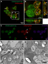
Figure 1
Figure 1
Chlamydiae infect Cladocopium sp. SCF049.01. (A–C) Chlamydiae located by FISH inside (white arrows) and on the cell wall (white arrowheads) of Cladocopium cells, observed by CLSM. Red: Chls523 probe (chlamydiae); green: Cladocopium autofluorescence; purple: nonEUB probe (antisense probe). (B) shows orthogonal projections of a Z-stack of four Cladocopium cells from (A), highlighting the presence of both intracellular and extracellular FISH signal. (D, E) Transmission electron micrographs showing chlamydial cells (chl) surrounded by inclusion membranes (inc) inside a Symbiodiniaceae cell. An overview image of the host cell containing the chlamydial cell in (E) is available as Fig. S4. Abbreviations: chl: chlamydial cell, inc: inclusion, nuc: nucleus, chr: chromosome, cp: chloroplast, st: starch granule. Scale bar: 500 nm.
Figure 2
Figure 2
Chlamydial infection of Cladocopium sp. is temporally stable. (A, B) Chlamydial abundance in culture supernatant (A) or cellular fraction (B), measured by digital PCR. For the cellular fraction (B), chlamydial gene copy data were normalized to either total Cladocopium cell numbers (i.e. every Cladocopium cell is infected) or 30% of Cladocopium cell numbers [i.e. only 30% of cells harbor chlamydiae; see (C)]. Boxes represent first quartile to third quartile for six independent replicates (also shown individually), the middle lines represent the medians, and the whiskers represent the minimum and maximum values. (C) Proportion of Cladocopium sp. cells stained by FISH with the chlamydiae-specific probe Chls523, as analyzed by flow cytometry. Each point represents one of three replicate flasks, with lines connecting the means. Gray-shaded areas represent dark time, while white areas represent light time. The effect of sampling day is indicated under the plot with its corresponding color, based on a Kruskal–Wallis test (ns: P > .05). (D, E) Relative abundance of chlamydial ASVs in the Cladocopium culture across 29 days (D, growth phase experiment) or 24 h (E, time series experiment, performed on Day 8 of the growth phase experiment—see black arrow in D), determined by 16S rRNA gene amplicon sequencing (see Fig. S2 for experimental design). Bacterial community profiling was performed in three fractions as previously described [7]: loosely associated bacteria (planktonic bacteria; blue), closely associated (intracellular and tightly attached to the cell wall; green) and intracellular bacteria (orange). Cladocopium cell counts across the growth phase experiments are also provided (dashed black line, D). Each point represents one of three replicate flasks, with lines connecting the means. For each fraction, the effect of sampling day or time on chlamydial relative abundance is indicated under the plot with its corresponding color, based on a Kruskal–Wallis test. ns: P > .05; *: P ≤ .05; **: P ≤ .01. Single replicate values are available in Table S5.
Figure 3
Figure 3
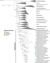
The chlamydial symbiont Candidatus Algichlamydia australiensis (Cla049 MAG) belongs to an undescribed, deep-branching Simkaniaceae genus. Chlamydial maximum likelihood phylogeny based on 15 conserved gene markers (Table S4) in 169 chlamydial genomes (Table S3). Confidence values based on 1000 ultrafast bootstrap replicates and 1000 replicates of the SH-like approximate likelihood ratio test are provided. Scale bar represents 1 nucleotide substitution per site. This tree was calculated using IQ-TREE 2 [51] with ModelFinder Plus under the LG + C20 + R4 model [52]; an additional tree calculated under the posterior mean site frequency (PMSF) model [55] using this tree as seed confirmed the phylogeny and is available in Fig. S6. MCF: metagenomic chlamydial family; CC-III: chlamydiae clade III.
Figure 4
Figure 4
Completeness, similarity, and synteny of the genomic regions encoding the T3SS (A, sct genes) and T4SS (B, tra genes) in Algichlamydia australiensis Cla049, compared with Simkania negevensis Z. Synteny is shown by lines joining the two genomes; protein identity is color-coded.

Similar articles

References

    1. Horn M. Chlamydiae as symbionts in eukaryotes. Ann Rev Microbiol 2008;62:113–31. 10.1146/annurev.micro.62.081307.162818 - DOI - PubMed
    1. Collingro A, Köstlbacher S, Horn M. Chlamydiae in the environment. Trends Microbiol 2020;28:877–88. 10.1016/j.tim.2020.05.020 - DOI - PubMed
    1. Dharamshi JE, Köstlbacher S, Schön MEet al. . Gene gain facilitated endosymbiotic evolution of Chlamydiae. Nat Microbiol 2023;8:40–54. 10.1038/s41564-022-01284-9 - DOI - PMC - PubMed
    1. Huang J, Gogarten JP. Did an ancient chlamydial endosymbiosis facilitate the establishment of primary plastids? Genome Biol 2007;8:R99. 10.1186/gb-2007-8-6-r99 - DOI - PMC - PubMed
    1. Cenci U, Bhattacharya D, Weber APMet al. . Biotic host–pathogen interactions As Major drivers of plastid endosymbiosis. Trends Plant Sci 2017;22:316–28. 10.1016/j.tplants.2016.12.007 - DOI - PubMed

LinkOut - more resources