. 2024 Jul 18;15(1):6068.
doi: 10.1038/s41467-024-50462-2.

Versatile nanobody-based approach to image, track and reconstitute functional Neurexin-1 in vivo

Affiliations

Versatile nanobody-based approach to image, track and reconstitute functional Neurexin-1 in vivo

Rosario Vicidomini et al. Nat Commun. .

Abstract

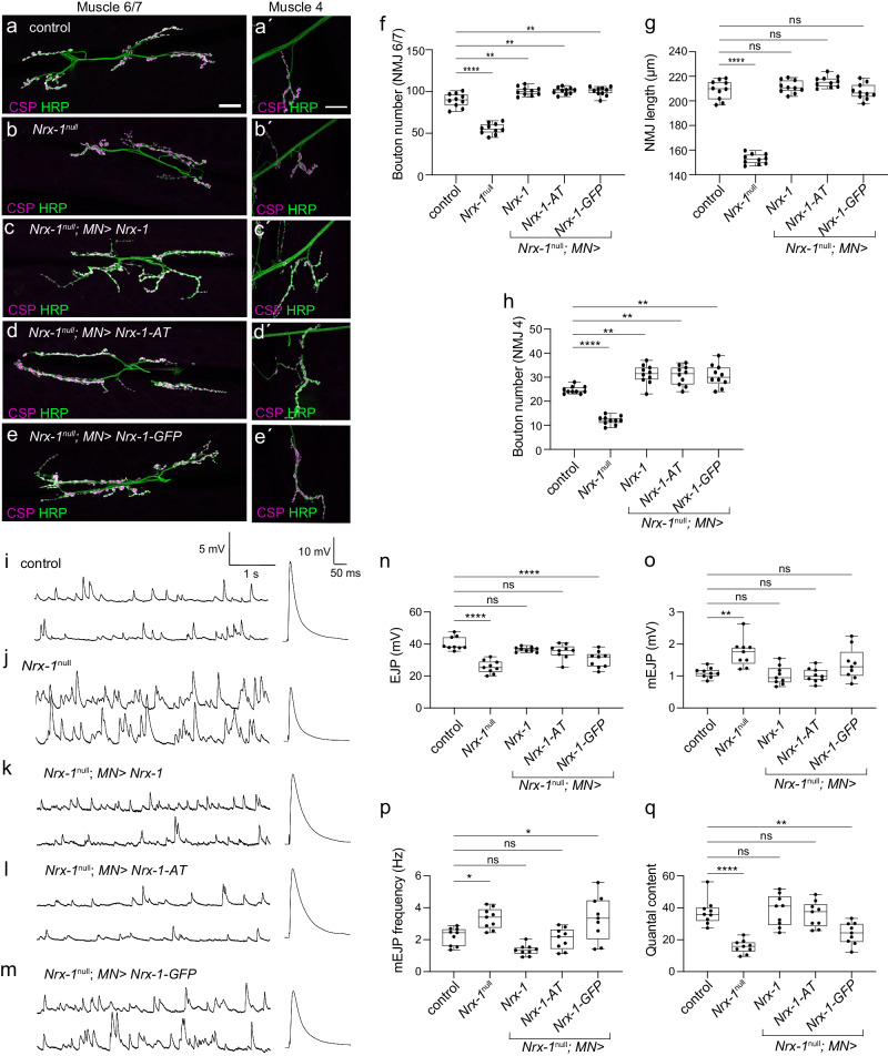
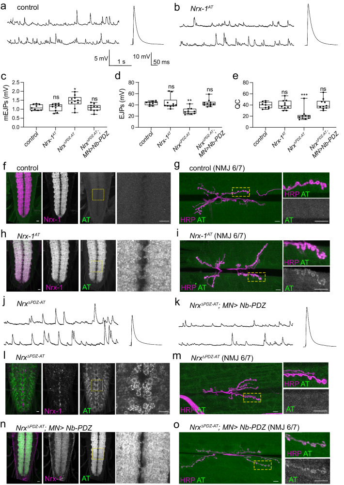
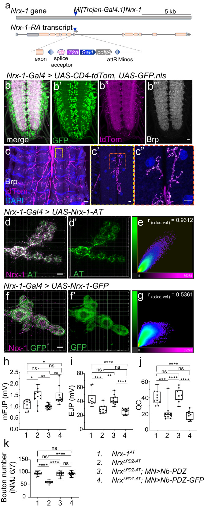
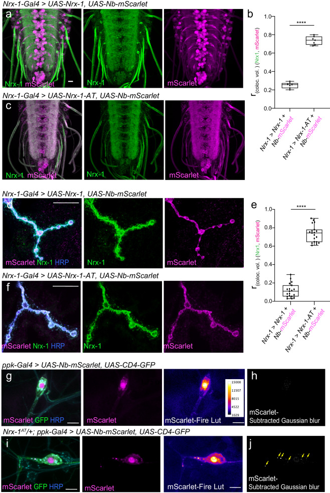
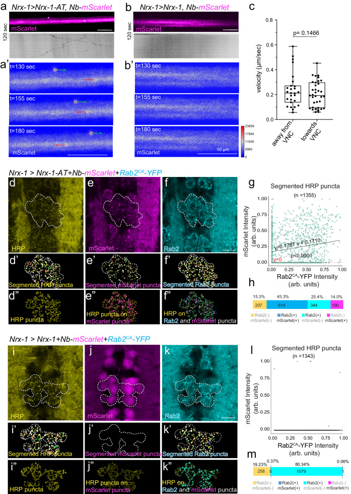
Neurexins are key adhesion proteins that coordinate extracellular and intracellular synaptic components. Nonetheless, the low abundance of these multidomain proteins has complicated any localization and structure-function studies. Here we combine an ALFA tag (AT)/nanobody (NbALFA) tool with classic genetics, cell biology and electrophysiology to examine the distribution and function of the Drosophila Nrx-1 in vivo. We generate full-length and ΔPDZ ALFA-tagged Nrx-1 variants and find that the PDZ binding motif is key to Nrx-1 surface expression. A PDZ binding motif provided in trans, via genetically encoded cytosolic NbALFA-PDZ chimera, fully restores the synaptic localization and function of NrxΔPDZ-AT. Using cytosolic NbALFA-mScarlet intrabody, we achieve compartment-specific detection of endogenous Nrx-1, track live Nrx-1 transport along the motor neuron axons, and demonstrate that Nrx-1 co-migrates with Rab2-positive vesicles. Our findings illustrate the versatility of the ALFA system and pave the way towards dissecting functional domains of complex proteins in vivo.


Conflict of interest statement

F.O. is an inventor on a pending patent by NanoTag Biotechnologies GmbH that covers the ALFA system and its use (application numbers: WO2020053239A1; US20220048947A1; EP3849996A1; CN113195516A; JP2022500076A). F.O. is a shareholder of NanoTag Biotechnologies GmbH. The remaining authors declare no competing interests.

Figures

Fig. 1
Fig. 1. ALFA tag site selection and in vivo detection.
a Domain organization of the Drosophila Nrx-1. The transgenes utilized in this study encode the predominant isoform (Nrx-1-RA) and the corresponding ALFA-tagged and GFP-tagged variants. b Alignment of Nrx-1 intracellular domains from various insect species indicates several blocks of conservation, including the C-terminal PDZ binding motif and the Diptera-specific G-rich expansions. The ALFA-tag insertion site is marked, together with other interaction domains of Drosophila Nrx-1: The Ephrin binding domain (residues 1760–1813) and the NSF binding site (residues 1788–1813). (Dm- Drosophila melanogaster, Md- Musca domestica, Cc- Ceratitis capita, Rc- Rhagoletis cerasi, Tc- Tribolium castaneum, Bm- Bombyx mori, Dp- Danaus Plexippus, Am- Apis mellifera, Hs- Harpegnathos saltator). cj Representative confocal images of third instar larvae VNC (c, d, e, g and i) and NMJs (f, h, and j) of the indicated genotypes labeled for Nrx-1 (magenta), GFP (green), and ALFA tag (cyan) showing the distribution of endogenous Nrx-1 and neuronally expressed Nrx-1 variants. These experiments were repeated three times with similar results. When overexpressed in the motor neurons, Nrx-1 decorates the presynaptic membrane and accumulates in distinct puncta at synaptic terminals. These puncta coincide with the ALFA-positive signals. In contrast, the GFP-positive signals are more diffuse and separated from the Nrx-1-positive puncta. The GFP and ALFA tag signals/channels are shown for all genotypes to illustrate the background signal. Scale bars: 10 µm. Genotypes: control (w1118), MN > UAS-Nrx-1 tag/no tag (BG380-Gal4/+; UAS-Nrx-1 tag/no tag/+), Nrx-1null (Nrx273/Df).
Fig. 2
Fig. 2. Neuronal expression of untagged and ALFA-tagged transgenes, but not Nrx-1-GFP, rescue the functional deficits at Nrx-1null NMJs.
ae Representative confocal images of NMJ 6/7 and NMJ4 from third instar larvae of the indicated genotypes stained for Horseradish peroxidase, HRP (green), which labels neuronal surface, and Cysteine-string protein, CSP (magenta), which labels presynaptic vesicles. These experiments were repeated three times with similar results. Scale bars, 20 µm. fh Quantification of bouton number (f and h) and NMJ length (g) in the indicated genotypes; n = 10 for each genotype. im Representative traces of miniature junctional potentials (mEJPs) and evoked junctional potentials (EJPs) in control, Nrx-1null mutant, and neuronally rescued larvae as indicated. EJPs are reduced at Nrx-1null mutant NMJs and are restored upon neuronal expression of Nrx-1 or Nrx-1-AT but not GFP-tagged transgenes. nq Quantification of mEJPs amplitude, mEJPs frequency, EJPs amplitude, and Quantal Content of the indicated genotypes; n = 9 for each genotype. Data are represented as mean ± SEM (one-way ANOVA with Tukey’s multiple comparisons); ****p < 0.0001, **p < 0.005, ns, p > 0.05. The boxes expand from the first to the third quartile, and the whiskers from minimum to maximum values; the center lines mark the mean values. Source data are provided as a Source Data file. Genotypes: control (w1118), Nrx-1null (Nrx273/Df), Nrx-1null; MN> Nrx-1tag/no tag (BG380-Gal4/+; UAS-Nrx-1tag/no tag/+; Nrx273/Df).
Fig. 3
Fig. 3. Detection and in vivo reconstitution of endogenously edited ALFA-tagged Nrx-1.
a, b Representative traces of mEJPs and EJPs in control and endogenously tagged Nrx-1AT third instar larvae. ce Quantification of mEJP amplitude, EJP amplitude, and Quantal Content in the indicated genotypes; n = 9 for the control and 10 for all the other genotypes. f, i Representative confocal images of VNCs or muscle 6/7 NMJs of third instar larvae of the indicated genotypes labeled for ALFA tag (green) and Nrx-1 or HRP (magenta). The images were acquired with the same confocal microscopy settings and scaled equally for direct visual comparison. These experiments were repeated three times with similar results; n = 15 for (fj) and 5 for (i). The edited Nrx-1AT shows the expected Nrx-1 accumulation in neurites. j, k Representative traces of mEJPs and EJPs recorded in NrxΔPDZ-AT third instar larvae alone (j) or in the presence of neuronally expressed Nb-PDZ chimera (k). Deletion of the PDZ binding motif of Nrx-1 causes loss-of-function phenotypes that resemble the Nrx-1null mutant deficits. These phenotypes are completely rescued by neuronal expression of the Nb-PDZ chimera. lo Representative confocal images of VNCs or muscle 6/7 NMJs of third instar larvae of the indicated genotypes labeled for ALFA tag (green) and Nrx-1 or HRP (magenta). These experiments were repeated three times with similar results; n = 15 for (ln) and 5 for (o). The absence of a PDZ binding motif shifts the distribution of NrxΔPDZ-AT from the neurites to the motor neuron soma, more specifically in the ER. The addition of the PDZ binding motif in trans restores the cell surface location and normal distribution of reconstituted Nrx-1 (NrxΔPDZ-AT/Nb-PDZ) to the neuropil. Scale bars: 10 µm. Data are represented as mean ± SEM (one-way ANOVA with Tukey’s multiple comparisons); ****p < 0.0001, **p < 0.005, ns, p > 0.05. The boxes expand from the first to the third quartile, and the whiskers from minimum to maximum values; the center lines mark the mean values. Source data are provided as a Source Data file. Genotypes: control (w1118), NrxΔPDZ-AT; MN > Nb-PDZ (BG380-Gal4/+; UAS-Nb-PDZ/+; NrxΔPDZ-AT).
Fig. 4
Fig. 4. Interfering with the PDZ binding motif disrupts Nrx-1 subcellular distribution.
a Diagram of Nrx-1-Gal4 organization. A MiMIC transposon, [MI10278], inserted right after exon 9 in the Nrx-1 gene, was converted with a Trojan T2A-Gal4 cassette inserted in the correct orientation and coding frame. The T2A-Gal4 in-frame insertion disrupts the expression of Nrx-1 and produces Gal4 instead. b, c Confocal micrographs of larval ventral cords (b) and synaptic terminals (c) of third instar larvae expressing UAS-CD4-tdTom and UAS-GFP.nls under Nrx-1-Gal4 control. Brp (white) marks the presynaptic active zones. The Nrx-1-Gal4 recapitulates the Nrx-1 endogenous expression pattern. dg Deconvolved STED images of NMJ of third instar larvae expressing Nrx-1-AT (d) or Nrx-1-GFP (f) and stained for Nrx-1 (magenta) and either ALFA tag or GFP (green). Scatter plots of Nrx-1 and ALFA tag (e) or GFP (g) signals. Pearson’s coefficient in the co-localization volume shows a very high correlation for Nrx-1 and ALFA tags, but a low correlation for Nrx-1 and GFP signals. These experiments were repeated three times with similar results. hj Summary bar graphs showing the mean amplitude of mEJPs (h), the mean amplitude of EJPs (i), and the QC (j) for the indicated genotypes; n = 10 for each genotype. k Quantification of bouton number for the indicated genotypes; n = 14 or more for each genotype. Scale bars: 100 µm (c), 10 µm (b”’, c’ and c”), and 2 µm (d and f). Data are represented as mean ± SEM (one-way ANOVA with Tukey’s multiple comparisons); ****p < 0.0001, ***p < 0.001, **p < 0.005, *p < 0.05, ns, p > 0.05. The boxes expand from the first to the third quartile, and the whiskers from minimum to maximum values; the center lines mark the mean values. Source data are provided as a Source Data file.
Fig. 5
Fig. 5. Compartment specific detection using genetically encoded cytosolic Nb-mScarlet.
ae Representative confocal images of VNCs (a, c) and NMJs (d, f) from third instar larvae expressing cytosolic Nb-mScarlet together with either untagged Nrx-1 or Nrx-1-AT, as indicated. All transgenes were expressed under the control of Nrx-1-Gal4, and the specimens were fixed and labeled for Nrx-1 (green) and mScarlet (magenta). These experiments were repeated three times with similar results; n = 7 (ac) or 14 (df) per genotype. Both Nrx-1 and Nrx-1-AT distribute to synaptic terminals. To quantify the co-localization between Nrx-1 and mScarlet channels, Pearson’s coefficient was calculated within an HRP-selected mask (b, e). In the absence of an ALFA tag, Nb-mScarlet does not co-localize with Nrx-1 and instead localizes to the neuron soma (see also Supplementary Fig. 10) and throughout the synaptic boutons. In larvae expressing Nrx-1-AT, Nb-mScarlet mirrors the Nrx-1 distribution. (gj) Representative confocal images of larval sensory neurons from control (g) or Nrx-1-AT/+ heterozygous (i) third instar larvae expressing cytosolic Nb-mScarlet and membrane-bound mCD4-GFP under the control of ppk-Gal4. These experiments were repeated three times with similar results; n = 7 for each genotype. The fillets were fixed and labeled for mScarlet (magenta), GFP (green), and HRP (blue). The mScarlet signals (shown in Fire Lut) were further filtered using Gaussian blur and image subtraction (h, j). Multiple mScarlet-positive vesicles, marked by yellow arrows, are detected in larvae with one copy of Nrx-1-AT but not in control. Additional examples are shown in Supplementary Fig. 14. Scale bars: 10 µm. Data are represented as mean ± SEM (unpaired t test); ****p < 0.0001. The boxes expand from the first to the third quartile, and the whiskers from minimum to maximum values; the center lines mark the mean values. Source data are provided as a Source Data file.
Fig. 6
Fig. 6. Cytosolic Nb-mScarlet captures Nrx-1-AT co-localization with Rab2 vesicles in the soma and trafficking along the motor neuron axons.
a, b Live imaging of Nrx-1-AT trafficking along the motor neuron axons. Representative snapshots and kymographs from time-lapse confocal imaging of axons co-expressing UAS-Nb-mScarlet with either UAS-Nrx-1-AT (a) or UAS-Nrx-1 (b) under the control of Nrx-1-Gal4. No mScarlet-positive puncta/vesicles are detectable in the absence of the ALFA tag. Vesicles move away (green) or towards the VNC (red) with similar mean velocities (c). N = 18 individual larvae were imaged over three different days; moving vesicles were detected and captured in two or three individual bundles per animal. Data are represented as mean ± SEM (unpaired t test). The boxes expand from the first to the third quartile, and the whiskers from minimum to maximum values; the center lines mark the mean values. dh Representative confocal images of VNC from third instar larvae expressing Nrx-1-AT, Nb-mScarlet, and Rab2CA-YFP under the control of Nrx-1-Gal4. The specimens were fixed and labeled for mScarlet (magenta), YFP (cyan), and HRP (yellow). Maximum projections of the z-planes containing the motor neurons soma are shown and the regions of interest comprising several motor neurons are marked. Vesicles identified by segmentation are shown in the lower panels and in the expanded Supplementary Fig. 17. Overlap of HRP-positive puncta on the combined mScarlet- and Rab2-positive puncta (f”) highlights the composition of HRP-positive puncta. g Scatter plot indicating mScarlet and Rab2 intensity in each HRP puncta enabled the analysis of puncta composition (h) 45% of the HRP puncta (614/1355 total) are positive for both mScarlet and Rab2. Linear regression and non-linear fit tests were used. The null hypothesis was tested using an F-test: F = 53.02 (Df = 1, Dfd = 1353), slope (0.1306 − 0.2269), Y-intercept (0.1507 − 0.1913) 95% confidence, p < 0.0001. im Confocal images and puncta analysis in VNCs from third instar larvae expressing untagged Nrx-1, Nb-mScarlet, and Rab2CA-YFP under the control of Nrx-1-Gal4. The analyses followed the workflow described above and revealed only 0.37% overlap among HRP, mScarlet, and Rab2 (5 vesicles /1343 total). F-test: F = 0.501 (Dfn = 1, Dfd = 1341), slope (− 0.006971 − 0.01484), Y-intercept (− 0.006971 − 0.01484) 95% confidence, p = 0.4331. Scale bars: 10 µm. Source data are provided as a Source Data file.

Similar articles

Cited by

References

    1. Gomez AM, Traunmuller L, Scheiffele P. Neurexins: molecular codes for shaping neuronal synapses. Nat. Rev. Neurosci. 2021;22:137–151. doi: 10.1038/s41583-020-00415-7. - DOI - PMC - PubMed
    1. Sudhof TC. Synaptic neurexin complexes: A molecular code for the logic of neural circuits. Cell. 2017;171:745–769. doi: 10.1016/j.cell.2017.10.024. - DOI - PMC - PubMed
    1. Owald D, et al. Cooperation of Syd-1 with Neurexin synchronizes pre- with postsynaptic assembly. Nat. Neurosci. 2012;15:1219–1226. doi: 10.1038/nn.3183. - DOI - PubMed
    1. Kurshan PT, et al. gamma-Neurexin and frizzled mediate parallel synapse assembly pathways antagonized by receptor endocytosis. Neuron. 2018;100:150–166 e154. doi: 10.1016/j.neuron.2018.09.007. - DOI - PMC - PubMed
    1. Biederer T, Sudhof TC. Mints as adaptors. Direct binding to neurexins and recruitment of munc18. J. Biol. Chem. 2000;275:39803–39806. doi: 10.1074/jbc.C000656200. - DOI - PubMed

MeSH terms

Substances

LinkOut - more resources