. 2024 Jul 17;12(1):41.
doi: 10.1038/s41413-024-00344-6.

PDZK1 protects against mechanical overload-induced chondrocyte senescence and osteoarthritis by targeting mitochondrial function

Affiliations

PDZK1 protects against mechanical overload-induced chondrocyte senescence and osteoarthritis by targeting mitochondrial function

Yan Shao et al. Bone Res. .

Abstract

Mechanical overloading and aging are two essential factors for osteoarthritis (OA) development. Mitochondria have been identified as a mechano-transducer situated between extracellular mechanical signals and chondrocyte biology, but their roles and the associated mechanisms in mechanical stress-associated chondrocyte senescence and OA have not been elucidated. Herein, we found that PDZ domain containing 1 (PDZK1), one of the PDZ proteins, which belongs to the Na+/H+ Exchanger (NHE) regulatory factor family, is a key factor in biomechanically induced mitochondrial dysfunction and chondrocyte senescence during OA progression. PDZK1 is reduced by mechanical overload, and is diminished in the articular cartilage of OA patients, aged mice and OA mice. Pdzk1 knockout in chondrocytes exacerbates mechanical overload-induced cartilage degeneration, whereas intraarticular injection of adeno-associated virus-expressing PDZK1 had a therapeutic effect. Moreover, PDZK1 loss impaired chondrocyte mitochondrial function with accumulated damaged mitochondria, decreased mitochondrion DNA (mtDNA) content and increased reactive oxygen species (ROS) production. PDZK1 supplementation or mitoubiquinone (MitoQ) application alleviated chondrocyte senescence and cartilage degeneration and significantly protected chondrocyte mitochondrial functions. MRNA sequencing in articular cartilage from Pdzk1 knockout mice and controls showed that PDZK1 deficiency in chondrocytes interfered with mitochondrial function through inhibiting Hmgcs2 by increasing its ubiquitination. Our results suggested that PDZK1 deficiency plays a crucial role in mediating excessive mechanical load-induced chondrocyte senescence and is associated with mitochondrial dysfunction. PDZK1 overexpression or preservation of mitochondrial functions by MitoQ might present a new therapeutic approach for mechanical overload-induced OA.


Conflict of interest statement

The authors declare no competing interests.

Figures

Fig. 1
Fig. 1
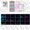
Chondrocyte PDZK1 is a potential candidate during chondrocyte senescence and OA progression responded to mechanical overload. a Quantitative PCR analysis of NHE and NHERF family members in mouse primary chondrocytes treated with 20% elongation strain loading for 24 h. n = 5. b Western blotting analysis and gray value of COL2A1, SOX9, PDZK1, p16ink4a and p21 in primary chondrocyte suffered from 20% elongation strain loading. n = 6 per group. c Representative images of safranin O/fast green staining, IHC staining of PDZK1 and quantification analysis of OARSI scale, PDZK1-positive chondrocytes as a proportion of the total chondrocytes in smooth cartilage and damaged cartilage of OA patients. Scale bars: 100 μm. n = 10 per group. d Representative images of safranin O/fast green staining, IHC staining of PDZK1 and quantitative analysis of OARSI scale, PDZK1-positive chondrocytes as a proportion of the total chondrocytes in articular cartilage of male mice aged 3 and 20 months. Scale bars: 50 μm. n = 10 per group. e Representative images HE staining, safranin O/fast green staining, IHC staining of PDZK1, immunofluorescence of p16ink4a and p21 and quantitative analysis of OARSI scale and positive chondrocytes in articular cartilage of mice treated with multiple loading episodes at peak loads of 4.0, 9.0 and 13.5 N. Scale bars: 50 μm. n = 5 per group Data are shown as mean + SD. ns not significant.*P < 0.05, **P < 0.01. NHE, sodium-hydrogen exchanger. NHERF NHE Regulatory Factor. IHC immunohistochemical, PDZK1 NHE Regulatory Factor 3, DMM destabilization of the medial meniscus, OA osteoarthritis, OARSI Osteoarthritis Research Society International
Fig. 2
Fig. 2
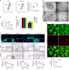
Pdzk1KO mice exhibit aggravated symptoms of mechanical load-induced OA. a Representative images of SA-β-Gal staining, immunofluorescence staining of γH2AX and quantification analysis of positive chondrocytes as a proportion of the total chondrocytes and average number of foci in passage 6 chondrocytes from Pdzk1KO mice and controls. Scale bars: 25 μm (SA-β-Gal) and 5 μm (γH2AX). n = 5 per group. b Representative images of safranin O/fast green staining, immunofluorescence of p16ink4a and p21 and quantification analysis of OARSI scale and positive chondrocytes as a proportion of the total chondrocytes in articular cartilage of Pdzk1KO and Control male mice aged 18 months. Scale bars: 50 μm. n = 5 per group. c Representative scanning electron microscopy micrographs show the damage of cartilage surface. d Representative images of safranin O/fast green staining, IHC staining of PDZK1, MMP13, immunofluorescence of Col2a1, p16ink4a, p21 and quantification analysis of OARSI scale and positive chondrocytes as a proportion of the total chondrocytes in articular cartilage of Pdzk1KO and Controls at 8 weeks after DMM surgery and Sham group. Scale bars: 50 μm. n = 10 per group. Data are shown as mean + SD. ns not significant; *P < 0.05. **P < 0.01. IHC immunohistochemical, DMM destabilization of the medial meniscus, OA osteoarthritis, OARSI Osteoarthritis Research Society International
Fig. 3
Fig. 3
Overexpression of PDZK1 defers chondrocytes senescence and protects against OA development. a Western blotting analysis of COL2A1, MMP13, PDZK1, p16ink4a and p21 in primary chondrocyte with or without adenovirus containing PDZK1 (Ad-Pdzk1) under 20% elongation strain loading for 24 h. b Gray value analysis of western blotting described in a. n = 3 per group. c Representative images of safranin O/fast green staining, HE staining and IHC staining of PDZK1 and quantification analysis of OARSI scale, HC/CC and PDZK1-positive chondrocytes as a proportion of the total chondrocytes in articular cartilage of PDZK1-expressing adeno-associated virus (AAV-Pdzk1) and control male mice at 4 weeks after loading of 13.5 N. Scale bars: 50 μm. n = 6 per group. d Representative images of immunofluorescence of Col2a1, MMP13, p16ink4a, p21, p53, γH2AX and quantitative analysis of positive chondrocytes as a proportion of the total chondrocytes in articular cartilage of AAV-Pdzk1 and control male mice at 4 weeks after loading of 13.5 N. Scale bars: 50 μm. n = 5 per group. ns not significant. *P < 0.05. **P < 0.01. NC negative control, IHC immunohistochemical, OA osteoarthritis, OARSI Osteoarthritis Research Society International
Fig. 4
Fig. 4
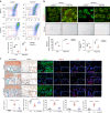
PDZK1 deficiency in chondrocytes inhibits mitochondrial functions. a Mitochondrial membrane potential was analyzed by flow cytometry using JC-1 dye in primary chondrocytes of Pdzk1KO and Control mice. Red box indicates the ratio of damaged mitochondria. n = 5 per group. b Mitochondrial DNA (mtDNA) content in chondrocytes of Pdzk1KO and Control mice was analyzed. Total DNA was extracted and amplified with the primer of mitochondrial cytochrome c oxidase 2 (COX2) and normalized to ribosomal protein s18 (RSP18). n = 6 per group. c Graphical representations of the relative abundance of mitochondrial‐encoded OXPHOS genes are presented. Cytb, ATP6, COX1, COX2 and COX3 mRNA levels in chondrocytes of Pdzk1KO and Control mice were assessed by real‐time PCR. n = 5 per group. d Transmission electron microscopy (TEM) micrographs show mitochondria ultrastructure morphology of primary chondrocytes of Pdzk1KO and Control mice. e Representative fluorescence of MitoTracker green and mitoSOX show mitochondria mass and ROS production of primary chondrocytes of Pdzk1KO and Control mice. Scale bars: 10 μm. Quantification analysis of mitoSOX mean fluorescence. n = 5 per group. f Representative images of immunofluorescence of TOMM20 and IHC of PINK1, PARKIN and quantitative analysis of positive chondrocytes as a proportion of the total chondrocytes in articular cartilage of Pdzk1KO and Controls at 8 weeks after DMM surgery and Sham group. Scale bars: 50 μm. n = 10 per group. ns not significant. *P < 0.05. **P < 0.01. IHC immunohistochemical, OA osteoarthritis
Fig. 5
Fig. 5
Mitoquinone (MitoQ) preserves mitochondrial functions and inhibits chondrocyte senescence induced by loss of PDZK1. a Mitochondrial membrane potential was analyzed by flow cytometry using JC-1 dye in passage 3 chondrocytes of Pdzk1KO and Control mice with or without MitoQ. Red box indicates the ratio of damaged mitochondria. n = 5 per group. b Representative fluorescence of MitoTracker green and mitoSOX and images of SA-β-Gal staining of primary chondrocytes described in a. Scale bar: 10 μm (MtioSOX) and 50 μm (SA-β-Gal).MitoSOX mean fluorescence and SA-β-Gal-positive chondrocytes as a proportion of the total chondrocytes were analyzed. n = 3 per group (MitoSOX) and n = 5 per group (SA-β-Gal). c Representative images of safranin O/fast green staining, immunofluorescence of Col2a1, MMP13, p16ink4a and p21 and quantification analysis of OARSI scale and positive chondrocytes as a proportion of the total chondrocytes in cartilage explants under the application of multiple loading episodes at a peak load of 4 N for 24 h with or without MitoQ. Scale bars: 50 μm. n = 6 per group. *P < 0.05. **P < 0.01
Fig. 6
Fig. 6
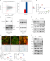
PDZK1 deficiency in chondrocytes interference with mitochondrial function through binding to Hmgcs2. a The mRNA expression profile of articular cartilage from Pdzk1KO mice and their littermate controls was analyzed. b Genes related to mitochondria function in Pdzk1KO mouse cartilage were assessed by real‐time PCR. n = 6 per group. c Hmgcs2 was immunoprecipitated from primary chondrocytes of C57 BL/6 J mice after stimulation with MG-132 and transfection with either Ad-Pdzk1 or si-Pdzk1. Western blotting detected the ubiquitination level of Hmgcs2. d Representative fluorescence of MitoTracker green and mitoSOX and images of SA-β-Gal staining of primary chondrocytes with or without Ad-Pdzk1 and si-Hmgcs2. Scale bar: 10 μm (MtioSOX) and 50 μm SA-β-Gal). MitoSOX mean fluorescence and SA-β-Gal-positive chondrocytes as a proportion of the total chondrocytes were analyzed. n = 5 per group. *P < 0.05. **P < 0.01. e Western blotting analysis of COL2A1, MMP13, p16ink4a and p21 in primary chondrocyte suffered from 20% elongation strain loading with or without BHB. f Western blotting analysis of COL2A1, MMP13, PDZK1, Hmgcs2, p16ink4a and p21 in primary chondrocytes treated with si-Pdzk1 or BHB. IHC immunohistochemical, OA osteoarthritis, DMM destabilization of the medial meniscus, Ad-Pdzk1 adenovirus containing PDZK1, BHB β-hydroxybutyric
Fig. 7
Fig. 7
PDZK1 protects against mechanical overload-induced chondrocyte senescence and osteoarthritis via targeting mitochondrial function

Similar articles

Cited by

References

    1. Hunter DJ, Bierma-Zeinstra S. Osteoarthritis. Lancet. 2019;393:1745–1759. doi: 10.1016/S0140-6736(19)30417-9. - DOI - PubMed
    1. Sharma L. Osteoarthritis of the Knee. N. Engl. J. Med. 2021;384:51–59. doi: 10.1056/NEJMcp1903768. - DOI - PubMed
    1. Glyn-Jones S, et al. Osteoarthritis. Lancet. 2015;386:376–387. doi: 10.1016/S0140-6736(14)60802-3. - DOI - PubMed
    1. Visser AW, et al. The relative contribution of mechanical stress and systemic processes in different types of osteoarthritis: the NEO study. Ann. Rheum. Dis. 2015;74:1842–1847. doi: 10.1136/annrheumdis-2013-205012. - DOI - PubMed
    1. Coryell PR, Diekman BO, Loeser RF. Mechanisms and therapeutic implications of cellular senescence in osteoarthritis. Nat. Rev. Rheumatol. 2021;17:47–57. doi: 10.1038/s41584-020-00533-7. - DOI - PMC - PubMed

LinkOut - more resources