. 2024 Jul 16;7(1):867.
doi: 10.1038/s42003-024-06559-x.

Specific glycine-dependent enzyme motion determines the potency of conformation selective inhibitors of threonyl-tRNA synthetase

Affiliations

Specific glycine-dependent enzyme motion determines the potency of conformation selective inhibitors of threonyl-tRNA synthetase

Hang Qiao et al. Commun Biol. .

Abstract

The function of proteins depends on their correct structure and proper dynamics. Understanding the dynamics of target proteins facilitates drug design and development. However, dynamic information is often hidden in the spatial structure of proteins. It is important but difficult to identify the specific residues that play a decisive role in protein dynamics. Here, we report that a critical glycine residue (Gly463) dominates the motion of threonyl-tRNA synthetase (ThrRS) and the sensitivity of the enzyme to antibiotics. Obafluorin (OB), a natural antibiotic, is a novel covalent inhibitor of ThrRS. The binding of OB induces a large conformational change in ThrRS. Through five crystal structures, biochemical and biophysical analyses, and computational simulations, we found that Gly463 plays an important role in the dynamics of ThrRS. Mutating this flexible residue into more rigid residues did not damage the enzyme's three-dimensional structure but significantly improved the thermal stability of the enzyme and suppressed its ability to change conformation. These mutations cause resistance of ThrRS to antibiotics that are conformationally selective, such as OB and borrelidin. This work not only elucidates the molecular mechanism of the self-resistance of OB-producing Pseudomonas fluorescens but also emphasizes the importance of backbone kinetics for aminoacyl-tRNA synthetase-targeting drug development.


Conflict of interest statement

The authors declare no competing interests.

Figures

Fig. 1
Fig. 1. Sequence alignment and structural analysis suggested that some residues may lead to OB resistance.
a Chemical structure of OB. b Superimposition of the structures of the catalytic domains of EcThrRS (gold, PDB code: 1EVK) and ObaO (white, predicted by AlphaFold). The RMSD is 0.740 Å over 280 Cα atoms. c Sequence alignment of ThrRS homologs. The alignment was performed by Clustal Omega and processed with Jalview. The alignment of Ala316, Gly463 and Leu489 corresponding to EcThrRS are highlighted in red boxes. d Zoomed-in view of the catalytic pocket (blue cartoon) of EcThrRS bound to OB (orange sticks). Tyr462 is shown as green sticks. Gly463 and Leu489 are shown as yellow sticks.
Fig. 2
Fig. 2. Ser463 confers the resistance of OB to EcThrRS rather than Met489.
a Diagram of the ΔTm values of EcThrRS_G463S and EcThrRS_L489M in the presence or absence of OB and 36j. Error bars represent the standard deviations (n  =  4, mean value  ±  SD). All the data points are shown as small circles. b Inhibitory curves of OB on the ATP hydrolysis activity of EcThrRS_G463S or EcThrRS_L489M. Error bars represent the standard deviations (n  =  4, mean value  ±  SD). All data points for EcThrRS_G463S and EcThrRS_L489M are shown as pink and green dots, respectively. c Superimposition of the structures of EcThrRS_WT–OB (marine cartoons) and EcThrRS_L489M–OB (green cartoons). The RMSD is 0.160 Å over 385 Cα atoms. d Zoomed-in view of the catalytic pocket of EcThrRS_G463S, which was crystallized in the presence of OB. The 2Fo-Fc electron map (blue meshes contoured at 1.0 σ) is shown together with the structure model. The Tyr462 residue and Ser463 residue are shown as green and cyan sticks, respectively. The OB binding site is circled with black dashed lines. In this state, the OB cannot form a covalent bond with Tyr462; hence, no density for the OB can be seen. The density of Tyr313 is also poor in this state.
Fig. 3
Fig. 3. Spatial interaction between Ser463 and Gln484 does not confer OB resistance.
a Zoomed-in view of the catalytic center of EcThrRS_G463S. Tyr462 is shown as green sticks, Ser463 is shown as slate sticks, and Gln484, Cys334, His385 and His511 are shown as pink sticks. b Superimposition of the structures of EcThrRS_G463S (pink cartoons) and EcThrRS_WT–OB (cyan cartoons). OB is shown as transparent orange sticks. c Diagram of the Tm values of EcThrRS_WT and EcThrRS_G463S_Q484A in the presence or absence of OB and 36j. Error bars represent the standard deviations (n  =  4, mean value  ±  SD). All the data points are shown as small circles. d Zoomed-in view of the catalytic pocket of EcThrRS_G463S_Q484A which was crystallized in the presence of OB. The 2Fo-Fc electron map (blue meshes contoured at 0.8 σ) is shown together with the structure model. Tyr462 is shown as green sticks. Ser463 and Ala484 are shown as cyan sticks. The OB binding site is circled with black dashed lines. In this state, the OB cannot form a covalent bond with Tyr462; hence, no density for OB can be seen.
Fig. 4
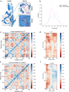
Fig. 4. The G463S mutation altered the dynamic properties of the active pocket.
a Arg363 and Ala460 are located on both sides of the inlet of the catalytic pocket where OB binds. These two residues are shown as magenta sticks. b The G463S mutation alters the distribution of the conformational states of the protein EcThrRS. The distance between the centers of mass of the residues Arg363 and Ala460 is defined as the width of the catalytic pocket. The conformational space shifts of WT and G463S are shown as cyan and pink curves, respectively. Eight sets of simulations with different random initial velocities were performed and data were collected from 9 to 23 Å per angstrom (n  =  8, mean value  ±  SD). c, d Dynamic cross-correlation maps (DCCM) of EcThrRS_WT. The dynamic cross-correlation matrix of Cα atoms around their mean positions is calculated. The extent of correlated motions and anticorrelated motions are color-coded from blue to red, which represents positive and negative correlations, respectively. (d) is a magnified view of the region in the red box in (c). e, f DCCM of EcThrRS_G463S. (f) is a magnified view of the region in the red box in (e).
Fig. 5
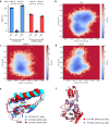
Fig. 5. The G463A mutation generates similar effects on the conformational change of ThrRS and blockage on the covalent binding of OB which were observed in the mutant G463S.
a Diagram of the ΔTm values of EcThrRS_WT and EcThrRS_G463A in the presence or absence of OB and 36j. Error bars represent the standard deviations (n  =  4, mean value  ±  SD). All the data points are shown as small circles. bd Free energy contour maps derived from the radius of gyration (Rg) and RMSD values, where the dark blue area indicates a lower energetic conformation state. The Rg was calculated for residues 419–467. EcThrRS_WT exhibited a different free energy landscape (FEL) from those of both G463S and G463A, especially possessing a larger conformational tether of Rg. e Superimposition of the structure of residues 419-467 of EcThrRS_WT (cyan cartoons), G463S (slate cartoons) and G463A (red cartoons) at the FEL nadir corresponding to Fig. 5b–d. Residues at positions 462 and 463 are shown as sticks and circled with black dashed lines. f Superimposition of the structures of EcThrRS_G463A (red cartoons) and EcThrRS_G463S (slate cartoons). The RMSD is 0.343 Å over 318 Cα atoms.
Fig. 6
Fig. 6. OB noncovalently binds to EcThrRS_G463A without inducing a conformational change.
a, b Zoomed-in view of the catalytic pocket of EcThrRS_G463A, which was crystallized in the presence of OB. The 2Fo-Fc electron densities of OB, Cys334, His385, His511, Tyr462 and Ala463 (contoured at 1.0 σ) are shown as transparent surfaces. OB does not form an ester bond with Tyr462. c Superimposition of the EcThrRS_Y462F–ATP structure (yellow cartoons, PDB code: 8H99) with the EcThrRS_G463A–OB structure (red cartoons). The nitrophenyl group of OB (in gray) binds between Phe379 and Arg520 where the adenine group of ATP (in purple blue) stacks. The orange parts in the middle of the panel are both terminal phosphate moieties of the cocrystallized ATP. d Superimposition of the EcThrRS_WT–OB structure (marine cartoons, PDB code: 8H98) with EcThrRS_G463A–OB (red cartoons). The ring-opened OB is shown as cyan sticks. The ring-closed OB is shown as gray sticks.
Fig. 7
Fig. 7. Gly463 mutations also confer resistance of EcThrRS to BN.
a Chemical structure of borrelidin (BN). b Superimposition of the structures of EcThrRS_WT–OB (PDB code: 8H98, marine cartoons) with EcThrRS_WT–BN (PDB code: 4P3P, gray cartoons). The RMSD is 0.396 Å over 345 Cα atoms. c Diagram of the ΔTm values of EcThrRS_WT/G463S/G463A in the presence or absence of BN. Error bars represent the standard deviations (n  =  4, mean value  ±  SD). All the data points are shown as small circles. d Inhibitory curves of BN on the ATP hydrolysis activity of EcThrRS_WT, G463S or G463A. Error bars represent the standard deviations (n  =  4, mean value  ±  SD). All the data points for EcThrRS_WT/G463S/G463A are shown as pale blue dots, pale pink square dots, and pale red triangle dots.

Similar articles

References

    1. Carter CW, Jr, Wills PR. The roots of genetic coding in aminoacyl-tRNA synthetase duality. Annu. Rev. Biochem. 2021;90:349–373. doi: 10.1146/annurev-biochem-071620-021218. - DOI - PubMed
    1. Rubio Gomez MA, Ibba M. Aminoacyl-tRNA synthetases. Rna. 2020;26:910–936. doi: 10.1261/rna.071720.119. - DOI - PMC - PubMed
    1. Pang, L., Weeks, S. D. & Van Aerschot, A. Aminoacyl-tRNA synthetases as valuable targets for antimicrobial drug discovery. Int. J. Mol. Sci.22 (2021). - PMC - PubMed
    1. Xie SC, Griffin MDW, Winzeler EA, Ribas de Pouplana L, Tilley L. Targeting aminoacyl tRNA synthetases for antimalarial drug development. Annu. Rev. Microbiol. 2023;77:111–129. doi: 10.1146/annurev-micro-032421-121210. - DOI - PubMed
    1. Kwon NH, Fox PL, Kim S. Aminoacyl-tRNA synthetases as therapeutic targets. Nat. Rev. Drug Discov. 2019;18:629–650. doi: 10.1038/s41573-019-0026-3. - DOI - PubMed

Publication types

MeSH terms

LinkOut - more resources