. 2024 Jul 4;25(13):7363.
doi: 10.3390/ijms25137363.

Arctigenin from Forsythia viridissima Fruit Inhibits the Replication of Human Coronavirus

Affiliations

Arctigenin from Forsythia viridissima Fruit Inhibits the Replication of Human Coronavirus

Jaeyeon So et al. Int J Mol Sci. .

Abstract

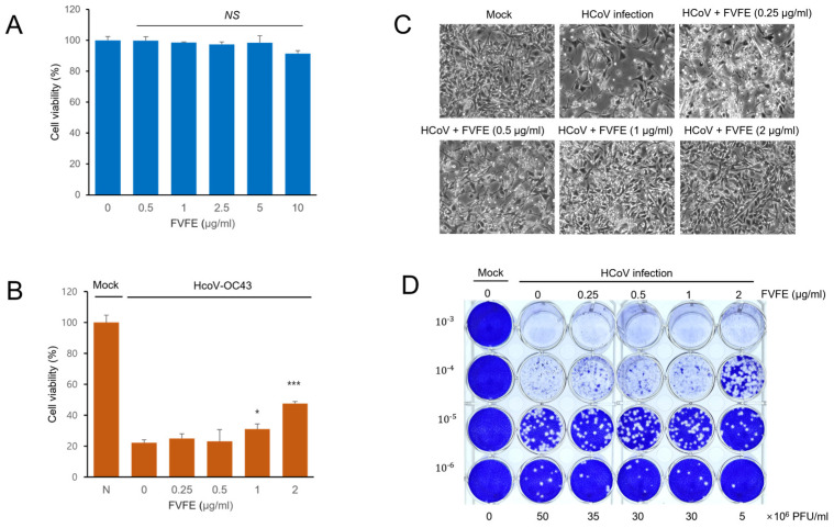
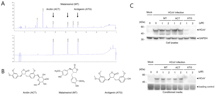
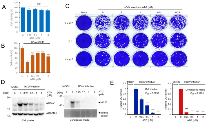
Coronavirus can cause various diseases, from mild symptoms to the recent severe COVID-19. The coronavirus RNA genome is frequently mutated due to its RNA nature, resulting in many pathogenic and drug-resistant variants. Therefore, many medicines should be prepared to respond to the various coronavirus variants. In this report, we demonstrated that Forsythia viridissima fruit ethanol extract (FVFE) effectively reduces coronavirus replication. We attempted to identify the active compounds and found that actigenin from FVFE effectively reduces human coronavirus replication. Arctigenin treatment can reduce coronavirus protein expression and coronavirus-induced cytotoxicity. These results collectively suggest that arctigenin is a potent natural compound that prevents coronavirus replication.

Keywords: antiviral; arctigenin; forsythia viridissima; human coronavirus; natural compound.


Conflict of interest statement

The authors declare no conflicts of interest.

Figures

Figure 1
Figure 1
Forsythia viridissima fruit ethanol extract (FVFE) treatment reduces the expression level of HCoV protein and RNA. (A) The fruit image of Forsythia viridissima. (B) FVFE treatment decreases the level of HCoV protein expression. RD cells were infected with HCoV and treated with FVFE for 72 h. Cells and the conditioned media were collected and the expression level of coronavirus proteins was examined by Western blot with an anti-HCoV antibody (upper panels). Glyceradehyde-3-phosphate dehydrogenase (GAPDH) was used as a loading control. The level of HCoV proteins was quantified and depicted in the graph (lower panel). The error bars represent the standard error. Experiments were repeated at least three times. Control vs. FVFE treatment, * p < 0.05, ** p < 0.01, *** p < 0.001. (C) RD cells were infected with HCoV and treated with the indicated concentration of FVFE. The infected cells were fixed and stained with anti-HCoV antibody (green). The immune-stained cells were counterstained with DAPI (blue). (D) FVFE treatment decreases the level of HCoV RNA expression. Total RNA was isolated from the cell lysates and the conditioned media. Quantitative RT-PCR (qRT-PCR) was used to evaluate the relative level of HCoV RNA. Expression levels of membrane protein (M), nucleoprotein (N), and RNA-dependent RNA polymerase (RdRp) mRNA were evaluated. The error bars represent the standard error. Experiments were repeated at least three times. Control vs. FVFE treatment, * p < 0.05, ** p < 0.01, *** p < 0.001. (E) Half-inhibitory concentration (IC50) was determined from qRT-PCR analysis.
Figure 2
Figure 2
FVFE treatment reduces coronavirus-induced cytopathic effect in RD cells. (A) RD cells were treated with the indicated concentration of FVFE for 24 h, and cell viability was examined by MTT assay. The error bars represent the standard error. (B) RD cells were infected with human coronavirus (HCoV-OC43). Three days after infection, cells were treated with the indicated concentration of FVFE and cell viability was evaluated. Control vs. FVFE treatment, * p < 0.05, *** p < 0.001, NS, not significant. (C) Microscopic images of coronavirus-infected cells with FVFE treatment were obtained. (D) RD cells were infected with coronavirus and treated with the indicated concentration of FVFE. The conditioned media were used for plaque formation assay to evaluate the amount of coronavirus in the media. The number in the Y-axis indicates dilutions of conditioned media.
Figure 3
Figure 3
Active compounds in FVFE were determined by HPLC analysis. (A) FVFE was analyzed using the well-known main components of FVFE by HPLC analysis. The signals of ethanol extract of the dried fruits of F. viridissima (upper panel). The mixture signals of arctiin, matairesinol, and arctigenin (lower panel) (B) The structure of arctiin (ACT), matairesinol (MT), and arctigenin (ATG) are shown. (C) ATG is effective in reducing the level of coronavirus protein. RD cells were infected with HCoV and treated with ACT, MT, and ATG for 72 h. Cells and conditioned media were collected and analyzed using Western blot with an anti-OC43 antibody.
Figure 4
Figure 4
ATG treatment effectively reduces HCoV infection. (A) RD cells were treated with the indicated concentration of ATG, and cell viability was examined to determine the cytotoxicity of ATG. (B) ATG treatment alleviates HCoV-induced cytotoxicity. RD cells were infected with HCoV and treated with ATG. Cell viability was determined to examine the antiviral effect of ATG. Experiments were repeated at least three times. Control vs. ATG treatment, *** p < 0.001, NS, not significant. (C) ATG treatment reduces plaque formation. RD cells were infected with HCoV and treated with ATG. The plaque formation assay was performed to examine the antiviral effects of ATG. (D) ATG treatment decreases the level of HCoV protein expression. RD cells were infected with HCoV and treated with ATG for 72 h. Cells and the conditioned media were collected and the expression level of coronavirus proteins was examined by Western blot with an anti-HCoV antibody. (E) Bar graphs show the relative FECV protein expression level (HCoV protein/GAPDH). The error bars represent the standard error (n = 3). Control vs. ATG treatment, ** p < 0.01, *** p < 0.001.

Similar articles

Cited by

References

    1. Paules C.I., Marston H.D., Fauci A.S. Coronavirus Infections—More Than Just the Common Cold. JAMA. 2020;323:707–708. doi: 10.1001/jama.2020.0757. - DOI - PubMed
    1. Harrison C.M., Doster J.M., Landwehr E.H., Kumar N.P., White E.J., Beachboard D.C., Stobart C.C. Evaluating the Virology and Evolution of Seasonal Human Coronaviruses Associated with the Common Cold in the COVID-19 Era. Microorganisms. 2023;11:445. doi: 10.3390/microorganisms11020445. - DOI - PMC - PubMed
    1. Cui J., Li F., Shi Z.-L. Origin and evolution of pathogenic coronaviruses. Nat. Rev. Microbiol. 2019;17:181–192. doi: 10.1038/s41579-018-0118-9. - DOI - PMC - PubMed
    1. Park R., Jang M., Park Y.-I., Park Y., Jung W., Park J., Park J. Epigallocatechin Gallate (EGCG), a Green Tea Polyphenol, Reduces Coronavirus Replication in a Mouse Model. Viruses. 2021;13:2533. doi: 10.3390/v13122533. - DOI - PMC - PubMed
    1. Jang M., Park R., Yamamoto A., Park Y.-I., Park Y., Lee S., Park J. AMPK inhibitor, compound C, inhibits coronavirus replication in vitro. PLoS ONE. 2023;18:e0292309. doi: 10.1371/journal.pone.0292309. - DOI - PMC - PubMed

LinkOut - more resources