. 2024 Jun 29;25(13):7205.
doi: 10.3390/ijms25137205.

Production of Spinocerebellar Ataxia Type 3 Model Mice by Intravenous Injection of AAV-PHP.B Vectors

Affiliations

Production of Spinocerebellar Ataxia Type 3 Model Mice by Intravenous Injection of AAV-PHP.B Vectors

Ayumu Konno et al. Int J Mol Sci. .

Abstract

We aimed to produce a mouse model of spinocerebellar ataxia type 3 (SCA3) using the mouse blood-brain barrier (BBB)-penetrating adeno-associated virus (AAV)-PHP.B. Four-to-five-week-old C57BL/6 mice received injections of high-dose (2.0 × 10¹¹ vg/mouse) or low-dose (5.0 × 10¹⁰ vg/mouse) AAV-PHP.B encoding a SCA3 causative gene containing abnormally long 89 CAG repeats [ATXN3(Q89)] under the control of the ubiquitous chicken β-actin hybrid (CBh) promoter. Control mice received high doses of AAV-PHP.B encoding ATXN3 with non-pathogenic 15 CAG repeats [ATXN3(Q15)] or phosphate-buffered saline (PBS) alone. More than half of the mice injected with high doses of AAV-PHP.B encoding ATXN3(Q89) died within 4 weeks after the injection. No mice in other groups died during the 12-week observation period. Mice injected with low doses of AAV-PHP.B encoding ATXN3(Q89) exhibited progressive motor uncoordination starting 4 weeks and a shorter stride in footprint analysis performed at 12 weeks post-AAV injection. Immunohistochemistry showed thinning of the molecular layer and the formation of nuclear inclusions in Purkinje cells from mice injected with low doses of AAV-PHP.B encoding ATXN3(Q89). Moreover, ATXN3(Q89) expression significantly reduced the number of large projection neurons in the cerebellar nuclei to one third of that observed in mice expressing ATXN3(Q15). This AAV-based approach is superior to conventional methods in that the required number of model mice can be created simply by injecting AAV, and the expression levels of the responsible gene can be adjusted by changing the amount of AAV injected. Moreover, this method may be applied to produce SCA3 models in non-human primates.

Keywords: AAV; AAV-PHP.B; Machado–Joseph disease; Purkinje cell; SCA3; Spinocerebellar ataxia type 3; adeno-associated virus; nuclear inclusion bodies.


Conflict of interest statement

The authors declare that the research was conducted in the absence of any commercial or financial relationships that could be construed as a potential conflict of interest.

Figures
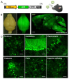
Figure 1
Figure 1
Broad and efficient transgene expression in the mouse brain by intravenous injection of AAV-PHP.B. (A) Expression cassette of the AAV genome. The ubiquitous CBh promoter drives GFP expression. WPRE and polyadenylation signal (pA) sequence were placed downstream of the GFP gene. (B) Bright field and fluorescent stereo microscopy of the whole brain (left). Right fluorescent image was obtained by cutting at the white line drawn on the left side of the brain. Scale bar, 1 mm. (C) Efficient GFP expression in different regions of the brain by intravenously injected AAV-PHP.B. Square regions on the sagittal section of the brain (right image in (B)) are enlarged. Scale bars; 50 µm.
Figure 2
Figure 2
Schema showing the experimental protocol. (A) Expression cassettes of the AAV genomes. The ubiquitous CBh promoter drives the expression of GFP-P2A-ATXN3(Q89) or -ATXN3(Q15). HA-tag was attached at the 5′-terminal of ATXN3 for immunolabeling the expressed ATXN3 protein by anti-HA antibody. (B) Schema showing experimental groups. Four-five-week wild-type mice were intravenously injected with high doses of BBB-permeable AAV-PHP.B encoding ATXN3(Q89), low-dose ATXN3(Q89) or high-dose ATXN3(Q15), or with PBS. (C) Diagram depicting the experimental protocol. A rotarod test was completed just before and every week up to 12 weeks after the AAV injection. After footprint analysis at 12 weeks, mice were sacrificed and subjected to immunohistochemistry (IHC).
Figure 3
Figure 3
Effects of AAV-mediated mutant ATXN3 expression on the survival and gaining body weight. (A) Survival curves of mice injected with high doses of AAV-PHP.B encoding ATXN3(Q89) or ATXN3(Q15), or low doses of AAV-PHP.B encoding ATXN3(Q89) or with PBS. **** p < 0.0001 by log-rank (Mantel–Cox) test. (B) Graph showing weight gain of mice. Measurement of mice injected with high doses of AAV-PHP.B encoding ATXN3(Q89) was stopped 4 weeks after injection as more than half of the mice died. n.s., not significant by one-way analysis of variance (ANOVA) followed by Tukey’s post hoc test at each time point.
Figure 4
Figure 4
Progressive motor deficits of mice injected with low doses of AAV-PHP.B encoding ATXN3(Q89). (A) Time course of rotarod performance of mice injected with AAV or PBS. Rotarod test was completed just before and every week post-injection. Mice were placed on a rod while accelerating from 4 to 40 rpm in 5 min, and the time it took for the mice to fall off the rod was measured. The average latency to fall of 4 trials was recorded. n = 7 [PBS, Q89(H)] or 8 [Q15, Q89(L)] mice, respectively. * p < 0.05, ** p < 0.01, *** p < 0.001 [vs. PBS], † p < 0.05, †† p < 0.01, ††† p < 0.001, †††† p < 0.0001 [vs. Q15(H)] by one-way analysis of variance (ANOVA) followed by Tukey’s post hoc test at each time point. (B,C) Footprint analysis performed 12 weeks after AAV injection. Mice with their hind limbs painted with a blue dye walked on a white paper for 30 cm (B). The average stride length was measured, excluding the beginning and end of the footprints (C). n = 4 mice, respectively. **, †† p < 0.01 by one-way analysis of variance (ANOVA) followed by Tukey’s post hoc test. n.s., not significant.
Figure 5
Figure 5
Dendrite atrophy and nuclear inclusion body formation in Purkinje cells. (A) Decreased molecular layer thickness in the cerebellar cortex of mice injected with low doses of AAV-PHP.B encoding ATXN3(Q89). Length depicted with double-headed arrows at the center of lobule 6 were measured using 4 to 6 sections per mouse. The mean value was used as a cerebellar molecular layer thickness of each mouse. Scale bars; 50 µm. (B) Quantitative analysis of the molecular layer thickness showing significantly smaller thickness in mice injected with low doses of AAV-PHP.B encoding ATXN3(Q89) compared with mice injected with high doses of AAV-PHP.B encoding ATXN3(Q15). n = 5 mice, respectively. *** p < 0.001 by unpaired t-test. (C) Nuclear inclusion body formation in Purkinje cells of mice injected with AAV-PHP.B injected with low doses of ATXN3(Q89). Cerebellar sections were double immunolabeled for HA (magenta) and calbindin (blue). Arrows indicate Purkinje cells contained nuclear inclusions. White arrowheads in the enlarged insert indicate nuclear inclusion body. Scale bars; 20 µm. (D) Graph showing quantitative analysis of ratios of nuclear inclusion (NI)-positive Purkinje cells (PC) to total Purkinje cells. n = 3 mice, respectively. **** p < 0.0001 by unpaired t-test.
Figure 6
Figure 6
Significant loss of large projection neurons in the cerebellar nuclei of mice expressing ATXN3(Q89). (A) Fluorescent images of the cerebellar nuclei double immunolabeled for HA (magenta) and Nissl (blue). Arrows indicate large projection neurons. Scale bar; 20 µm. (B) Graph showing quantitative analysis of number of large neurons per 106 cubic micrometers of cerebellar nucleus. n = 3 mice, respectively. ** p < 0.01 by unpaired t-test.

Similar articles

Cited by

References

    1. McLoughlin H.S., Moore L.R., Paulson H.L. Pathogenesis of SCA3 and implications for other polyglutamine diseases. Neurobiol. Dis. 2020;134:104635. doi: 10.1016/j.nbd.2019.104635. - DOI - PMC - PubMed
    1. Li X., Liu H., Fischhaber P.L., Tang T.S. Toward therapeutic targets for SCA3: Insight into the role of Machado-Joseph disease protein ataxin-3 in misfolded proteins clearance. Prog. Neurobiol. 2015;132:34–58. doi: 10.1016/j.pneurobio.2015.06.004. - DOI - PubMed
    1. Ren H., Hao Z., Wang G. Autophagy and Polyglutamine Disease. Adv. Exp. Med. Biol. 2020;1207:149–161. - PubMed
    1. Ingram M.A., Orr H.T., Clark H.B. Genetically engineered mouse models of the trinucleotide-repeat spinocerebellar ataxias. Brain Res. Bull. 2012;88:33–42. doi: 10.1016/j.brainresbull.2011.07.016. - DOI - PMC - PubMed
    1. Nóbrega C., Nascimento-Ferreira I., Onofre I., Albuquerque D., Conceição M., Déglon N., de Almeida L.P. Overexpression of mutant ataxin-3 in mouse cerebellum induces ataxia and cerebellar neuropathology. Cerebellum. 2013;12:441–455. doi: 10.1007/s12311-012-0432-0. - DOI - PubMed

LinkOut - more resources