Review
. 2024 Aug 7;227(4):iyae072.
doi: 10.1093/genetics/iyae072.

The Caenorhabditis elegans cuticle and precuticle: a model for studying dynamic apical extracellular matrices in vivo

Affiliations
Review

The Caenorhabditis elegans cuticle and precuticle: a model for studying dynamic apical extracellular matrices in vivo

Meera V Sundaram et al. Genetics. .

Abstract

Apical extracellular matrices (aECMs) coat the exposed surfaces of animal bodies to shape tissues, influence social interactions, and protect against pathogens and other environmental challenges. In the nematode Caenorhabditis elegans, collagenous cuticle and zona pellucida protein-rich precuticle aECMs alternately coat external epithelia across the molt cycle and play many important roles in the worm's development, behavior, and physiology. Both these types of aECMs contain many matrix proteins related to those in vertebrates, as well as some that are nematode-specific. Extensive differences observed among tissues and life stages demonstrate that aECMs are a major feature of epithelial cell identity. In addition to forming discrete layers, some cuticle components assemble into complex substructures such as ridges, furrows, and nanoscale pillars. The epidermis and cuticle are mechanically linked, allowing the epidermis to sense cuticle damage and induce protective innate immune and stress responses. The C. elegans model, with its optical transparency, facilitates the study of aECM cell biology and structure/function relationships and all the myriad ways by which aECM can influence an organism.

Keywords: C. elegans; ZP; collagen; cuticle; extracellular matrix.


Conflict of interest statement

Conflicts of interest The author(s) declare no conflict of interest.

Figures
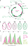
Fig. 1.
Fig. 1.
aECM dynamics across the C. elegans life cycle. a) The molt cycle. Assembly and removal/shedding of the precuticle (pink) and cuticle (green) are indicated by the inner bars. In larvae, the precuticle and then the new cuticle are assembled underneath the pre-existing cuticle before it is shed. Contrary to cuticle, precuticle is endocytosed and cleared before hatch/molt. Note that precise timing varies among different matrix components and cartoons illustrate only general trends. b) Dynamics of the first precuticle and cuticle in the embryo, based on Birnbaum et al. (2023). Cuticle collagens (DPY-17, SQT-3) are initially present in the extraembryonic space, outside of the precuticle (NOAH-1, LPR-3). The precuticle is endocytosed (puncta) after cuticle collagens assemble at the membrane. c) Oscillation of matrix gene expression during the molt cycle. Different groups of precuticle and cuticle genes cycle differently, with precuticle and furrow collagens cycling earlier than annuli collagens; adapted from Meeuse et al. (2020).
Fig. 2.
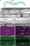
Fig. 2.
Cuticle substructures. a) Cartoon diagram of adult body cuticle, showing circumferential furrows and longitudinal alae. b) Longitudinal TEM slice of the adult epidermis, showing cuticle annuli/furrows and layers. f, furrow. s, struts. Image credit: Nicolas Brouilly & N.P. c) Longitudinal TEM slice of the epidermis in a L4.7 stage larva. Note the presumed precuticle matrix present between the old and new cuticles. Image credit: Nicolas Brouilly & N.P. d and e) Confocal images of tagged matrix proteins at mid-L4. d) Precuticle examples (SfGFP::LPR-3, left and GRL-7::mCherry, right). e) Cuticle examples (strut collagen BLI-1::mNG, left and furrow collagen DPY-7::SfGFP, right). Image credits: Nicholas Serra, Trevor Barker, and Susanna Birnbaum.
Fig. 3.
Fig. 3.
Cuticle-lined epithelia in the adult hermaphrodite. a) The epidermis. Most of the epidermis consists of syncytia generated by cell–cell fusion during embryogenesis (Podbilewicz and White 1994). hyp7 is the epidermal syncytium that encloses most of the body, whereas hyp1–6 are at the anterior and hyp8–11 are at the posterior. The lateral seam epidermis exists as individual cells until the early L4 stage, when these too fuse to form left and right seam syncytia. b) Interfacial tubes. During larval molting, the cuticle linings of these tubes are also shed and replaced. c) Sensilla consist of socket pore (yellow) and sheath (gray) glial cells that form tube-like compartments that surround the distal portions of ciliated sensory neurons, allowing them to access the environment. Socket glia are lined by precuticle and cuticle (green) but also share sensory matrices with the sheath glia. These different matrices help shape the tube channel and connect it to the epidermal cuticle (Perens and Shaham 2005; Heiman and Shaham 2009; Oikonomou et al. 2011; Low et al. 2019) (The Precuticle). In males, some socket glia produce specialized cuticular mating structures (Sulston et al. 1980; Jiang and Sternberg 1999) (The Cuticle).
Fig. 4.
Fig. 4.
aECM proteins with their domains. Only a few representatives of each matrix protein family are shown. See text and Supplementary Table 1 for further information.
Fig. 5.
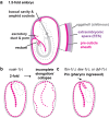
Fig. 5.
The embryonic sheath precuticle. a) Schematic of 1.5-fold embryo, with sheath (pink), extraembryonic space (purple), and eggshell (gray). At this stage, the extraembryonic space contains a mix of secreted precuticle and cuticle proteins that have not yet assembled (Birnbaum et al. 2023). Precuticle lined interfacial tubes are also shown. b) Defective elongation in a noah-1 mutant (adapted from Vuong-Brender et al. (2017)). Dashed lines indicate abnormal sheath. c) Pharynx ingressed (Pin) phenotype observed with incomplete penetrance in fbn-1, dex-1, and dyf-7 mutants. While FBN-1 is a component of the epidermal sheath (Kelley et al. 2015; Balasubramaniam et al. 2023), DEX-1 and DYF-7 contribute to buccal precuticle and amphid glia sensory matrices (Heiman and Shaham 2009; Cohen et al. 2019). It therefore appears that several different matrices contribute to buccal integrity.
Fig. 6.
Fig. 6.
Excretory duct tube defects of precuticle mutants. a) the duct lumenal precuticle (pink) expands lumen diameter (white bars) and counters morphogenetic forces (arrows) involved in lumen elongation and narrowing. b) In let-653(−) or other mutants with an abnormal precuticle (light pink), the duct lumen fragments during elongation and fluid then accumulates in the “upstream” region nearest the canal cell. Adapted from Gill et al. (2016).
Fig. 7.
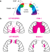
Fig. 7.
Precuticle ZP protein localization in the developing vulva lumen (mid-L4). a) EGF and Notch signaling promote 1˚ and 2˚ lineages that give rise to seven different vulva cell types (vulA-vulF) (Sharma-Kishore et al. 1999). b) The vulva lumen central cone matrix is marked by the plasminogen/apple-like (PAN) domains of LET-653, but mostly excludes FBN-1. The LET-653 ZP domain and NOAH-1 mark specific vulva cell surfaces (Gill et al. 2016; Cohen, Sparacio et al. 2020).
Fig. 8.
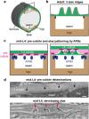
Fig. 8.
Adult alae form via AFB-dependent precuticle delamination. a) Schematic diagram of the adult worm; box indicates region of interest in b–d. b) Adult alae consist of three vertical cuticle ridges (green) above the seam. The seam is linked to the surrounding hyp7 syncytium by apical junctions (black). c) Arrangement of AFBs and precuticle factors as alae develop underneath the L4 cuticle (Katz et al. 2022). LPR-3 (magenta) marks future ridges, while LET-653 (light pink) marks future valleys. Cortical AFBs (blue) in the underlying seam and hyp7 also correspond to future locations of valleys. Left panel depicts an earlier stage (L4.4) corresponding to the top TEM image in (d), while right panel depicts a later stage (L4.6) when alae have begun to form. d) TEM images. In top image (L4.4), black arrows point to four small slits or delaminations in the precuticle underneath the L4 cuticle. These correlate with locations of AFBs, which are needed to pattern LPR-3 and the alae (Katz et al. 2022). In bottom image (L4.6), pink arrows point to newly formed alae tips, which are regions that have remained adherent to the L4 cuticle. Image credits: Ken Nguyen and David Hall.
Fig. 9.
Fig. 9.
The adult male tail ventral view, showing cuticular structures (green) including the tail fan and the hook and spicules. The gubernaculum (not shown) is located on the dorsal side. Drawing based on Jiang and Sternberg (1999) and Lints and Hall (2009).
Fig. 10.
Fig. 10.
Furrows. a) The cuticle is decorated with periodic circumferential indentations called furrows, separating annuli. Several collagens mark specifically each structure. Actin and microtubules (MT) align transiently with furrows before each molt in the main lateral epidermis. b) A DPY-7::SfGFP L2 larvae is molting, detaching from its old L1 cuticle (arrow), scale bar 10 µm. Image credit: N.P. c) and d) are AFM topography of the cuticle of wild-type (c) and furrow-less dpy-2 mutants (d) (from Aggad et al. (2023)).
Fig. 11.
Fig. 11.
Adult cuticle layers and struts. a) Ultrastructural layer appearance, adapted from Adams et al. (2023). Some known contents are indicated at right; see text for details. b) Strut. BLI collagens define hollow cylinders by superresolution microscopy, suggesting other contents may fill the strut interior (Adams et al. 2023). Epicuticlins also localize to struts as well as to a cortical or epicuticle layer above them (Pooranachithra et al. 2024). c) Blistered mutants facilitate assessment of cuticle layer contents, since cortical and basal layers become widely separated as the medial layer expands (Adams et al. 2023). In this case, SQT-3::mNG is clearly in a basal cuticle layer below the blisters (asterisks). Image credit: M.V.S.
Fig. 12.
Fig. 12.
Cuticle attachment structures: HDs and meisosomes. a) The epidermal syncytium can be separated into two structurally different regions. The first one is dorso- and ventro-lateral, above the muscles where the epidermis is really thin and rich in hemidesmosomes (blue). The second includes the lateral epidermis and the ventral and dorsal ridges; it contains the main cytoplasm and is rich in meisosomes (yellow). b) A transverse section of an adult without the internal organ highlight the body wall muscles in red, with the two different regions of the epidermis (sand) and the seam cell (white). Note that the cortical cytoskeleton (brown) is disorganized in the lateral epidermis (yellow), but organized below the circumferential furrows in the dorso and ventral lateral epidermis (blue). c) Hemidesmosomes attach the muscles to the cuticle through basal and apical components linked by intermediate filaments (IFs). d) Meisosomes are specific to the lateral epidermis and ventral and dorsal ridges; they are parallel stacks of folded plasma membrane (pm); mvb, multivesicular bodies. e) TEM image of a longitudinal section showing the different layers of the cuticle and meisosomes (m) at the junction with the epidermal cell (epi); cuticle (cut), furrow (f), mitochondria (mit). Scale bar 200 nm. From Aggad et al. (2023).
Fig. 13.
Fig. 13.
Collagen maturation steps in C. elegans and vertebrate collagens. a) Major steps of cuticle collagen maturation, with cellular sites indicated. See text and Supplementary Table 3 for specific genes/enzymes involved. There is still much debate in the literature about where in (or out of) the cell N- and C-terminal cleavage of COL1 occurs (Hellicar et al. 2022), nor do we know this information for cuticle collagens. b) Cuticle collagen maturation is temporally controlled. SQT-3::mNG(int) is secreted into the extraembryonic space before the 1.5-fold stage, but it only detectably assembles into cuticle ∼3 h later (Birnbaum et al. 2023). The mNeonGreen tag is located internally, soon after the BLI-4/PCSK-dependent cleavage site. c) Structural comparison of C. elegans and vertebrate collagens and their proteolytic cleavage sites. Ce DPY-17 is an example of a conventionally secreted cuticle collagen with a signal peptide (SP), while Ce SQT-3 is a predicted type II transmembrane (TM) protein. Human COL-13A is a transmembrane MACIT collagen (Tu et al. 2015) and COL1A is the classic type I fibrillar collagen (Revell et al. 2021). Gray boxes indicate the Gly-X-Y motifs, which are interrupted in cuticle collagens and MACITs. Black boxes indicate noncollagenous domains. Both DPY-17 and COL13A have predicted coiled-coil (CC, yellow) domains N-terminal to their collagen domains (Ludwiczak et al. 2019), and both and SQT-3 have sites for furin/PCSK-dependent cleavage (orange), whereas COL1A is cleaved by ADAM-TS. Only SQT-3 and COL1A have evidence for astacin protease cleavage (blue) at their C-termini. Note that proteins and domains are not drawn to scale. Genbank accession #s: DPY-17: NP_498086.1; SQT-3: NP_001256412.1; COL13A; NP_001123575; COL1A1, P02452. d) In the embryo, loss of DPY-17 collagen, BLI-4/PCSK, or DPY-31/astacin disrupts SQT-3 maturation prior to ER export, apical secretion, or cuticle assembly, respectively (Birnbaum et al. 2023). Note that these are temporary or partial blocks, because by hatch some SQT-3 does incorporate into the cuticle of these mutants.
Fig. 14.
Fig. 14.
Models for the relationship between the first precuticle and cuticle. a) Is the precuticle a porous structure that lets collagens pass through it and assemble within it? b) Or is the precuticle a separate layer that corrals new collagens below it? (It is also possible that, through phase separation, the aECM transforms from an initial arrangement as in (a) to a later arrangement as in (b). While (a) seems most compatible with subsequent precuticle endocytosis, (b) could explain how the precuticle disrupts interactions between the new cuticle and preexisting matrices (such as the eggshell in embryos or the old cuticle in larvae) to promote successful hatching and molting. EES, extraembryonic space.

Similar articles

Cited by

References

    1. Abete-Luzi P, Eisenmann DM. 2018. Regulation of C. elegans L4 cuticle collagen genes by the heterochronic protein LIN-29. Genesis. 56(5). doi:10.1002/dvg.23106. - DOI - PMC - PubMed
    1. Abolafia J. 2006. Order Rhabditida: suborder Rhabditina. In: Eyualem-Abebe EA, Traunspurger W, Andrássy I, editors. Freshwater Nematodes: Ecology and Taxonomy. Wallingford (UK): CAB International. p. 696–721. doi:10.1079/9780851990095.0696. - DOI
    1. Adams JRG, Pooranachithra M, Jyo EM, Zheng SL, Goncharov A, Crew JR, Kramer JM, Jin Y, Ernst AM, Chisholm AD. 2023. Nanoscale patterning of collagens in C. elegans apical extracellular matrix. Nat Commun. 14(1):7506. doi:10.1038/s41467-023-43058-9. - DOI - PMC - PubMed
    1. Aggad D, Brouilly N, Omi S, Essmann CL, Dehapiot B, Savage-Dunn C, Richard F, Cazevieille C, Politi KA, Hall DH, et al. 2023. Meisosomes, folded membrane microdomains between the apical extracellular matrix and epidermis. Elife. 12:e75906. doi:10.7554/eLife.75906. - DOI - PMC - PubMed
    1. Agudelo CW, Samaha G, Garcia-Arcos I. 2020. Alveolar lipids in pulmonary disease. A review. Lipids Health Dis. 19(1):122. doi:10.1186/s12944-020-01278-8. - DOI - PMC - PubMed

MeSH terms

Substances

Grants and funding

LinkOut - more resources