Review
. 2024 Jun 30;13(13):1133.
doi: 10.3390/cells13131133.

Spatio-Temporal Regulation of Notch Activation in Asymmetrically Dividing Sensory Organ Precursor Cells in Drosophila melanogaster Epithelium

Affiliations
Review

Spatio-Temporal Regulation of Notch Activation in Asymmetrically Dividing Sensory Organ Precursor Cells in Drosophila melanogaster Epithelium

Mathieu Pinot et al. Cells. .

Abstract

The Notch communication pathway, discovered in Drosophila over 100 years ago, regulates a wide range of intra-lineage decisions in metazoans. The division of the Drosophila mechanosensory organ precursor is the archetype of asymmetric cell division in which differential Notch activation takes place at cytokinesis. Here, we review the molecular mechanisms by which epithelial cell polarity, cell cycle and intracellular trafficking participate in controlling the directionality, subcellular localization and temporality of mechanosensitive Notch receptor activation in cytokinesis.

Keywords: Drosophila melanogaster; Notch signaling; asymmetric cell division; cell fate determinants; cell polarity; epithelial cells.


Conflict of interest statement

The authors declare no conflicts of interest.

Figures

Figure 1
Figure 1
Overview of the Notch pathway in Drosophila. The Notch pathway is a paracrine communication pathway involving Delta (red), the transmembrane ligand expressed and localized at the plasma membrane of the so-called signal-emitting cell, depicted in red. Delta interacts in trans with the Notch receptor (blue), localized at the plasma membrane of the signal-receiving cell, depicted in blue. Following ligand binding, Notch undergoes two successive proteolytic cleavages. The first cleavage at S2 is ligand-induced and generates an activated membrane form of Notch, which is then processed at S3 by the γ-secretase complex. This leads to the release of NICD, its translocation into the nucleus, and association with the transcriptional co-activator Mastermind and the DNA-binding transcription factor Suppressor of Hairless (Su(H)), resulting in transcriptional activation.
Figure 2
Figure 2
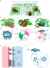
Asymmetric division of the SOP and site of Notch activation in cytokinesis. (A) Three-dimensional view of the dorsal thorax pupal notum, a single-layer epithelium composed of epidermal cells and SOPs (red cells). Adherens junctions labelled by E-cadherin (green) are localized apically and delimit the contour of epithelial cells. SOPs are regularly spaced thanks to Notch-dependent lateral inhibition. (BD) High magnifications of the SOP in interphase (B), during mitosis (C) and at mitosis exit once the SOP daughters, the anterior pIIb (red) and posterior pIIa (blue) cells, are formed (D). (B′) Schematic representation of the SOP in interphase illustrating the position of cell–cell junctions, adherens junctions (AJs, green) and septate junctions SJs (blue). PCP-dependent planar asymmetry of Baz-aPKC-Par6 (magenta) is initiated prior to mitosis. (C′) Schematic representation of the SOP in prometaphase. The PCP complex (Fz-Dsh) localized at the posterior apical cortex, together with the Baz-aPKC-Par6 polarity complex, (magenta) while Pins-Dlg co-localize with Numb and Neur at the antero-basal cortex (green). Mud interacts with Pins at the anterior basal cortex, and with Fz-Dsh at the antero-posterior cortex. In turn, Mud, by interacting with the Dynein complex, causes the spindle to orient along the anterior–posterior axis with a slight apical–basal tilt of the spindle. (D′) schematic representation of the pIIa-pIIb SOP daughter cells. The apical–basal polarity of the SOP daughter is preserved despite the polarity remodeling that has taken place in mitosis. However, specific to pIIa-pIIb cells (not seen following cytokinesis of epidermal cells) is the assembly of Baz clusters at the apical and the lateral interface. (E) Baz clusters contain Notch, Delta and Spdo, and Baz activity is required for efficient Notch signaling. (F) Photo-tracking of NICD revealed that the main contributors in Notch signaling originates from the basal pool of Notch (arrow). While the apical pool also contributes to Notch signaling, it remains to be determined if proteolytic activation occurs directly at the apical plasma membrane or if it is relocated from the apical to basal plasma membrane (dotted arrow).
Figure 3
Figure 3
Numb regulates the recycling of Notch-Sanpodo in the pIIb cell. (A) During SOP division, Numb (green) localizes at the anterior cortex and promotes the asymmetric distribution of a-Adaptin (α-Ada, orange), one of the subunits of the AP-2 complex, regulating Clathrin-mediated endocytosis. (B) During cytokinesis, Numb is unequally partitioned in the pIIb cell, where it is relocated from the cortex towards apical endosomes in an a-Ada-dependent manner. There, Numb prevents the recycling of Notch-Spdo (blue/purple) to the plasma membrane in favor of late endosomal transport. In contrast, in the pIIa cell, in the absence of Numb, Notch-Spdo can be recycled to the plasma membrane. According to this model, where Numb negatively regulates the recycling of Notch-Spdo, Numb, by reducing the level of Notch present at the plasma membrane of the pIIb cell, generates the asymmetrical low Notch/high Notch levels in pIIb and pIIa cells, respectively.
Figure 4
Figure 4
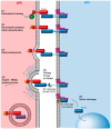
Neur regulates Delta endocytosis in the pIIb cell to trigger mechanosensitive activation of Notch in the pIIa cell. Activation of Notch (blue in the pIIa cell) is a multi-step process. It requires first the binding of the ligand Delta (red in pIIb) in trans (1). Then, Neur (green) ubiquitinates Delta (post-translational modification of the cytoplasmic domain of Delta depicted in yellow) (2). The ubiquitin moiety serves as a signal triggering Delta internalization (3). Arp2/3- and WASp-dependent polymerization of branched actin promotes extra pushing forces on the forming endocytic vesicle (4). Delta is bound to Notch, and Delta endocytosis exerts pulling forces on Notch, (5) thereby causing a conformational change in the Notch extracellular domain which reveals the S2 cleavage site for ADAM secretase. The second proteolytic cleavage triggered by g-secretase (cleavage S3) occurs, releasing into the cytoplasm of the pIIa cell the Notch intracellular domain, which can then be translocated into the nucleus.

Similar articles

References

    1. Artavanis-Tsakonas S., Muskavitch M.A. Notch: The past, the present, and the future. Curr. Top. Dev. Biol. 2010;92:1–29. doi: 10.1016/S0070-2153(10)92001-2. - DOI - PubMed
    1. Liu J., Sato C., Cerletti M., Wagers A. Notch signaling in the regulation of stem cell self-renewal and differentiation. Curr. Top. Dev. Biol. 2010;92:367–409. doi: 10.1016/S0070-2153(10)92012-7. - DOI - PubMed
    1. Koch U., Radtke F. Notch signaling in solid tumors. Curr. Top. Dev. Biol. 2010;92:411–455. doi: 10.1016/S0070-2153(10)92013-9. - DOI - PubMed
    1. Masek J., Andersson E.R. The developmental biology of genetic Notch disorders. Development. 2017;144:1743–1763. doi: 10.1242/dev.148007. - DOI - PubMed
    1. Zhou B., Lin W., Long Y., Yang Y., Zhang H., Wu K., Chu Q. Notch signaling pathway: Architecture, disease, and therapeutics. Signal Transduct. Target. Ther. 2022;7:95. doi: 10.1038/s41392-022-00934-y. - DOI - PMC - PubMed

Publication types

Grants and funding

This research was funded in part by a research grant from the Agence Nationale de la Recherche (ANR) programme PRC Vie, santé et bien être ACTriCE (ANR-20-CE13-0015), and Fondation ARC (PJA 20191209388).

LinkOut - more resources