Review
. 2024 Aug 21;35(8):1089-1115.
doi: 10.1021/acs.bioconjchem.4c00253. Epub 2024 Jul 11.

Targeted Protein Degradation (TPD) for Immunotherapy: Understanding Proteolysis Targeting Chimera-Driven Ubiquitin-Proteasome Interactions

Affiliations
Review

Targeted Protein Degradation (TPD) for Immunotherapy: Understanding Proteolysis Targeting Chimera-Driven Ubiquitin-Proteasome Interactions

Rajamanikkam Kamaraj et al. Bioconjug Chem. .

Abstract

Targeted protein degradation or TPD, is rapidly emerging as a treatment that utilizes small molecules to degrade proteins that cause diseases. TPD allows for the selective removal of disease-causing proteins, including proteasome-mediated degradation, lysosome-mediated degradation, and autophagy-mediated degradation. This approach has shown great promise in preclinical studies and is now being translated to treat numerous diseases, including neurodegenerative diseases, infectious diseases, and cancer. This review discusses the latest advances in TPD and its potential as a new chemical modality for immunotherapy, with a special focus on the innovative applications and cutting-edge research of PROTACs (Proteolysis TArgeting Chimeras) and their efficient translation from scientific discovery to technological achievements. Our review also addresses the significant obstacles and potential prospects in this domain, while also offering insights into the future of TPD for immunotherapeutic applications.


Conflict of interest statement

The authors declare no competing financial interest.

Figures
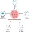
Figure 1
Figure 1
Types of Immunotherapies in the treatment of cancer. Checkpoint Inhibitors: Drugs that unleash the immune system by blocking proteins that prevent it from attacking cancer cells. Monoclonal Antibodies: Laboratory-produced proteins that target specific molecules in cancer cells, marking them for destruction or delivering toxic substances. Adoptive Cell Transfer: Collecting and modifying immune cells in the lab before reintroducing them into the patient’s body to enhance the immune response against cancer. Cancer Vaccines: Stimulate the immune system’s response against cancer cells, either preventing certain types of cancer or targeting existing cancer. Cytokines: Small proteins that activate immune cells and enhance their anticancer activity, used to boost the immune response against cancer. Oncolytic virus therapy for cancer: Oncolytic viruses are delivered to the patient. They infect and kill cancer cells, releasing viral particles and tumor antigens. The viral particles infect more cancer cells, while the tumor antigens trigger an immune response. The immune system clears the remaining cancer cells and prevents relapse.
Figure 2
Figure 2
a) The structure and function of PROTACs involve the combination of two ligands: one specific to the POI and the other targeting an E3 ligase. These ligands are connected by a linker, which facilitates the proximity of the POI to the E3 ligase. Subsequently, the target protein undergoes polyubiquitination, where ubiquitin molecules are attached, mediated by an E2 conjugating enzyme. The proteasome then degrades the polyubiquitinated target protein. Notably, the PROTAC itself remains intact throughout this process and can be reused in subsequent cycles, akin to an enzyme’s catalytic cycle. b) Crystal structure-based representation for substrate (BRD4-POI) recruitment to the E3 ligase cereblon (CRBN/DDB1 complex) by a heterobifunctional proteolysis-targeting chimera (PROTAC-dBET23). Ligands are shown as ball and stick representations (Protein Data Bank (PDB) code: 6BN7). DDB1, DNA damage-binding protein 1.
Figure 3
Figure 3
PROTAC-mediated modulation of the immune response against cancer cells. Tumor cell killing is selectively carried out by T cells, which recognize T cell receptor (TCR) antigens produced from PROTAC-induced proteolysis. The cell surface displays new MHC-I peptides derived from cancer cell-specific antigens via PROTAC-induced protein degradation. PROTACs specifically target proteins expressed in cancer settings, resulting in the generation of unique MHC-I complexes. These complexes can be recognized by TCRs on T cells. The proteins of interest (POIs), WT1 and BET, have confirmed protein peptides that serve as surface antigens for T cell recognition.,,
Figure 4
Figure 4
Model illustrating the control of CAR T cell activity through CAR degradation. A novel CAR T cell safety strategy specifically targets the CAR protein rather than the CAR T cell itself. The PROTAC compound, directed against the bromodomain (BD), degrades the BD-containing CAR protein. Notably, CAR expression is restored upon removal of the PROTAC compound from the cell or system, demonstrating its reversibility.
Figure 5
Figure 5
PROTAC-based cancer immunotherapy aims to transform the immunosuppressive tumor microenvironment into an immunoactive state through three distinct pathways. First, the application of PROTACs leads to the elimination of oncogenic proteins that are crucial for the growth and survival of cancer cells, thereby inducing immunogenic cell death. Second, PROTACs disrupt the immune checkpoint present on cancer cells, rendering them susceptible to immune attack by cytotoxic T cells. Lastly, PROTACs effectively eradicate immunosuppressive signal-associated cytokines, thus reducing the population of regulatory immune cells within tumor tissues.
Figure 6
Figure 6
Chemical structures of PD-L1 degraders. The figure shows the chemical structures of three PD-L1 degraders: 21a, BMS-37-C3, and P22. The structures are drawn using ChemDraw. In the top right corner, a 3D representation of a PD-L1 bound BMS-8 inhibitor is shown. Red arrows show the optimal linker attachment sites.
Figure 7
Figure 7
Flowchart showing different steps in PROTAC development using AI.
Figure 8
Figure 8
Advancing ligand discovery with proteomics and computational approaches is a field that encompasses various methodologies, including AI/ML-based methods. These methods can be categorized into three distinct groups (I–III) based on the type of input information they utilize. Adapted with permission from ref (50) under CCBY 4.0 License.
Figure 9
Figure 9
DMPK Optimization Cascade for Identifying Orally Bioavailable Degraders. This schematic depicts the key steps involved in the preclinical DMPK (drug metabolism and pharmacokinetics) optimization cascade used to identify PROTAC degraders suitable for oral administration. The cascade focuses on optimizing various properties of the degrader molecule to ensure successful oral delivery: 1. Compound Characterization: Initial evaluation of the degrader molecule’s physicochemical properties, including permeability and protein binding. 2. Compound Optimization: Iterative cycles of testing and refinement to improve the degrader’s metabolic stability and solubility, both crucial factors for oral bioavailability. 3. Compound Assessment: Evaluation of the optimized degrader’s oral pharmacokinetics (PK) in rodent models, often accompanied by PK modeling to understand absorption mechanisms. 4. Identification of Orally Efficacious Degraders: Selection of degrader candidates demonstrating both potency and good oral bioavailability for further PK/PD studies. Investigation of the relationship between degrader exposure and its pharmacodynamic (PD) effects, such as tumor growth inhibition. By navigating this cascade, researchers can identify promising PROTAC degraders with the potential to be developed into orally administered therapeutics.
Figure 10
Figure 10
A diagram illustrating different formulation strategies to tackle the physicochemical challenges related to PROTACs is shown below. Adapted with permission from under CCBY 4.0 License.

Similar articles

References

    1. Conforti L. The ion channel network in T lymphocytes, a target for immunotherapy. Clinical Immunology 2012, 142 (2), 105–106. 10.1016/j.clim.2011.11.009. - DOI - PubMed
    1. Syn N. L.; Teng M. W. L.; Mok T. S. K.; Soo R. A. De-novo and acquired resistance to immune checkpoint targeting. Lancet Oncology 2017, 18 (12), e73110.1016/S1470-2045(17)30607-1. - DOI - PubMed
    1. Barbari C.; Fontaine T.; Parajuli P.; Lamichhane N.; Jakubski S.; Lamichhane P.; Deshmukh R. R. Immunotherapies and Combination Strategies for Immuno-Oncology. International Journal of Molecular Sciences 2020, 21, 5009.10.3390/ijms21145009. - DOI - PMC - PubMed
    1. Sadeghi Rad H.; Monkman J.; Warkiani M. E.; Ladwa R.; O’Byrne K.; Rezaei N.; Kulasinghe A. Understanding the tumor microenvironment for effective immunotherapy. Medicinal Research Reviews 2021, 41 (3), 1474–1498. 10.1002/med.21765. - DOI - PMC - PubMed
    1. Seidel J. A.; Otsuka A.; Kabashima K.. Anti-PD-1 and Anti-CTLA-4 Therapies in Cancer: Mechanisms of Action, Efficacy, and Limitations. Frontiers in Oncology 2018, 8,10.3389/fonc.2018.00086. - DOI - PMC - PubMed

Publication types

Substances

LinkOut - more resources