. 2024 Jul 16;121(29):e2404349121.
doi: 10.1073/pnas.2404349121. Epub 2024 Jul 10.

IFIH1 (MDA5) is required for innate immune detection of intron-containing RNA expressed from the HIV-1 provirus

Affiliations

IFIH1 (MDA5) is required for innate immune detection of intron-containing RNA expressed from the HIV-1 provirus

Mehmet Hakan Guney et al. Proc Natl Acad Sci U S A. .

Abstract

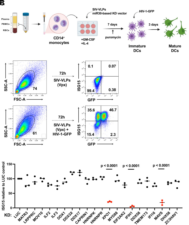
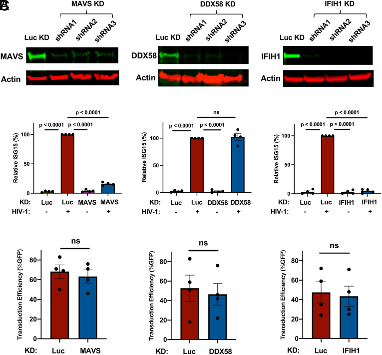
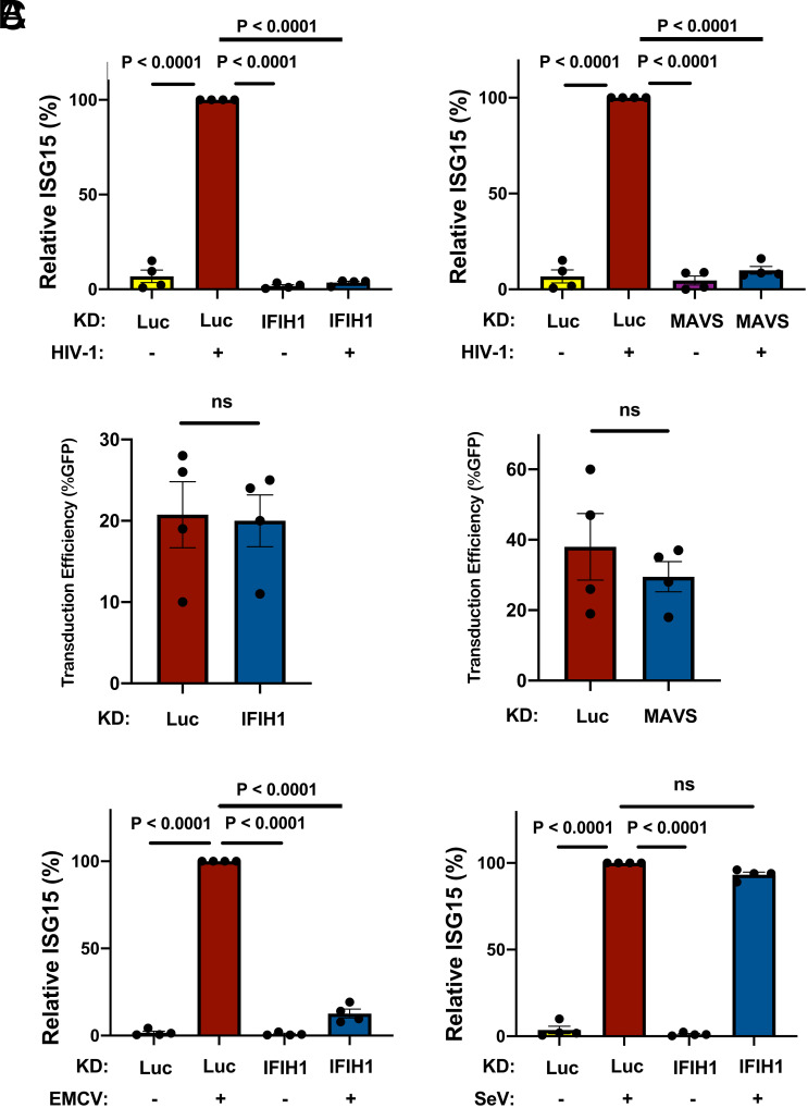
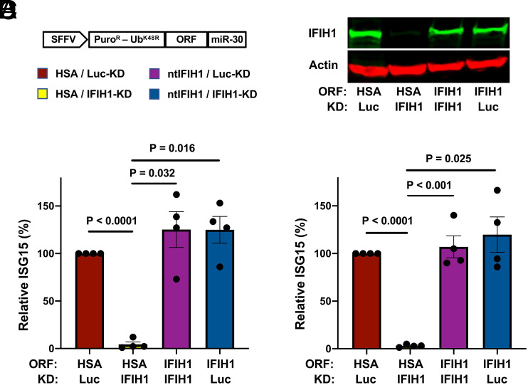
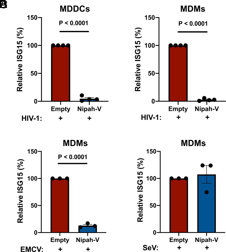
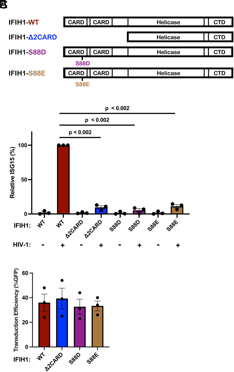
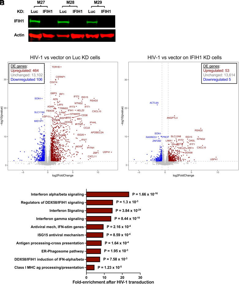
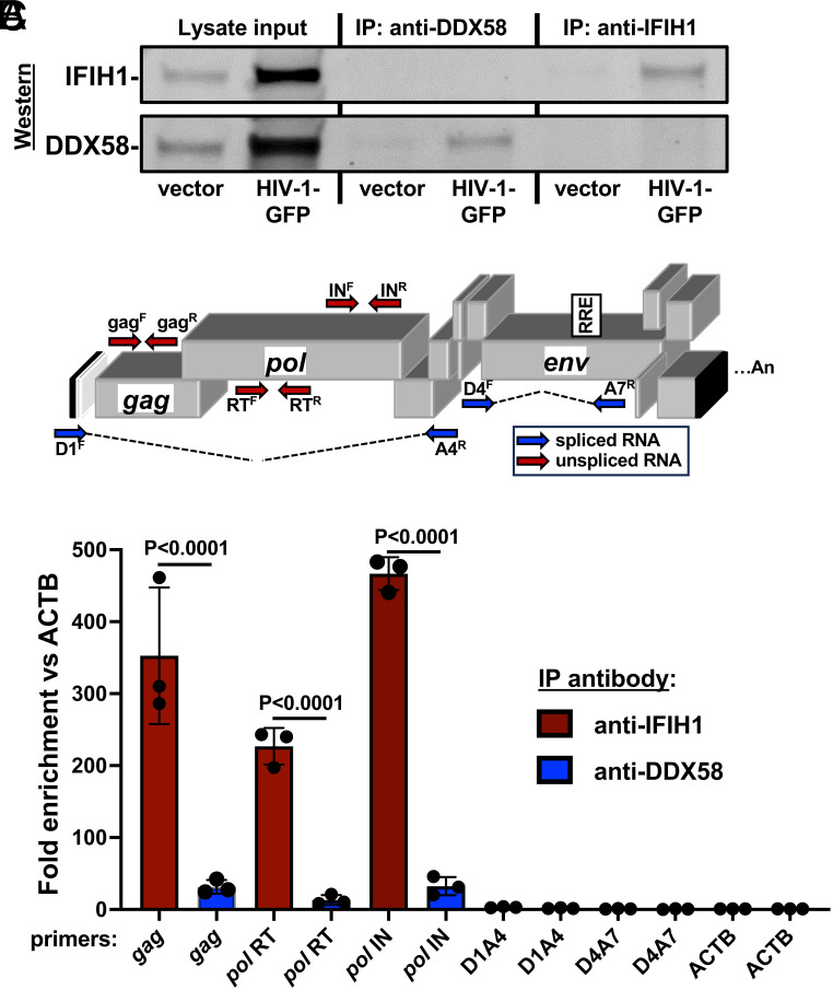
Intron-containing RNA expressed from the HIV-1 provirus activates type 1 interferon in primary human blood cells, including CD4+ T cells, macrophages, and dendritic cells. To identify the innate immune receptor required for detection of intron-containing RNA expressed from the HIV-1 provirus, a loss-of-function screen was performed with short hairpin RNA-expressing lentivectors targeting twenty-one candidate genes in human monocyte-derived dendritic cells. Among the candidate genes tested, only knockdown of XPO1 (CRM1), IFIH1 (MDA5), or MAVS prevented activation of the interferon-stimulated gene ISG15. The importance of IFIH1 protein was demonstrated by rescue of the knockdown with nontargetable IFIH1 coding sequence. Inhibition of HIV-1-induced ISG15 by the IFIH1-specific Nipah virus V protein, and by IFIH1-transdominant 2-CARD domain-deletion or phosphomimetic point mutations, indicates that IFIH1 (MDA5) filament formation, dephosphorylation, and association with MAVS are all required for innate immune activation in response to HIV-1 transduction. Since both IFIH1 (MDA5) and DDX58 (RIG-I) signal via MAVS, the specificity of HIV-1 RNA detection by IFIH1 was demonstrated by the fact that DDX58 knockdown had no effect on activation. RNA-Seq showed that IFIH1 knockdown in dendritic cells globally disrupted the induction of IFN-stimulated genes by HIV-1. Finally, specific enrichment of unspliced HIV-1 RNA by IFIH1 (MDA5), over two orders of magnitude, was revealed by formaldehyde cross-linking immunoprecipitation (f-CLIP). These results demonstrate that IFIH1 is the innate immune receptor for intron-containing RNA from the HIV-1 provirus and that IFIH1 potentially contributes to chronic inflammation in people living with HIV-1, even in the presence of effective antiretroviral therapy.

Keywords: HIV-1; MDA5; dendritic cells; type 1 interferon; unspliced RNA.


Conflict of interest statement

Competing interests statement:The authors declare no competing interest.

Figures

Fig. 1.
Fig. 1.
Targeted loss-of-function screen in dendritic cells identified IFIH1, MAVS, and XPO1 as required for ISG15-induction by HIV-1 transduction. (A) Schematic showing the experimental design of the knockdown screen in monocyte-derived dendritic cells. CD14+ monocytes transduced with shRNA-puromycin resistance vectors were differentiated into dendritic cells and selected with puromycin. Differentiated cells were transduced with HIV-1-GFP on day 7. Three days later, cells were stained for intracellular ISG15 and analyzed by flow cytometry. (B) Representative gating strategy for flow cytometry data presented in this manuscript. (C) Loss-of-function screen in dendritic cells targeting 21 genes encoding HIV-1 RNA binding proteins or innate immune receptors. The plot depicts ISG15 signal upon HIV-1-GFP transduction after suppressing the indicated candidate gene relative to ISG15 signal in Luciferase control knockdown cells (n = 3 shRNA target sites, n = 2 donors). P values were determined by one-way ANOVA with Dunnett’s multiple comparisons test, relative to luciferase knockdown control.
Fig. 2.
Fig. 2.
Knockdown of IFIH1 or MAVS in dendritic cells prevents ISG15-induction by HIV-1. (A) CD14+ monocytes were transduced with shRNA-puromycin resistance vectors targeting MAVS, DDX58, IFIH1, or luciferase (Luc) control, as indicated, and cells were differentiated into dendritic cells under puromycin selection as in Fig. 1A. Representative immunoblots show the protein signal in the indicated samples. (B) Differentiated cells were transduced with HIV-1-GFP for 3 d, and then assessed by flow cytometry for intracellular ISG15. Plots show relative ISG15 signal in the indicated knockdown cells, with and without HIV-1-GFP transduction (mean ± SEM, n = 4 donors). P values were determined by one-way ANOVA with Tukey’s multiple comparisons test. (C) Percentage GFP+ cells after HIV-1-GFP transduction of the dendritic cells in B (mean ± SEM, n = 4 donors). P values were determined by the two-tailed, paired t test.
Fig. 3.
Fig. 3.
IFIH1 or MAVS knockdown prevents ISG15-induction by HIV-1 in macrophages. (A) CD14+ monocytes were transduced with shRNA-puromycin resistant vectors targeting IFIH1, MAVS, or LUC control, as indicated. Cells were selected with puromycin for 3 d, and differentiated into macrophages in the presence of GM-CSF. Cells were transduced with HIV-1-GFP for 3 d and then assessed by flow cytometry for intracellular ISG15. Plots show relative ISG15 signal in the indicated knockdown cells, with and without HIV-1-GFP transduction (mean ± SEM, n = 4 donors). P values were determined by one-way ANOVA with Tukey’s multiple comparisons test. (B) Quantification of GFP+ cell populations in donors used in the experiments for A (mean ± SEM, n = 4 donors). P values were determined by the two-tailed, paired t test. (C) As in A, except that macrophages were infected with either encephalomyocarditis virus (EMCV) or Sendai virus (SeV), instead of with HIV-1-GFP (mean ± SEM, n = 4 donors). P values were determined by one-way ANOVA with Tukey’s multiple comparisons test.
Fig. 4.
Fig. 4.
IFIH1 (MDA5) protein is required for ISG15-induction by HIV-1 in myeloid cells. (A) Schematic representation of all-in-one shRNA-rescue lentivector, in which the SFFV promoter expresses a tripartite fusion of puromycin N-acetyltransferase (puroR), the K48R mutant of ubiquitin (UbK48R), and an ORF encoding the gene of interest, as well as a modified miR30-based shRNA (miR-30). (BD) All-in-one lentivectors encoding HSA/CD24, or nontargetable, shRNA-resistant IFIH1 coding sequence (ntIFIH1), along with shRNA targeting Luc or IFIH1 as indicated in A, were used to transduce dendritic cells (B and C), or macrophages (D). (B) Representative immunoblot for quantification of IFIH1 (MDA5) protein levels in indicated samples. (C and D) The percentage of ISG15+ cells was quantified by flow cytometry 3 d after HIV-1-GFP transduction and normalized to 100% for HSA ORF/Luc-knockdown cells (mean ± SEM, n = 4 donors for each). P values were determined by one-way ANOVA with Tukey’s multiple comparisons test.
Fig. 5.
Fig. 5.
Nipah virus V protein prevents ISG15-induction by HIV-1. (A and B) Nipah virus V protein was expressed from a lentivector in dendritic cells (A), or macrophages (B), and cells were subsequently transduced with HIV-1-GFP for 3 d. The percentage of ISG15+ cells was quantified by flow cytometry and normalized to the values for cells expressing the empty vector (mean ± SEM, n = 4 donors for each). P values were determined by the two-tailed, paired t test. (C and D) Nipah virus V protein was expressed in macrophages and subsequently either infected with EMCV (C) or SeV (D). The percentage of ISG15+ cells was measured by flow cytometry and normalized to the values for cells expressing the empty vector (mean ± SEM, n = 3 donors for each). P values were determined by the two-tailed, paired t test.
Fig. 6.
Fig. 6.
IFIH1 mutants defective for interaction with MAVS act in trans to abrogate ISG15-induction by HIV-1 in dendritic cells. (A) Schematic of the IFIH1 domain structure and mutants generated here. (B) Dendritic cells were transduced with either the vector expressing human codon optimized WT IFIH1 or one of the three indicated IFIH1 mutants. After selection with puromycin, cells were transduced with HIV-1-GFP for 3 d. The percentage of ISG15+ cells was quantified by flow cytometry and normalized to the values for cells expressing WT IFIH1 (mean ± SEM, n = 3 donors for each). P values were determined by one-way ANOVA with Dunnett’s multiple comparisons test. (C) Quantification of GFP+ cell populations in donors used in the experiments for B, mean ± SEM, n = 3 donors.
Fig. 7.
Fig. 7.
Effect of HIV-1 transduction and of IFIH1 knockdown on the dendritic cell transcriptome. CD14+ monocytes from three blood donors (coded M27, M28, and M29) were transduced with shRNA-puromycin resistance vectors targeting either IFIH1 or Luc control, as indicated. Cells were selected with puromycin for 3 d, and differentiated into dendritic cells. (A) Immunoblot shows steady-state levels of IFIH1 (MDA5) protein for the indicated samples. Then, dendritic cells were either transduced with HIV-1-GFP or left untransduced. At 48 h, RNA was isolated from cells, and poly(A)-selected libraries were generated for RNA-Seq. Volcano plot depicting differentially expressed genes after HIV-1 challenge in Luc knockdown control cells (B), or in IFIH1-knockdown cells (C), as determined by DESeq2 combining RNA-Seq data from the three blood donors. Red dots indicate genes with log2 fold change of normalized counts >1; P < 0.01. Blue dots indicate the log2 fold change of normalized counts <−1; P < 0.01. Gray dots failed to achieve either criterion. (D) Reactome pathway analysis based on 276 differentially expressed genes in HIV-1 transduced Luc-knockdown cells in comparison to HIV-1 transduced IFIH1-knockdown cells. Gene Ontology-produced P values (as determined by Fisher’s exact test) with FDR correction (Benjamini–Hochberg method) are shown. SI Appendix, Tables S1 and S2 show differentially expressed genes in B and C, respectively.
Fig. 8.
Fig. 8.
Unspliced HIV-1 RNA expressed from the provirus in dendritic cells associates specifically with IFIH1. Dendritic cells generated from three blood donors were transduced with HIV-1 GFP, or with minimal HIV-1 vector. At 72 h, cells were cross-linked with formaldehyde and proteins were immunoprecipitated using affinity-purified rabbit IgG specific for IFIH1 (IP:anti-IFIH1) or DDX58 (IP: anti-DDX58), as indicated. (A) Western blot of cell lysate used for immunoprecipitation, anti-DDX58 immunoprecipitate, and anti-IFIH1 immunoprecipitate, probed using antibody targeting IFIH1 (Upper row) or DDX58 (Lower row). (B) Schematic of the HIV-1 genome showing the location of qPCR primers that detect unspliced (red) or spliced (blue) transcripts. (C) Fold enrichment of HIV-1 RNA in immunoprecipitates obtained with anti-IFIH1 antibody (red bars) or anti-DDX58 antibody (blue bars), as determined by RT-qPCR using primers specific for the RNAs indicated across the X axis. Each bar shows the mean value relative to ACTB for dendritic cells obtained from three individual blood donors, ±the SEM. SI Appendix, Table S4 shows the primers used in RT-qPCR here.

Update of

Similar articles

Cited by

References

    1. Wong J. K., et al. , Recovery of replication-competent HIV despite prolonged suppression of plasma viremia. Science 278, 1291–1295 (1997). - PubMed
    1. McMyn N. F., et al. , The latent reservoir of inducible, infectious HIV-1 does not decrease despite decades of antiretroviral therapy. J. Clin. Invest. 133, e171554 (2023). - PMC - PubMed
    1. Hunt P. W., et al. , T cell activation is associated with lower CD4+ T cell gains in human immunodeficiency virus-infected patients with sustained viral suppression during antiretroviral therapy. J. Infect. Dis. 187, 1534–1543 (2003). - PubMed
    1. Li J. Z., et al. , Antiretroviral therapy reduces T-cell activation and immune exhaustion markers in human immunodeficiency virus controllers. Clin. Infect. Dis. 70, 1636–1642 (2020). - PMC - PubMed
    1. Douek D. C., Roederer M., Koup R. A., Emerging concepts in the immunopathogenesis of AIDS. Annu. Rev. Med. 60, 471–484 (2009). - PMC - PubMed

MeSH terms