. 2024 Jul 16;121(29):e2320709121.
doi: 10.1073/pnas.2320709121. Epub 2024 Jul 10.

STING trafficking activates MAPK-CREB signaling to trigger regulatory T cell differentiation

Affiliations

STING trafficking activates MAPK-CREB signaling to trigger regulatory T cell differentiation

Wei Lin et al. Proc Natl Acad Sci U S A. .

Abstract

The Type-I interferon (IFN-I) response is the major outcome of stimulator of interferon genes (STING) activation in innate cells. STING is more abundantly expressed in adaptive T cells; nevertheless, its intrinsic function in T cells remains unclear. Intriguingly, we previously demonstrated that STING activation in T cells activates widespread IFN-independent activities, which stands in contrast to the well-known STING-mediated IFN response. Here, we have identified that STING activation induces regulatory T cells (Tregs) differentiation independently of IRF3 and IFN. Specifically, the translocation of STING from the endoplasmic reticulum to the Golgi activates mitogen-activated protein kinase (MAPK) activity, which subsequently triggers transcription factor cAMP response element-binding protein (CREB) activation. The activation of the STING-MAPK-CREB signaling pathway induces the expression of many cytokine genes, including interleukin-2 (IL-2) and transforming growth factor-beta 2 (TGF-β2), to promote the Treg differentiation. Genetic knockdown of MAPK p38 or pharmacological inhibition of MAPK p38 or CREB markedly inhibits STING-mediated Treg differentiation. Administration of the STING agonist also promotes Treg differentiation in mice. In the Trex1-/- autoimmune disease mouse model, we demonstrate that intrinsic STING activation in CD4+ T cells can drive Treg differentiation, potentially counterbalancing the autoimmunity associated with Trex1 deficiency. Thus, STING-MAPK-CREB represents an IFN-independent signaling axis of STING that may have profound effects on T cell effector function and adaptive immunity.

Keywords: Innate immunity; STING; T cells; autoimmunity; type-I interferon.


Conflict of interest statement

Competing interests statement:The authors declare no competing interest.

Figures

Fig. 1.
Fig. 1.
STING activation induces Treg differentiation independently of IRF3 and IFN. (AC) DMXAA or cGAMP treatment induces Treg differentiation. Wild-type (WT) naïve CD4 T cells were treated with mock, DMXAA (2.5 μg/mL), or cGAMP (10 μg/mL) for 3 d, and Treg differentiation was analyzed by intracellular staining of Foxp3 and FACS analysis. Representative FACS plots are shown in (A), Treg percentages (B) and cell numbers (C) are shown in bar graphs. (D and E) IRF3 or IFNAR1 deficiency does not affect STING-mediated Treg differentiation. WT, Sting−/−, StingS365A/S365A, Irf3−/−, and Ifnar1−/− naïve CD4 T cells were treated with mock or DMXAA (2.5 μg/mL) for 3 d, and Treg differentiation was analyzed by intracellular staining of Foxp3 and FACS analysis. Representative FACS plots are shown in (D), and statistics are shown in (E). (FH) IFNGR1 deficiency does not affect STING-mediated Treg differentiation. WT and Ifngr1−/− naïve CD4 T cells were treated with mock, DMXAA (2.5 μg/mL), or cGAMP (20 μg/mL) for 3 d, and Treg differentiation was analyzed. Representative FACS plots are shown in (F), Treg percentages (G), and cell numbers (H) are shown in bar graphs. Error bars: SEM; ****P < 0.0001; *P < 0.05; **P < 0.01; ***P < 0.001; ns, not significant. Two-way ANOVA test. Data shown are representative of at least two independent experiments.
Fig. 2.
Fig. 2.
STING induces TGF-β2 and IL-2 expression to promote Treg differentiation. (A and B) TGF-β inhibitor treatment inhibits STING-mediated Treg differentiation. WT naïve CD4+ T cells were treated with DMXAA (2.5 μg/mL) in the presence or absence of TGF-β inhibitor SB 431542 (5 μM) for 3 d. Treg differentiation was analyzed by intracellular staining of Foxp3 and FACS analysis. Representative FACS plots are shown in (A), and statistics are shown in (B). (C) STING induces Tgfβ2 and Foxp3 expression. WT and Sting−/− CD4+ T cells were treated with mock, DMXAA (2.5 μg/mL), or cGAMP (10 μg/mL) for 3 d. Tgfβ1, Tgfβ2, Tgfβ3, and Foxp3 expression were detected by qRT-PCR. (D and E) The deficiency of IRF3 or IFNAR1 does not affect STING-mediated Tgfβ2 and Foxp3 expression. WT, Sting−/−, StingS365A/S365A, Irf3−/−, and Ifnar1−/− CD4+ T cells were treated with mock or DMXAA (2.5 μg/mL) for 3 d. Tgfβ2 (D) and Foxp3 (E) expression were detected by qRT-PCR. (F) STING activation induces Il2 expression. WT, Sting−/−, and StingS365A/S365A CD4+ T cells were treated with mock or DMXAA (2.5 μg/mL), and Il2 expression was detected by qRT-PCR. (G and H) IL-2 neutralization inhibits STING-mediated Treg differentiation. WT naïve CD4+ T cells were treated with cGAMP (10 μg/mL) in the presence or absence of IgG control, anti-IL2 neutralizing antibody JES6-1A12 (10 μg/mL) or S4B6-1 (10 μg/mL) for 3 d. Treg differentiation was analyzed. Representative FACS plots are shown in (G), and statistics are shown in (H). Error bars: SEM; **P < 0.01, ***P < 0.001, ****P < 0.0001; ns, not significant. Two-way ANOVA test. Data shown are representative of at least two independent experiments.
Fig. 3.
Fig. 3.
STING activates CREB1 to induce TGF-β2 and IL-2 expression in T cells. (A) IPA prediction of transcription factor activation by STING. WT, Sting−/−, and StingS365A/S365A T cells were treated with mock or DMXAA, followed by RNA-sequencing analysis (7). The RNA-seq data were analyzed by IPA to predict transcription factor activation by STING. (B) CREB1 binding site is predicted to exist in the Tgfβ2 and Il2 promoter sequence. Tgfβ2 and Il2 promoter sequences were scanned by JASPAR (https://jaspar2020.genereg.net/) and found to contain a CREB1 binding motif, which is highlighted in red. (C and D) STING activates CREB1. WT and Sting−/− CD4+ T cells were treated with DMXAA for the indicated hours, and CREB and STING signaling were detected by Western blotting (C). Densitometry analysis is performed and normalized to the mock sample (D). The red arrow indicates the p-CREB band. Data shown are representative of at least two independent experiments.
Fig. 4.
Fig. 4.
STING trafficking activates MAPK–CREB signaling in T cells. (A and B) Naïve CD4 T cells from WT, Sting−/−, StingS365A/S365A, Irf3−/−, and Ifnar1−/− mice were activated with anti-CD3 + anti-CD28 antibodies for 2 d, followed by treatment with mock or DMXAA (2.5 μg/mL) overnight. Cell lysates were then collected for the detection of MAPK–CREB signaling. Densitometry analysis is performed and normalized to the mock sample (B). (C) The schematic representation of STING mutations designed to selectively block STING signaling at specific stages. (D and E) Reconstitution of Sting−/− CD4+ T cells with STING mutants. Sting−/− CD4+ T cells were reconstituted with STING WT or various mutants, followed by stimulation with mock or DMXAA (2.5 μg/mL) overnight. MAPK–CREB and STING signaling were assessed by Western blotting. Densitometry analysis is shown in (E). The data presented are representative of at least three independent experiments.
Fig. 5.
Fig. 5.
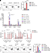
MAPK–CREB is required for STING-mediated Treg differentiation. (AC) WT Naïve CD4 T cells were activated with anti-CD3 + anti-CD28 antibodies for 2 d, followed by treatment with mock or DMXAA (2.5 μg/mL) for indicated hours. Cell lysates were then collected for the detection of MAPK–CREB signaling. Densitometry analysis was performed and normalized to the mock sample (B and C). (D and E) WT naïve CD4+ T cells were stimulated with mock or DMXAA (2.5 μg/mL) for 1 to 3 d, followed by Treg analysis. Representative FACS plots are shown in (D), and statistics are shown in (E). (F and G) Knockdown of p38 inhibits STING-mediated Treg differentiation. WT naïve CD4+ T cells were transduced with lentiviral-mediated sh-luciferase control or sh-p38, followed by stimulation with mock, DMXAA (2.5 μg/mL), or cGAMP (20 μg/mL) for 3 d. Representative FACS plots are shown in (F), and statistics are shown in (G). (H and I) p38 or CREB inhibitor treatment inhibits STING-mediated Treg differentiation. WT naïve CD4+ T cells were treated with mock or cGAMP (20 μg/mL) in the presence or absence of p38 inhibitors sb203850 (10 μM) or sb202190 (10 μM), or CREB inhibitor 666-15 (1 μM), for 3 d. Treg differentiation was analyzed by intracellular staining of Foxp3 and FACS analysis. Representative FACS plots are shown in (H), and statistics are shown in (I). Error bars: SEM; *P < 0.05, ***P < 0.001, ****P < 0.0001; ns, not significant. Two-way ANOVA test. Data shown are representative of two independent experiments.
Fig. 6.
Fig. 6.
STING activation promotes Treg differentiation in mice. Six-week-old mice were i.p. injected with vehicle or DMXAA (10 mg/kg) for 10 consecutive days. Splenocytes were harvested and stained for CD4, CD8, CD44, CD62L, and Foxp3. (AC) Analysis of Treg cells. Representative FACs plots are shown in (A). Treg cell percentages and numbers are shown in (B and C). (D) The ratio of effector T cells to Treg cells. (EK) Analysis of naïve, central memory, and effector CD4+ T cells. Representative FACs plots are shown in (E). The percentage (FH) and cell number (IK) of naïve, central memory, and effector CD4+ T cells are shown in bar graphs. (LR) Analysis of naïve, central memory, and effector CD8+ T cells. Representative FACs plots are shown in (L). The percentage (MO) and cell number (PR) of naïve, central memory, and effector CD8+ T cells are shown in bar graphs. Error bars: SEM; *P < 0.05, ***P < 0.001, ****P < 0.0001; ns, not significant. Two-way ANOVA test.
Fig. 7.
Fig. 7.
CD4+ T cell-intrinsic STING activation induces Treg differentiation in Trex1−/−autoimmune mice. (A) A schematic representation of constitutively activated STING mouse models. (BG) CD4+ T cell and Treg analysis of WT, Sting−/−, StingS365A/S365A, StingN153S/+, and Trex1−/− mice. Splenocytes from the indicated mice were stained for Foxp3 and CD4. Representative FACS plots are shown in (B), statistics for CD4+ T cells and Tregs are shown in (CF), and the ratio of effector T cells to Tregs is shown in (G). (H and I) WT and Trex1−/− naïve CD4 T cells were treated with mock, DMXAA (2.5 μg/mL), or cGAMP (20 μg/mL) for 3 d, and Treg differentiation was analyzed. Representative FACS plots (H) and statistics (I) are shown. (J and K) Treg analysis of Trex1−/− and Trex1−/− Sting−/− mice. Splenocytes were stained for Foxp3 and CD4. Representative FACS plots are shown in (J), and statistics of Tregs are shown in (K). (L) A schematic representation of adoptive transfer of naïve CD4+CD25- T cells into Rag1−/− mice. (M) Tregs were depleted in the isolated naïve CD4+ T cells from WT, Trex1−/−, and Trex1−/− Sting−/− mice. (NP) Treg analysis of adoptively transferred CD4+ T cells. Naïve CD4+ T cells were adoptively transferred into Rag1−/− mice for 1 mo, and Tregs were analyzed in spleens and lymph nodes. Representative dot plots are shown in (N), and Treg percentages are shown in (O and P). Error bars: SEM; **P < 0.01, ****P < 0.0001; ns, not significant. Two-Way ANOVA test. Data shown are representative of at least two independent experiments.

Similar articles

References

    1. Sun L., Wu J., Du F., Chen X., Chen Z. J., Cyclic GMP-AMP synthase is a cytosolic DNA sensor that activates the type I interferon pathway. Science 339, 786–791 (2013). - PMC - PubMed
    1. Wu J., et al. , Cyclic GMP-AMP is an endogenous second messenger in innate immune signaling by cytosolic DNA. Science 339, 826–830 (2013). - PMC - PubMed
    1. Ishikawa H., Barber G. N., STING is an endoplasmic reticulum adaptor that facilitates innate immune signalling. Nature 455, 674–678 (2008). - PMC - PubMed
    1. Ishikawa H., Ma Z., Barber G. N., STING regulates intracellular DNA-mediated, type I interferon-dependent innate immunity. Nature 461, 788–792 (2009). - PMC - PubMed
    1. Jeltema D., Abbott K., Yan N., STING trafficking as a new dimension of immune signaling. J. Exp. Med. 220, e20220990 (2023). - PMC - PubMed

MeSH terms

Substances

LinkOut - more resources