. 2024 Jul 9;15(1):5766.
doi: 10.1038/s41467-024-50197-0.

Neutrophil extracellular traps promote immunopathogenesis of virus-induced COPD exacerbations

Affiliations

Neutrophil extracellular traps promote immunopathogenesis of virus-induced COPD exacerbations

Orestis Katsoulis et al. Nat Commun. .

Abstract

Respiratory viruses are a major trigger of exacerbations in chronic obstructive pulmonary disease (COPD). Airway neutrophilia is a hallmark feature of stable and exacerbated COPD but roles played by neutrophil extracellular traps (NETS) in driving disease pathogenesis are unclear. Here, using human studies of experimentally-induced and naturally-occurring exacerbations we identify that rhinovirus infection induces airway NET formation which is amplified in COPD and correlates with magnitude of inflammation and clinical exacerbation severity. We show that inhibiting NETosis protects mice from immunopathology in a model of virus-exacerbated COPD. NETs drive inflammation during exacerbations through release of double stranded DNA (dsDNA) and administration of DNAse in mice has similar protective effects. Thus, NETosis, through release of dsDNA, has a functional role in the pathogenesis of COPD exacerbations. These studies open up the potential for therapeutic targeting of NETs or dsDNA as a strategy for treating virus-exacerbated COPD.


Conflict of interest statement

S.L.J. has personally received consultancy fees from AstraZeneca, Bioforce, Enanta and GlaxoSmithKline. S.L.J. is an inventor on patents on the use of inhaled interferons for treatment of exacerbations of airway diseases and on rhinovirus vaccines. S.L.J. is Director and shareholder of Virtus Respiratory Research Ltd. JDC has received research grants from AstraZeneca, Boehringer Ingelheim, GlaxoSmithKline, Gilead Sciences, Grifols, Novartis, Insmed and Trudell; and received consultancy or speaker fees from Antabio, AstraZeneca, Boehringer Ingelheim, Chiesi, GlaxoSmithKline, Insmed, Janssen, Novartis, Pfizer, Trudell and Zambo. A.S. has received honoraria for speaking from AstraZeneca. A.D.A. is Chief Medical Officer at Santersus AG. The remaining authors declare no competing interests.

Figures

Fig. 1
Fig. 1. NETosis is increased during human RV infection in COPD.
A Experiment schematic. 9 participants with chronic obstructive pulmonary disease (COPD), 10 healthy smokers and 11 healthy non-smokers underwent sampling at baseline, day 5, day 9 and convalescence during experimental RV-A16 challenge. B Sputum DNA/elastase complexes (C) total intact (H3.1) and (D) citrullinated (H3.R8) nucleosomes were quantified by ELISA. ††P < 0.01 COPD group day 9 vs baseline; P < 0.05 COPD group day 5 or 42 vs baseline; ***P < 0.001 COPD vs. non-smokers **P < 0.01 COPD vs. non-smokers. *P < 0.05 COPD vs. non-smokers. ###P = 0.001 COPD vs smokers. #P < 0.05 COPD vs smokers. Data are presented as mean values ± SEM. Data analysed by two-tailed Wilcoxon matched pairs, signed-rank test to compare baseline to post-infection or two-tailed Mann–Whitney test to compare between experimental groups. n = 9 in each group in (B) due to sample availability. Source data are provided as a Source Data file.
Fig. 2
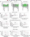
Fig. 2. Airway NETs correlate with immunological and clinical COPD exacerbation severity.
Correlation of sputum DNA-elastase complexes, total intact (H3.1) and citrullinated (H3.R8) nucleosomes with (AC) cellular airway inflammation, cytokines, MUC5AC and neutrophil elastase. DF sputum virus loads. GI lower respiratory tract symptoms scores. JL PEFR change. Correlation analysis used was non-parametric (Spearman’s correlation) performed on COPD (n = 9), healthy smoker (n = 10) and healthy non-smoker (n = 11) participants pooled into a single group ***P < 0.001, **P < 0.01, *P < 0.05. Source data are provided as a Source Data file.
Fig. 3
Fig. 3. Airway NETs are increased during naturally occurring virus associated exacerbations.
A Experiment schematic. 18 individuals with chronic obstructive pulmonary disease (COPD) were monitored prospectively. Sputum samples were taken during stable state (baseline), at presentation with an exacerbation associated with positive virus detection, and 2 weeks after exacerbation presentation. Sputum concentrations of (B) DNA-elastase complexes, (C) total intact (H3.1) and (D) citrullinated (H3.R8) nucleosomes were measured at stable-state and following virus-induced exacerbation. Data are presented as mean values ± SEM. *P < 0.05 ***P < 0.001,. Data analysed by two-tailed Wilcoxon matched pairs, signed-rank test to compare steady-state with exacerbation onset or 2 weeks after exacerbation. Source data are provided as a Source Data file.
Fig. 4
Fig. 4. Elastase treatment combined with rhinovirus infection models exacerbated neutrophils and NETs in mice.
A Experimental schematic. Mice were treated with intranasal porcine pancreatic elastase or PBS control. 10 days later, mice were inoculated with RVA1 or UV-inactivated virus control. B Total neutrophil numbers in lung tissue (C) % CD63+ (D) % CD64+ (E) Lamp-1 and (F) CXCR4 expressing neutrophils were characterised by flow cytometry. Bronchoalveolar lavage (BAL) concentrations of (G) total intact (H3.1) (H) citrullinated (H3.R8) nucleosomes and (I) lactate dehydrogenase (LDH) were quantified by ELISA. n = 4–6 mice/group, representative of at least two independent experiments. Data presented as mean ± SEM. Data analysed by two-tailed Mann–Whitney U test. *P < 0.05, **P < 0.01,. Source data are provided as a Source Data file.
Fig. 5
Fig. 5. Pharmacological neutrophil elastase inhibition reduces immunopathology in a mouse model of virus-exacerbated COPD.
A Experimental schematic. Mice were treated with intranasal porcine pancreatic elastase or PBS control. 10 days later, mice received i.p. injection of the Neutrophil elastase inhibitor (NEi; GW311616A) or vehicle control 12 h before RVA1 inoculation. B Bronchoalveolar lavage (BAL) concentrations of Total intact (H3.1) and citrullinated (H3.R8) nucleosomes were quantified by ELISA. C Histological lung inflammation scores. D BAL total cells, neutrophils, macrophages and lymphocytes were enumerated by cytospin. BAL concentrations of (E) chemokines CXCL10/IP-10 and CCL5/RANTES (F) pro-inflammatory cytokines TNF, IL-1β and IL-6, (G) Muc5ac and (H) secretory leucocyte protease inhibitor (SLPI) were quantified by ELISA. (I) Airway hyperresponsiveness (enhanced pause [Penh]) to methacholine challenge was measured by whole body plethysmography. n = 4-5 mice/group, representative of at least two independent experiments. Data presented as mean ± SEM. Data analysed by two-tailed Mann–Whitney U test. *P < 0.05, **P < 0.01,. Source data are provided as a Source Data file.
Fig. 6
Fig. 6. Host dsDNA induction is increased during human RV infection in COPD and correlates with immunopathology and clinical exacerbation severity.
A Experimental schematic. 9 participants with chronic obstructive pulmonary disease (COPD), 10 healthy smokers and 11 healthy non-smokers underwent sampling at baseline, day 5, day 9 and convalescence during experimental rhinovirus infection. B Sputum dsDNA concentrations following RV-A16 challenge in 9 COPD, 10 healthy smoker and 10 healthy non-smoker control participants. Correlation of peak sputum dsDNA concentrations with (C) sputum concentrations of Total intact (H3.1) and citrullinated (H3.R8) nucleosomes, (D) sputum Virus loads (E) sputum total and neutrophil cell counts (F) sputum cytokine concentrations and (G) sputum MUC5AC concentrations. Correlation of peak sputum dsDNA with (H) Upper and Lower respiratory tract symptoms. In (B) ††P < 0.01 COPD group day 9 vs baseline. Data in (B) are presented as mean values ± SEM and analysed by two-tailed Wilcoxon matched pairs, signed-rank test to compare baseline to post-infection or two-tailed Mann–Whitney test to compare between experimental groups. **P < 0.01 COPD vs. non-smokers #p < 0.05 COPD vs smoker. In (CI) correlation analysis used was non-parametric (Spearman’s correlation) performed on COPD (n = 9), healthy smoker (n = 10) and healthy non-smoker (n = 11) participants pooled into a single group. Source data are provided as a Source Data file.
Fig. 7
Fig. 7. DNAse treatment reduces immunopathology in a mouse model of virus-exacerbated COPD.
A Experimental schematic. Mice were treated by DNaseI (or vehicle control) by i.p. injection 4 h before inoculation and 1 and 2 days post-infection and by the intranasal route 8 h and 1 and 2 days post-infection (B) Bronchoalveolar lavage (BAL) concentrations of dsDNA was quantified by ELISA. C Histological lung inflammation scores (D) BAL total cells, neutrophils, macrophages and lymphocytes were enumerated by cytospin. BAL concentrations of (E) chemokines CXCL10/IP-10 and CCL5/RANTES and CCL2 (F) pro-inflammatory cytokines TNF, IL-1β and IL-6 and (G) mucin glycoprotein Muc5ac and (H) secretory leucocyte protease inhibitor (SLPI) were quantified by ELISA. I Airway hyperresponsiveness (enhanced pause [Penh]) to methacholine challenge was measured by whole body plethysmography. n = 4–5 mice/group representative of at least two independent experiments. Data are presented as mean values ± SEM. Data analysed by two-tailed Mann–Whitney U test. *P < 0.05, **P < 0.01,. Source data are provided as a Source Data file.
Fig. 8
Fig. 8. Pharmacological inhibition of NETosis using a PAD inhibitor reduces dsDNA and immunopathology in a mouse model of virus-exacerbated COPD.
A Experimental schematic. Mice were treated with intranasal porcine pancreatic elastase or PBS control. 10 days later, mice received i.p. injection of a PAD inhibitor PADi; BB-CL-Amidine or vehicle control 12 h before RVA1 inoculation. B Bronchoalveolar lavage (BAL) concentrations of Total intact (H3.1) and citrullinated (H3.R8) nucleosomes were quantified by ELISA. C BAL concentrations of dsDNA quantified by ELISA. D BAL total cells, neutrophils, macrophages and lymphocytes. BAL concentrations of (E) chemokines CXCL10/IP-10 and CCL5/RANTES (F) pro-inflammatory cytokines IL-6, IL-1β,TNF and (G) Muc5ac were quantified by ELISA. n = 5 mice/group. Data expressed as mean ± SEM. Data analysed by two-tailed Mann–Whitney U test. *P < 0.05, **P < 0.01, ***P < 0.001. Source data are provided as a Source Data file.

Similar articles

References

    1. Singanayagam A, Joshi PV, Mallia P, Johnston SL. Viruses exacerbating chronic pulmonary disease: the role of immune modulation. BMC Med. 2012;10:27. doi: 10.1186/1741-7015-10-27. - DOI - PMC - PubMed
    1. Mallia P, et al. Experimental rhinovirus infection as a human model of chronic obstructive pulmonary disease exacerbation. Am. J. Respir. Crit. Care Med. 2011;183:734–742. doi: 10.1164/rccm.201006-0833OC. - DOI - PMC - PubMed
    1. Footitt J, et al. Oxidative and nitrosative stress and histone Deacetylase-2 activity in exacerbations of COPD. Chest. 2016;149:62–73. doi: 10.1378/chest.14-2637. - DOI - PMC - PubMed
    1. Lonergan M, et al. Blood neutrophil counts are associated with exacerbation frequency and mortality in COPD. Respir. Res. 2020;21:166. doi: 10.1186/s12931-020-01436-7. - DOI - PMC - PubMed
    1. Moermans C, et al. Local and systemic cellular inflammation and cytokine release in chronic obstructive pulmonary disease. Cytokine. 2011;56:298–304. doi: 10.1016/j.cyto.2011.07.010. - DOI - PubMed

MeSH terms