. 2024 Jul 9;148(1):2.
doi: 10.1007/s00401-024-02763-5.

Cleavage site-directed antibodies reveal the prion protein in humans is shed by ADAM10 at Y226 and associates with misfolded protein deposits in neurodegenerative diseases

Affiliations

Cleavage site-directed antibodies reveal the prion protein in humans is shed by ADAM10 at Y226 and associates with misfolded protein deposits in neurodegenerative diseases

Feizhi Song et al. Acta Neuropathol. .

Abstract

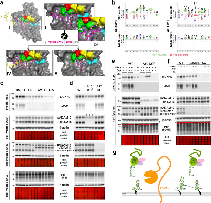
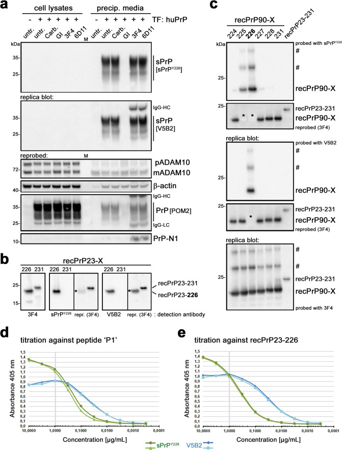
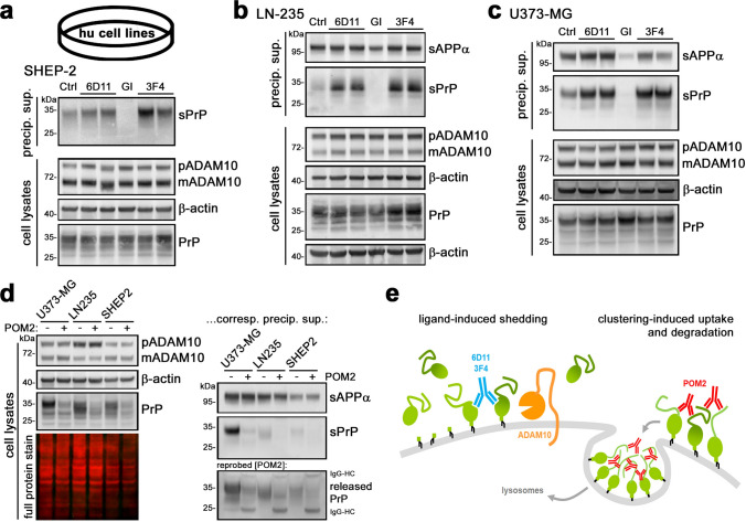
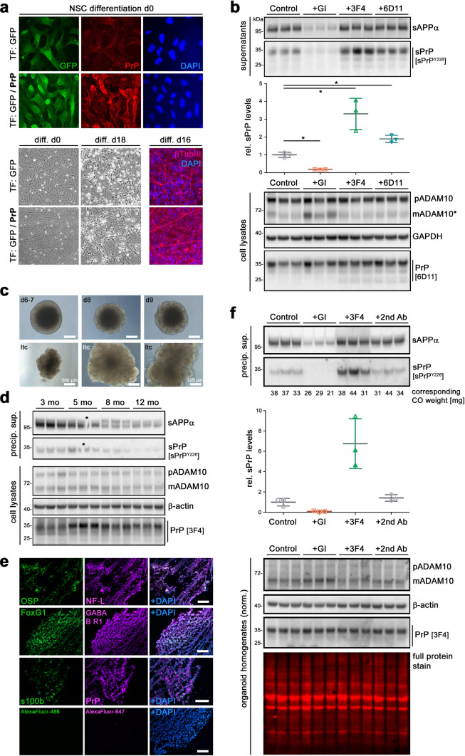
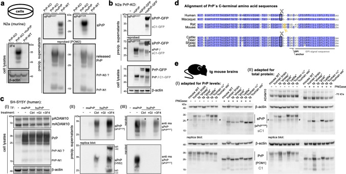
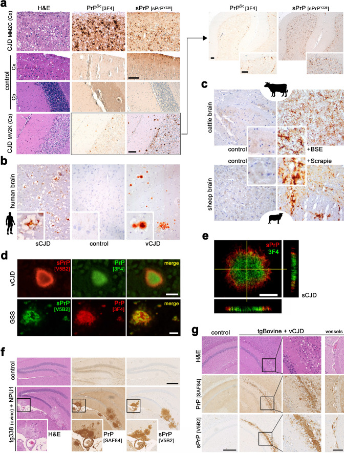
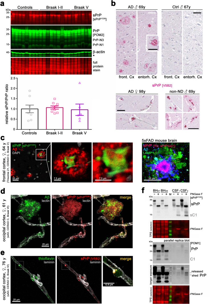
Proteolytic cell surface release ('shedding') of the prion protein (PrP), a broadly expressed GPI-anchored glycoprotein, by the metalloprotease ADAM10 impacts on neurodegenerative and other diseases in animal and in vitro models. Recent studies employing the latter also suggest shed PrP (sPrP) to be a ligand in intercellular communication and critically involved in PrP-associated physiological tasks. Although expectedly an evolutionary conserved event, and while soluble forms of PrP are present in human tissues and body fluids, for the human body neither proteolytic PrP shedding and its cleavage site nor involvement of ADAM10 or the biological relevance of this process have been demonstrated thus far. In this study, cleavage site prediction and generation (plus detailed characterization) of sPrP-specific antibodies enabled us to identify PrP cleaved at tyrosin 226 as the physiological and apparently strictly ADAM10-dependent shed form in humans. Using cell lines, neural stem cells and brain organoids, we show that shedding of human PrP can be stimulated by PrP-binding ligands without targeting the protease, which may open novel therapeutic perspectives. Site-specific antibodies directed against human sPrP also detect the shed form in brains of cattle, sheep and deer, hence in all most relevant species naturally affected by fatal and transmissible prion diseases. In human and animal prion diseases, but also in patients with Alzheimer`s disease, sPrP relocalizes from a physiological diffuse tissue pattern to intimately associate with extracellular aggregated deposits of misfolded proteins characteristic for the respective pathological condition. Findings and research tools presented here will accelerate novel insight into the roles of PrP shedding (as a process) and sPrP (as a released factor) in neurodegeneration and beyond.

Keywords: Alzheimer’s disease; Dementia; Extracellular vesicles; Neuroprotection; Prions; Proteolytic processing.


Conflict of interest statement

The authors have no conflict of interest to declare.

Figures

Fig. 1
Fig. 1
Cleavage site prediction using structural models and pharmacological/genetic proof of human PrP shedding being ADAM10-dependent. a I: Proteolytic domain of ADAM10 (based on [114]) with Zn2+ coordinated in the catalytic center. Key residues of substrate-binding pockets highlighted for S1 (yellow; V297/F323/D325/V327), S1′ (cyan; V376/I379/T380/I416/T422) and S3 (green; L301/L330/W332). Overlaid extracellular C-terminal sequence 642FMRCRLVDADGPLG655 (yellow) of another ADAM10 molecule (crystal structure PDB: 6BE6) and C-terminal end of PrP 217YERESQAYYQRGS230 (purple). Magnification (II) and detail (III) of PrP’s C-terminal sequence within ADAM10`s catalytic domain suggesting formation of a salt bridge (SB; PrPR228-ADAM10E298) and close proximity of the suspected cleavage site (Y226↓Q227) and Zn2+ within catalytic cave. IV: N-terminal parts (C-termini of sPrP or soluble ADAM10) are released after cleavage. V: Remaining C-terminal PrP residues may be freed from catalytic domain (possibly regulated by SB) and stay at the membrane or be endocytosed/degraded. b IceLogos: preferred and disfavored aa in different positions to the potential cleavage site (P1↓P1′) based on various peptide/protein substrates of ADAM10/ADAM17 using PICS (modified from [127]) and TAILS (modified from [110]). Favored (green background) and disfavored residues (red background) for putative PrP shedding. c WB of sPrP/sAPPα (media) and PrP/ADAM10/ADAM17 (lysates) of A549 cells treated with metalloprotease inhibitors GI254023X (GI) or/and GW280264X (GW). β-actin and total protein stain (TPS): loading controls. d Assessment in wild-type, ADAM10-knockout and ADAM17-knockout cells. e WB of WT and A10KO cells treated or not with ADAM-stimulating PMA and/or inhibitor GW. Two different A10KO lines were used (d: A10KOa; e: A10KOb; hence different inactive mutant bands #). f Analysis in WT and A17KO cells with/without PMA and/or GW/GI. Red saturated bands (e, f) result from residual β-actin signal (reprobing for PrP). g Model of membrane-proximate PrP shedding. With the recent suggestion of G229 (instead of previously assumed C-terminal serine) as actual GPI-attachment site in human PrP [51], distance between cleavage site and membrane would be preserved between mice and humans
Fig. 2
Fig. 2
Direct comparison of polyclonal sPrPY226 and monoclonal V5B2 antibodies. a Human neuroblastoma (SH-SY5Y) cells almost lacking endogenous PrP expression (see signal in lysates of non-transfected (−) cells; left lane) transfected (+) with a human PrP-coding plasmid, untreated (untr.) or treated with carbachol (Carb.), ADAM10 inhibitor GI, or PrP-directed antibodies 3F4 or 6D11. Cell lysates (left half of blots) and precipitated media supernatants (right half) loaded on two replica blots and initially detected with either sPrPY226 or V5B2 yielding comparable signals (note that heavy chains (HC) of the treatment antibodies are also detected with the (anti-mouse) secondary antibody used for V5B2 detection). Re-probing with pan-PrP antibody POM2 confirmed overexpression of PrP in transfected cells (note that this cell-associated PrP was neither detected with sPrPY226 nor with V5B2). Levels of premature (p) and mature/active (m) ADAM10, β-actin (loading control) and N-terminal PrP fragment N1 were also assessed. Actin in media indicates some transfection-induced cell death (note the comparably weak signal in untransfected cells). Dominance of mADAM10 in media likely associated with extracellular vesicles. Detectability of soluble PrP-N1 is rescued in the presence of 3F4 and 6D11 antibodies, protecting this instable fragment from proteolytic degradation [92]. M = marker lane. sPrPY226 and V5B2 were also compared on replica immunoblots of recPrP23-226 (mimicking sPrP) versus recPrP23-231 (full-length) (b) or of N-terminally (i.e., at aa 90) truncated recPrP with different C-termini (X, as indicated) as well as full-length recPrP (23–231) (c). Blots were re-probed and an additional replica blot directly detected with the pan-PrP antibody 3F4. Asterisks in re-probed blots (b and c) mark “burned” signals resulting from overexposure during the previous detection shown above. ‘#’ indicates SDS-stable dimers/oligomers of recPrP. Comparison of sPrPY226 (green) and V5B2 (blue) in ELISA against the peptide ‘P1’ used for immunization of mice to generate V5B2 (d) or against human recPrP23-226 (e)
Fig. 3
Fig. 3
PrP shedding, ADAM10 inhibition, and effects of PrP-directed antibodies in human CNS cancer cell lines. Representative WB showing basal levels of sPrP (Ctrl; left lane of each blot) detected with the sPrPY226 antibody in precipitated overnight media supernatants of neuroblastoma cells (SHEP-2; in a), astrocytoma cells (LN-235; in b) and glioblastoma cells (U373-MG; in c). In all cell lines, shedding is increased upon treatment with PrP-directed antibodies 6D11 and 3F4 and abolished when treated with an ADAM10 inhibitor (GI). sAPPα was detected in media (in b and c) as another cleavage product generated by ADAM10. Corresponding cell lysates assessed for levels of PrP, premature (p) and mature/active (m) ADAM10, and β-actin (serving as loading control) are shown underneath (ac). d Treatment with the antibody POM2 in all three cell lines results in the reduction of cell-associated PrP levels (left panel) as well as sPrP and released PrP in corresponding media samples (right panel). e Scheme showing the shedding-stimulating effect of PrP-directed antibodies and the exceptional reduction in total PrP levels caused by POM2 IgG (illustration modified from [71])
Fig. 4
Fig. 4
PrP shedding in human neuronally differentiated stem cells and iPSC-derived cerebral organoids. a IF analysis of embryonic stem cell-derived NSC (upon lentiviral transfection to express either GFP (green) or GFP and exogenous PrP (red)) at day 0 of neuronal differentiation (upper panel). Bright field microscopy (lower left panel) showing morphological differences between day 0 and 18. IF analysis at day 16 (lower right panel) reveals neuronal marker β-tubulin III. b Immunoblot of sPrP and sAPPα in conditioned media (supernatants; upper panel), quantification of relative sPrP levels (diagram; middle panel), and cellular levels of ADAM10, GAPDH and PrP (lysates; lower panel) following 30 days of differentiation and 18 h treatment with ADAM10 inhibitor GI (a lower concentration [6 µM] was used here, hence the residual signal for sPrP) or PrP-directed IgGs (3F4/6D11). DMSO-treated controls served as reference (set to 1). n = 3 wells per condition; mean ± SE; Student’s t test with *p < 0.05. c iPSC-derived cerebral organoids (CO) at different days of differentiation and after neuroepithelial bud expansion ready for long-term culture (ltc). Scale bar 250 µm unless indicated. d Levels of sPrP and sAPP (conditioned media) and PrP, ADAM10 and β-actin (loading control) in CO homogenates after 3–12 months in culture. e Different cell types detected in differentiated organoids by IF analysis of typical markers (OSP = oligodendrocyte-specific protein; NF-L = neurofilament light chain (neurons); GABABR1 = γ-aminobutyric acid type B receptor subunit 1 (inhibitory neurons); s100b = S100 calcium-binding protein B (astrocytes)). PrP expression was also detected. DAPI used to stain nuclei. Controls with only fluorescently labeled 2nd antibodies (AlexaFluor) revealed no signals. Scale bars 200 µm. f Treatment of CO with GI (inhibition), 3F4 antibody (stimulation) and a non-specific secondary antibody (negative control). sPrP and sAPP in precipitated media (sPrP quantification shown below) and ADAM10 and PrP in respective CO homogenates (individual CO weights shown below lanes) assessed by WB. TPS and β-actin: loading controls. We refrained from statistical analysis considering variation in CO weights
Fig. 5
Fig. 5
Heterologous cleavage and species-specificity of sPrP-directed antibodies. a Shedding in murine (ms) PrP-KO N2a cells overexpressing human (hu) PrP (PrP-KO and WT-N2a were controls for no and endogenous PrP expression, respectively). HuPrP overexpression confirmed by a 3F4-positive signal. Replica blots of precipitated media detected with either sPrPG227 (ms sPrP) or sPrPY226 (hu sPrP). Presence of released PrP fragments was confirmed by re-probing with POM2. b WB of PrP-KO cells transfected with huPrP or GFP-tagged versions of hu or ms PrP (GFP located within the C-terminal half of PrP). sPrPG227 exclusively detects ms sPrP-GFP and shed C1-GFP, whereas re-probing with sPrPY226 reveals hu sPrP, sPrP-GFP and shed C1-GFP in media samples. Expression of respective cell-associated PrP forms in lysates (using pan-PrP antibody POM1) shown below. c WB of hu SH-SY5Y cells transfected (TF) with msPrP or huPrP (the latter treated or not with GI or 3F4-IgG). (I) Lysates; (II) replica blots of precipitated media probed with either polyclonal sPrPY226 (top) or monoclonal V5B2 (bottom) detecting hu sPrP (basal (Ctrl), inhibited (GI) or increased (3F4)); (III) re-probing with sPrPG227 reveals ms sPrP (* indicates signals from the initial detection due to primary/secondary antibody combination). d C-terminal aa sequences of PrP in different species including GPI-anchor signal sequence and attachment site. The ADAM10 cleavage site is marked in yellow for rats and mice, in black for human and monkey PrP. Note the sequence similarity of the latter with cattle, deer, sheep and goat. e Assessment of sPrP and PrP in brains of transgenic (tg) mice expressing PrP of different species. WT mouse and a human brain homogenate included as controls. PNGase F digestion performed for deglycosylation (shown on the right side of each blot). Protein amounts were either roughly adapted to PrP expression (I) or normalized for total protein (II). Actin: loading control, ADAM10 levels are also shown in II. # indicates an unspecific band detected with sPrPY226
Fig. 6
Fig. 6
Redistribution of sPrP and association with prion deposits in prion diseases of humans and animals. a (Immuno)histochemical (IHC) assessment of PrPSc (3F4 antibody upon harsh tissue pre-treatment) and sPrP (polycl. antibody sPrPY226) in two CJD cases compared to a control without diagnosed neurodegeneration. Coarse-grained and perivacuolar PrPSc deposits present in frontal cortex [Cx] of a MM2C case, while the cerebellum [Cb] of a MV2K case shows typical Kuru-like plaques (note that tissue disruption in control is due to pre-treatment prior to PrPSc detection). Shed PrP shows a diffuse distribution in the control and re-distributes into an aggregated appearance in brains affected by CJD (scale bars 100 µm). In MV2K, more sPrP clusters appear than actual PrPSc plaques, which is further supported by an overview comparison (upper right panel). b Monocl. antibody V5B2 used in IHC to detect sPrP in brain sections of a sCJD and a vCJD patient (compared to a control). c Detection of sPrP (V5B2) in the brains of cattle affected or not with BSE (upper panel) and sheep with or without Scrapie (lower panel). d, e IF analyses showing association of sPrP (V5B2) with extracellular PrP aggregates (3F4) in acquired (vCJD) and genetic prion diseases (GSS) (d; standard fluorescence microscopy; scale bars 20 µm) and sCJD (e; z-stacks with side projections; scale bar 5 µm). f, g Histological analyses of large PrPSc deposits (here: SAF84 antibody without harsh pre-treatment) and sPrP (V5B2) in hippocampal areas of prion-infected transgenic mice expressing ovine (tg338; f) or bovine PrP (g). Tg338 mice infected with NPU1 prions present with large and dense amyloid-like plaques (f). TgBov mice infected with vCJD show extended prion deposition along the corpus callosum. Boxes indicate position of magnified areas (g). In both models, association of aggregates with brain vessels is observed. Non-infected mice of the respective genotype served as controls. Scale bars 250 µm (and 100 µm for the ‘vessels’ panel in g)
Fig. 7
Fig. 7
Shed PrP analysis in AD and CAA, and sensitive detectability of sPrP in human CSF. a WB analysis of sPrP and total PrP in cortex of AD patients (Braak stage I–II (n = 8), Braak stage V (n = 9)) compared to non-neurodegeneration controls (n = 6). Actin and TPS: loading controls. Asterisk indicates signals from previous PrP detection. Besides inter-individual alterations in sPrP, quantification (below) of the sPrP/PrP ratio reveals no significant differences between groups. b IHC of sPrP in AD and controls with sPrP showing both diffuse and dense (birefringent) plaque-like pattern reminiscent of bona fide Aβ deposits (upper panel). Dense vessel-associated sPrP signal can be found in some AD cases and controls (lower panel). Scale bars 50 µm. c Closer inspection by IF microscopy in brain sections of a patient with AD/Trisomy 21 reveals sPrP in the center of some (yet not all) Aβ plaques, as reported earlier in mouse models ([71]; on the right: sPrP detection in 5xFAD brain with sPrPG227 antibody; LAMP1 indicates dystrophic neurites or microglial lysosomes). d Plaque-like clusters (highlighted by dotted line in merge picture) of Aβ and sPrP co-purified during isolation of microvessels from AD brain. Co-localization of both molecules was also found at/in vessels. Lectin: endothelial marker. Orthogonal projection of this picture presented in Supplementary Fig. 9a). e Association of amyloid and sPrP in/at brain vessels was verified in another AD case using another set of stainings (V5B2 for sPrP, thioflavin for (Aβ) aggregates, anti-laminin as endothelial/vessel marker). Another vessel of this sample shown in orthogonal view in Supplementary Fig. 9b). Scale bars as indicated. f WB of sPrP and total PrP in brain homogenates (BH) and CSF samples (patients not diagnosed with neurodegeneration). Deglycosylation (+ PNGase F) performed for better detection of (shed) C1 fragment (resulting from shedding after α-cleavage). 20 µg of protein were loaded for BH, whereas CSF samples had only 1 or 3 µg of total protein
Fig. 8
Fig. 8
Graphical summary of sPrP-specific antibodies and PrP shedding in humans*. a The widely expressed metalloprotease ADAM10 (orange) is the functionally relevant sheddase of PrP (green) in the human body and constitutively releases shed PrP (sPrP) into the extracellular space, from where it is also drained into body fluids such as CSF (not depicted to simplify matters). C = cytoplasm; PM = plasma membrane. We here identified the cleavage site between PrP’s tyrosine 226 and glutamine 227. b We generated cleavage site-specific antibodies against this neo-C-terminus (Y226). The sPrP-specific poly- and monoclonal antibodies do not detect full-length membrane-bound forms of PrP and can now be used in several routine methods, such as immunoblotting (WB), ELISA, and immunohistochemistry (IHC), to analyse a wide range of biological samples in basic science and diagnostics. c As shown before in mice, we demonstrate that PrP shedding can also be stimulated in the human system by PrP-directed ligands (e.g., antibodies), a mechanism of potential therapeutic value. d *We also found that the cleavage site in human PrP is shared by other mammals including sheep/goats, cattle and deer. Hence, the sPrP-specific antibodies presented here will also foster analyses in the most relevant species (naturally) affected by prion diseases. e Among other findings, we show that sPrP redistributes from a diffuse pattern (in healthy brain) to markedly cluster with extracellular deposits of misfolded proteins in neurodegenerative diseases of humans and animals, possibly pointing towards a protective sequestrating activity of sPrP (containing all relevant binding sites) against toxic diffusible conformers in the extracellular space

Similar articles

References

    1. Aguilar-Calvo P, Sevillano AM, Bapat J, Soldau K, Sandoval DR, Altmeppen HC, et al. Shortening heparan sulfate chains prolongs survival and reduces parenchymal plaques in prion disease caused by mobile, ADAM10-cleaved prions. Acta Neuropathol. 2020;139:527–546. doi: 10.1007/s00401-019-02085-x. - DOI - PMC - PubMed
    1. Aguzzi A, Baumann F, Bremer J. The prion's elusive reason for being. Annu Rev Neurosci. 2008;31:439–477. doi: 10.1146/annurev.neuro.31.060407.125620. - DOI - PubMed
    1. Aguzzi A, Calella AM. Prions: protein aggregation and infectious diseases. Physiol Rev. 2009;89:1105–1152. doi: 10.1152/physrev.00006.2009. - DOI - PubMed
    1. Altmeppen HC, Prox J, Krasemann S, Puig B, Kruszewski K, Dohler F, et al. The sheddase ADAM10 is a potent modulator of prion disease. Elife. 2015 doi: 10.7554/eLife.04260. - DOI - PMC - PubMed
    1. Altmeppen HC, Prox J, Puig B, Kluth MA, Bernreuther C, Thurm D, et al. Lack of a-disintegrin-and-metalloproteinase ADAM10 leads to intracellular accumulation and loss of shedding of the cellular prion protein in vivo. Mol Neurodegener. 2011;6:36. doi: 10.1186/1750-1326-6-36. - DOI - PMC - PubMed