. 2024 Aug 14;15(8):e0166124.
doi: 10.1128/mbio.01661-24. Epub 2024 Jul 9.

Alternative ergosterol biosynthetic pathways confer antifungal drug resistance in the human pathogens within the Mucor species complex

Affiliations

Alternative ergosterol biosynthetic pathways confer antifungal drug resistance in the human pathogens within the Mucor species complex

María Isabel Navarro-Mendoza et al. mBio. .

Abstract

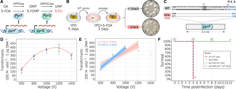
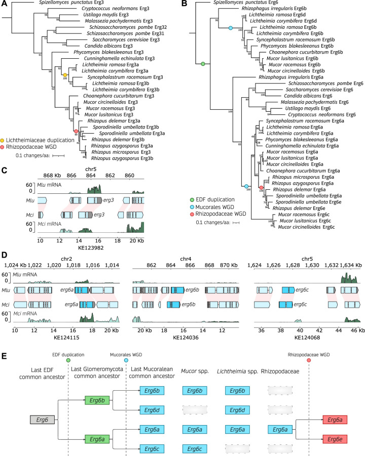
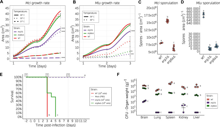
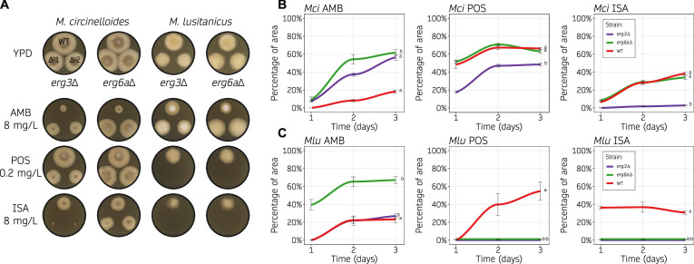
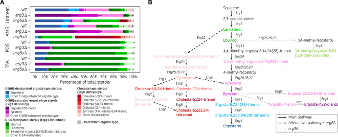
Mucormycoses are emerging fungal infections caused by a variety of heterogeneous species within the Mucorales order. Among the Mucor species complex, Mucor circinelloides is the most frequently isolated pathogen in mucormycosis patients and despite its clinical significance, there is an absence of established genome manipulation techniques to conduct molecular pathogenesis studies. In this study, we generated a spontaneous uracil auxotrophic strain and developed a genetic transformation procedure to analyze molecular mechanisms conferring antifungal drug resistance. With this new model, phenotypic analyses of gene deletion mutants were conducted to define Erg3 and Erg6a as key biosynthetic enzymes in the M. circinelloides ergosterol pathway. Erg3 is a C-5 sterol desaturase involved in growth, sporulation, virulence, and azole susceptibility. In other fungal pathogens, erg3 mutations confer azole resistance because Erg3 catalyzes the production of a toxic diol upon azole exposure. Surprisingly, M. circinelloides produces only trace amounts of this toxic diol and yet, it is still susceptible to posaconazole and isavuconazole due to alterations in membrane sterol composition. These alterations are severely aggravated by erg3Δ mutations, resulting in ergosterol depletion and, consequently, hypersusceptibility to azoles. We also identified Erg6a as the main C-24 sterol methyltransferase, whose activity may be partially rescued by the paralogs Erg6b and Erg6c. Loss of Erg6a function diverts ergosterol synthesis to the production of cholesta-type sterols, resulting in resistance to amphotericin B. Our findings suggest that mutations or epimutations causing loss of Erg6 function may arise during human infections, resulting in antifungal drug resistance to first-line treatments against mucormycosis.

Importance: The Mucor species complex comprises a variety of opportunistic pathogens known to cause mucormycosis, a potentially lethal fungal infection with limited therapeutic options. The only effective first-line treatments against mucormycosis consist of liposomal formulations of amphotericin B and the triazoles posaconazole and isavuconazole, all of which target components within the ergosterol biosynthetic pathway. This study uncovered M. circinelloides Erg3 and Erg6a as key enzymes to produce ergosterol, a vital constituent of fungal membranes. Absence of any of those enzymes leads to decreased ergosterol and consequently, resistance to ergosterol-binding polyenes such as amphotericin B. Particularly, losing Erg6a function poses a higher threat as the ergosterol pathway is channeled into alternative sterols similar to cholesterol, which maintain membrane permeability. As a result, erg6a mutants survive within the host and disseminate the infection, indicating that Erg6a deficiency may arise during human infections and confer resistance to the most effective treatment against mucormycoses.

Keywords: Erg3; Erg6; Mucorales; antifungal resistance; ergosterol; fungi; genetic tranformation.


Conflict of interest statement

The authors declare no conflict of interest.

Figures

Fig 1
Fig 1
Uracil auxotrophy enables M. circinelloides transformation based on a pyrF selectable marker. (A) Role of PyrF (orotate phosphoribosyltransferase, OPRTase) and PyrG (orotidine monophosphate decarboxylase, OMPDCase) in the uracil biosynthetic pathway, converting orotic acid (OA) to orotidine monophosphate (OMP), and OMP into uridine monophosphate (UMP), respectively. Alternatively, the same enzymes catalyze the conversion of 5-fluoroorotic acid (5-FOA) into fluoro-OMP (FOMP), and FOMP into 5-fluorouracil (5-FU), respectively. (B) Experimental design followed to isolate uracil auxotrophic strains. Briefly, wild-type spores were collected from YPD medium, counted, and one million spores were plated onto YPD containing 5-FOA. After 3 days, 5-FOA resistant colonies were isolated and transferred to new 5-FOA containing medium to confirm the resistance and then inoculated onto minimal medium either with or without uracil to confirm the auxotrophy. (C) pyrF sequences from the wild-type (WT) and uracil auxotrophic (Ura-, MIN6) strains, focusing on the 5′-region of the gene. The TATA box motif (gray background around position −25), transcription start site (arrow, position +1), pyrF coding sequence (blue font), and encoded one-letter amino acids are shown. (D, E) Transformation efficiency is measured as transformants per 100 million protoplasts per microgram of DNA using different voltages in the electroporation procedure. Individual values (dots) from biological triplicates were used to determine the mean and SD values as black lines. In (D), a smoothed curve is drawn as a red line. Significant differences among voltages were determined by a one-way ANOVA and Tukey HSD test, and voltages showing different letters indicate significant differences (P-value ≤ 0.05). In (E), two linear regression models are displayed showing a positive correlation for voltages from 600 to 1,000 V (blue line) or to 1,200 V (red line). Pearson’s correlation coefficients (R) are shown for each regression. (F) Kaplan-Meier survival curves of immunosuppressed mice after infection with M. circinelloides wildtype and transformable strains (color-coded). Intraperitoneal immunosuppressive (IS) treatments are shown. Strains showing significant differences in virulence are indicated as different letters (P-value ≤ 0.0001), assessed by a Log-rank test.
Fig 2
Fig 2
Erg3 and Erg6 evolutionary history across the Mucoromycota. (A, B) Phylogenetic tree depicting the Erg3 (A) and Erg6 (B) amino acid sequence alignments in clinically relevant fungi, with a specific focus on Mucorales. The Erg protein sequences from the chytrid Spizellomyces punctatus were employed as outgroups to root the trees. Additionally, the Erg6 sequence from Rhizophagus irregularis, a member of the Glomeromycota and the closest non-Mucoromycota sequence, was included to provide clarity on duplication events in (B). Noteworthy branching events, including whole-genome duplications (WGD) or gene duplications, are denoted by color-coded circles. Node support is represented as a percentage based on 1,000 bootstrapping iterations. The branch length is scaled to 0.1 substitutions per amino acid, as indicated in the legend. (C, D) Plots illustrating gene transcription across M. lusitanicus (Mlu) and M. circinelloides (Mci) genomic regions containing erg3 (C) and erg6 orthologs (D). The genomic coordinates for both species are displayed above (Mlu) and below (Mci) each plot. Gene annotation is depicted as arrowed blocks, indicating the direction of transcription and the boundaries of exons and introns. The coloring scheme assigns cyan blue to erg coding sequences, light blue to the remaining coding sequences, and gray to untranslated regions. Interspecies synteny among genes is depicted as pink shading. Gene transcription is shown as rRNA-depleted RNA read coverage, with the forward strand represented in dark green and the reverse strand in light green, enabling assessment of sense and antisense transcription. In some instances, the x-axes were inverted, and in those cases, the forward and reverse strand orientations were also inverted to enhance visualization and clarification. (E) Model illustrating the inferred evolutionary history of Erg6 across the early-diverging fungi (EDF).
Fig 3
Fig 3
Alterations in the ergosterol biosynthetic pathway result in decreased growth rate, sporulation, and virulence. (A, B) Growth rate of M. circinelloides (A) and M. lusitanicus (B) erg3Δ and erg6aΔ (shown as color-coded lines) on YPD medium at different temperatures (shown as distinct line types), measured as colony area across 24 hour intervals. Individual values were determined either in six biological replicates for the wild type or in three biological replicates of two independently generated mutants (six total values, grouped together for simplicity as no significant differences were detected), and used to plot a smoothed curve and SD values as black lines. Significant area differences in depicted strains compared to the wild-type growth rate across the whole time-course were determined by a one-way ANOVA and Tukey HSD test for each temperature group and shown as asterisks (**P-value ≤ 0.01, ***P-value ≤ 0.001, and ****P-value ≤ 0.0001). (C, D) Sporulation rate of M. circinelloides (C) and M. lusitanicus (D) erg3Δ and erg6aΔ after a 72 hour incubation at room temperature on YPG medium, determined as the number of spores per cm2. Values from individual measurements were obtained from three biological replicates of either the wild-type strain or two independently generated mutants (grouped together as no significant differences were found). These values are depicted in a dot plot, with standard deviation (SD) values represented as black lines. Strains are grouped by letters showing significant sporulation differences determined by a one-way ANOVA and Tukey HSD test (P-value ≤ 0.01). (E) Kaplan-Meier survival curves of immunosuppressed mice after infection with M. circinelloides wild-type and ergosterol mutants (color-coded). For erg3Δ and erg6aΔ, two independently generated mutants were injected into groups of five mice. For simplicity, after observing no significant differences in the results, survival curves were grouped together per gene deletion in groups of 10. Intraperitoneal immunosuppressive (IS) treatments are shown. Strains showing significant differences in virulence are indicated as different letters (P-value ≤ 0.01), assessed by a Log-rank test. (F) Fungal burden at 3 days post-infection in immunosuppressed mice, measured as colony-forming units (CFU) per gram of five different organs. SD and mean (black lines) were assessed from three biological triplicates. For each organ category, strains are grouped by letters showing significant fungal burden differences (one-way ANOVA and Tukey HSD test, P-value ≤ 0.01).
Fig 4
Fig 4
Loss of Erg3 or Erg6a function leads to changes in azole and polyene susceptibility. (A, B, C) Amphotericin B (AMB, 8 mg/L), posaconazole (POS, 0.2 mg/mL), and isavuconazole (ISA, 8 mg/mL) susceptibility testing of wild type and ergosterol mutants on solid YPD media. Each species was assayed at its optimal temperature, 30°C and 26°C for M. circinelloides (Mci) and M. lusitanicus (Mlu), respectively. In (A), representative images of growth after 48 hours in all of the drug containing media and the control medium without drug (YPD). Two independently generated mutants were assayed for each gene deletion (Δ−1 and Δ−2). In (B, C), Mci (B) and Mlu (C) growth rates from each mutant across time (24 hour intervals) were determined as the percentage of growth area from the same strain cultured on YPD medium with and without drug. Individual values were determined either in six biological replicates for the wild type or in three biological replicates from two independently generated mutants (six total values, grouped together for simplicity as no significant differences were detected) and used to plot a smoothed curve and SD values as black lines. Strains were grouped by letters showing significant growth rate differences across the whole time-course (one-way ANOVA and Tukey HSD test, P-value ≤ 0.01).
Fig 5
Fig 5
Ergosterol composition upon exposure to first-line mucormycosis treatments is altered by Erg3 and Erg6a deficiencies. (A) Percentage of total sterols in M. circinelloides wildtype (WT), erg3Δ, and erg6aΔ strains when exposed to different antifungal treatments (amphotericin B, posaconazole, and isavuconazole) and untreated RPMI cultures, represented in a stacked bar plot. Mean and SD (only upper bound is shown) values were determined from six replicates in the WT strain; for each mutation and condition, two independently generated mutants were tested in triplicate (total of 6 replicates). Sterols are classified into four main categories that reflect the expected end products from the main ergosterol pathway [C-5(6)-desaturated sterols], Erg3 deficiency [C-5(6)-saturated sterols], Erg6 deficiency (cholesta-type sterols), and Erg11 inhibition due to azole exposure (14-methylated sterols). Within each category, the most abundant sterols are color-coded to facilitate visualization. (B) Model of the ergosterol biosynthetic pathway in Mucorales. The main pathway is depicted by straight, black arrows. Alternative pathways that are relevant in erg3 (dashed, black arrows) or erg6a deletions (dotted, gray arrows) are also shown. For better illustration, sterol compounds are classified and color-coded as in (A).

Update of

Similar articles

Cited by

References

    1. Petrikkos G, Skiada A, Lortholary O, Roilides E, Walsh TJ, Kontoyiannis DP. 2012. Epidemiology and clinical manifestations of mucormycosis. Clin Infect Dis 54 Suppl 1:S23–34. doi:10.1093/cid/cir866 - DOI - PubMed
    1. Cornely OA, Alastruey-Izquierdo A, Arenz D, Chen SCA, Dannaoui E, Hochhegger B, Hoenigl M, Jensen HE, Lagrou K, Lewis RE, et al. . 2019. Global guideline for the diagnosis and management of mucormycosis: an initiative of the European confederation of medical mycology in cooperation with the mycoses study group education and research consortium. Lancet Infect Dis 19:e405–e421. doi:10.1016/S1473-3099(19)30312-3 - DOI - PMC - PubMed
    1. Hoenigl M, Seidel D, Carvalho A, Rudramurthy SM, Arastehfar A, Gangneux J-P, Nasir N, Bonifaz A, Araiza J, Klimko N, Serris A, Lagrou K, Meis JF, Cornely OA, Perfect JR, White PL, Chakrabarti A, ECMM and ISHAM collaborators . 2022. The emergence of COVID-19 associated mucormycosis: a review of cases from 18 countries. Lancet Microbe 3:e543–e552. doi:10.1016/S2666-5247(21)00237-8 - DOI - PMC - PubMed
    1. Gold JAW, Adjei S, Gundlapalli AV, Huang Y-LA, Chiller T, Benedict K, Toda M. 2023. Increased hospitalizations involving fungal infections during COVID-19 pandemic, United States, January 2020–December 2021. Emerg Infect Dis 29:1433–1437. doi:10.3201/eid2907.221771 - DOI - PMC - PubMed
    1. World Health Organization . 2022. WHO fungal priority pathogens list to guide research, development and public health action. Geneva.

MeSH terms

Supplementary concepts

LinkOut - more resources