. 2024 Jun 21:12:1422978.
doi: 10.3389/fcell.2024.1422978. eCollection 2024.

Acute activation of hemichannels by ethanol leads to Ca2+-dependent gliotransmitter release in astrocytes

Affiliations

Acute activation of hemichannels by ethanol leads to Ca2+-dependent gliotransmitter release in astrocytes

Gonzalo I Gómez et al. Front Cell Dev Biol. .

Abstract

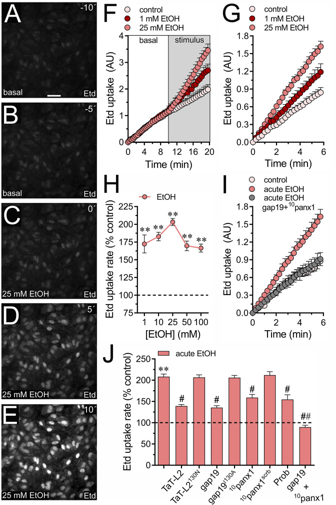
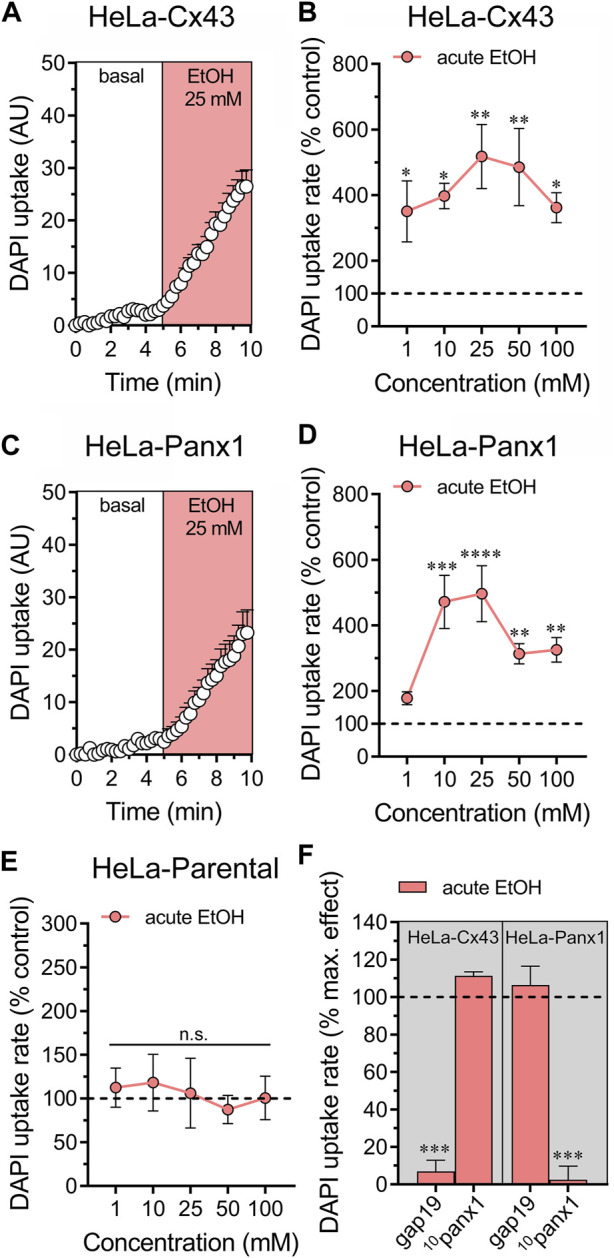
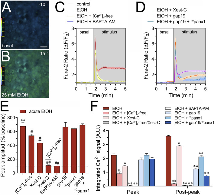
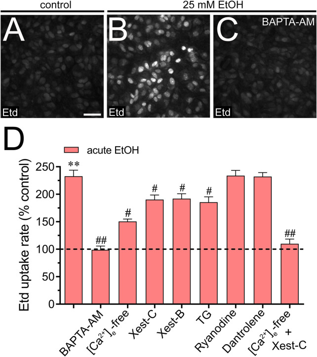
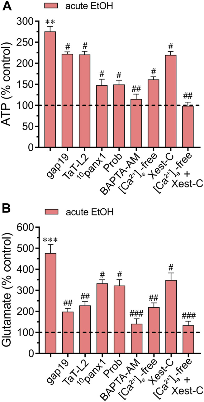
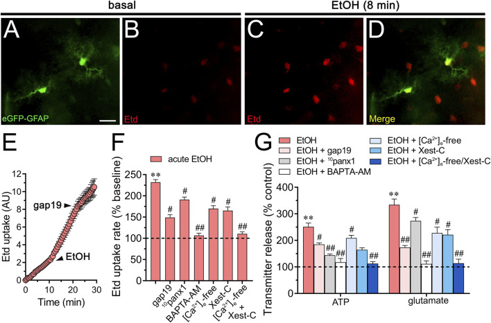
Multiple studies have demonstrated that acute ethanol consumption alters brain function and cognition. Nevertheless, the mechanisms underlying this phenomenon remain poorly understood. Astrocyte-mediated gliotransmission is crucial for hippocampal plasticity, and recently, the opening of hemichannels has been found to play a relevant role in this process. Hemichannels are plasma membrane channels composed of six connexins or seven pannexins, respectively, that oligomerize around a central pore. They serve as ionic and molecular exchange conduits between the cytoplasm and extracellular milieu, allowing the release of various paracrine substances, such as ATP, D-serine, and glutamate, and the entry of ions and other substances, such as Ca2+ and glucose. The persistent and exacerbated opening of hemichannels has been associated with the pathogenesis and progression of several brain diseases for at least three mechanisms. The uncontrolled activity of these channels could favor the collapse of ionic gradients and osmotic balance, the release of toxic levels of ATP or glutamate, cell swelling and plasma membrane breakdown and intracellular Ca2+ overload. Here, we evaluated whether acute ethanol exposure affects the activity of astrocyte hemichannels and the possible repercussions of this phenomenon on cytoplasmatic Ca2+ signaling and gliotransmitter release. Acute ethanol exposure triggered the rapid activation of connexin43 and pannexin1 hemichannels in astrocytes, as measured by time-lapse recordings of ethidium uptake. This heightened activity derived from a rapid rise in [Ca2+]i linked to extracellular Ca2+ influx and IP3-evoked Ca2+ release from intracellular Ca2+ stores. Relevantly, the acute ethanol-induced activation of hemichannels contributed to a persistent secondary increase in [Ca2+]i. The [Ca2+]i-dependent activation of hemichannels elicited by ethanol caused the increased release of ATP and glutamate in astroglial cultures and brain slices. Our findings offer fresh perspectives on the potential mechanisms behind acute alcohol-induced brain abnormalities and propose targeting connexin43 and pannexin1 hemichannels in astrocytes as a promising avenue to prevent deleterious consequences of alcohol consumption.

Keywords: alcoholism; connexin 43; connexins; glia; hemichannels; pannexin-1; pannexins.


Conflict of interest statement

The authors declare that the research was conducted in the absence of any commercial or financial relationships that could be construed as a potential conflict of interest. The author(s) declared that they were an editorial board member of Frontiers, at the time of submission. This had no impact on the peer review process and the final decision

Figures

FIGURE 1
FIGURE 1
Acute ethanol exposure increases the activity of Cx43 and Panx1 hemichannels in cultured astrocytes. (A–E) Representative fluorescence micrographs illustrating time-lapse recordings of Etd uptake by astrocytes for 10 (A) and 5 (B) seconds before and after stimulation with 25 mM ethanol (EtOH) for 0 (C), 5 (D), or 10 (E) seconds. (F) Time-lapse measurements of Etd uptake by astrocytes under control conditions (pale pink circles) or after the acute treatment with 1 mM (dark red circles) or 25 mM (salmon red circles) ethanol. (G) Time-lapse measurements of Etd uptake by astrocytes under control conditions (pale pink circles) or treated for 1 min before and during recordings with 1 mM (dark red circles) or 25 mM (salmon red circles) ethanol. (H) Averaged Etd uptake rate normalized with the control condition (dashed line) by astrocytes acutely treated with different concentrations of ethanol (salmon red circles). **p < 0.005, ethanol treatment compared to control conditions (one-way ANOVA followed by Tukey’s post hoc test). (I) Time-lapse measurements of Etd uptake by astrocytes under control conditions (pale pink circles) or treated for 1 min before and during recordings with 25 mM ethanol alone (salmon red circles) or in combination with 50 µM gap19 + 50 µM 10panx1 (gray circles). (J) Averaged Etd uptake rate normalized with control condition (dashed line) by astrocytes acutely treated with 25 mM ethanol alone or in combination with the following blockers: 50 µM TaT-L2, 50 µM TaT-L2H126K/I130N, 50 µM gap19, 50 µM gap19I130A, 50 µM 10panx1, 50 µM 10panx1scrb, 500 µM Probenecid (Prob), and 50 µM gap19 + 50 µM 10panx1. **p < 0.0005, ethanol compared to control; # p < 0.05, ## p < 0.005; effect of pharmacological agents compared to ethanol treatment (one-way ANOVA followed by Tukey’s post hoc test). Data were obtained from at least three independent experiments with three or more repeats each one (≥25 cells analyzed for each repeat). Calibration bar = 150 μm.
FIGURE 2
FIGURE 2
Ethanol acutely augments the activity of hemichannels in HeLa cells transfected with Cx43 or Panx1. (A) Time-lapse measurements of DAPI uptake by HeLa-Cx43EGFP cells under basal conditions and then acutely exposed to 25 mM ethanol. (B) Averaged DAPI uptake rate normalized with the control condition (dashed line) by HeLa-Cx43EGFP cells acutely treated with different concentrations of ethanol (salmon red circles). (C) Time-lapse measurements of DAPI uptake by HeLa-Panx1EGFP cells under basal conditions and then exposed to 25 mM ethanol. (D) Averaged DAPI uptake rate normalized with the control condition (dashed line) by HeLa-Panx1EGFP cells acutely treated with different concentrations of ethanol (salmon red circles). (E) Averaged DAPI uptake rate normalized with the control condition (dashed line) by HeLa parental cells acutely treated with different concentrations of ethanol (salmon red circles). (F) Averaged DAPI uptake rate normalized to the maximun effect evoked by 25 mM ethanol (dashed line) by HeLa-Cx43EGFP or HeLa-Panx1EGFP cells pretreated with the following blockers: 50 µM gap19 or 50 µM 10panx1. ***p < 0.0001, effect of pharmacological agents compared to ethanol treatment (two-tailed Student’s unpaired t-test). Data were obtained from at least three independent experiments with three or more repeats each one (≥15 cells analyzed for each repeat).
FIGURE 3
FIGURE 3
Cx43 and Panx1 hemichannels contribute to the acute ethanol-induced rise on [Ca2+]i by cultured astrocytes. (A–B) Representative photomicrographs of Ca2+ signal (340/380 nm ratio) by astrocytes for 10 s before (A) and after stimulation with 25 mM ethanol for 15 s (B). (C–D) Representative plots of relative changes in Ca2+ signal over time by astrocytes under control conditions (black line) or after the acute treatment with 25 mM ethanol (red line) alone or in combination with the following agents or conditions: [Ca2+]i-free bath solution (yellow line), 10 µM BAPTA-AM (purple line), 5 µM Xest-C (magenta line), 50 µM gap19 (orange line) and 50 µM gap19 + 50 µM 10panx1 (light blue line). (E) Averaged data of ethanol-induced peak amplitude by astrocytes normalized to basal Fura-2a.m. ratio. In addition, the effect of the following agents or conditions are shown: [Ca2+]i-free bath solution, 5 µM Xest-C, [Ca2+]i-free bath solution + 5 µM Xest-C, 10 µM BAPTA-AM, 50 µM gap19, 50 µM 10panx1, or 50 µM gap19 + 50 µM 10panx1. **p < 0.0005, ethanol compared to basal condition; # p < 0.05, ## p < 0.005; effect of pharmacological agents compared to ethanol treatment (one-way ANOVA followed by Tukey’s post hoc test). (F) Averaged data of the area under the curve during and after the ethanol-induced peak of Ca2+ signal by astrocytes. In addition, the effect of the following agents or conditions are shown: [Ca2+]i-free bath solution, 5 µM Xest-C, [Ca2+]i-free bath solution + 5 µM Xest-C, 10 µM BAPTA-AM, 50 µM gap19, 50 µM 10panx1, or 50 µM gap19 + 50 µM 10panx1. *p < 0.05, **p < 0.005; effect of pharmacological agents compared to ethanol treatment (one-way ANOVA followed by Tukey’s post hoc test). Data were obtained from at least three independent experiments with three or more repeats each one (≥25 cells analyzed for each repeat). Calibration bar: 150 μm.
FIGURE 4
FIGURE 4
Extracellular Ca2+ influx and Ca2+ release from intracellular stores contribute to the acute ethanol-induced activation of Cx43 and Panx1 hemichannels in cultured astrocytes. (A–C) Representative fluorescence images depicting Etd uptake measurements (5 min of recording) in astrocytes under control conditions (A) or acutely treated with 25 mM ethanol (B) alone or plus 10 µM BAPTA-AM (C). (D) Averaged Etd uptake rate normalized with control condition (dashed line) by astrocytes acutely treated with 25 mM ethanol alone or in combination with the following blockers or conditions: 10 µM BAPTA-AM, [Ca2+]i-free bath solution, 5 µM Xest-C, 5 µM Xest-B, 2 µM thapsigargin (TG), 100 µM ryanodine, 50 µM dantrolene or [Ca2+]i-free bath solution + 5 µM Xest-C. **p < 0.0005, ethanol compared to control; # p < 0.05, ## p < 0.005; effect of pharmacological agents compared to ethanol treatment (one-way ANOVA followed by Tukey’s post hoc test). Data were obtained from at least three independent experiments with three or more repeats each one (≥25 cells analyzed for each repeat). Calibration bar = 150 μm.
FIGURE 5
FIGURE 5
Acute ethanol exposure increases the release of gliotransmitters via the opening of Cx43 and Panx1 hemichannels. Averaged data of ATP (A) or glutamate (B) normalized to control conditions (dashed line) by astrocytes acutely treated with 25 mM ethanol alone or in combination with the following agents or conditions: 50 µM gap19, 50 µM TaT-L2, 50 µM 10panx1, 500 µM Probenecid (Prob), 10 µM BAPTA-AM, [Ca2+]i-free bath solution, 5 µM Xest-C or [Ca2+]i-free bath solution + 5 µM Xest-C. **p < 0.0005, ***p < 0.0001, ethanol compared to control; # p < 0.05, ## p < 0.005, ### p < 0.001; effect of pharmacological agents compared to ethanol treatment (one-way ANOVA followed by Tukey’s post hoc test). Data were obtained from at least three independent experiments with three or more repeats each one.
FIGURE 6
FIGURE 6
Acute ethanol exposure increases the release of ATP and glutamate in brain slices by a mechanism involving the activation of Cx43 and Panx1 hemichannels in a [Ca2+]i-dependent manner. (A–D) Representative fluorescence micrographs of Etd uptake (red) in hippocampal eGFP-GFAP astrocytes (green) from acute brain slices under basal conditions (A, B) and after the acute treatment with 25 mM ethanol for 8 min (C, D). (E) Time-lapse measurements of Etd uptake by eGFP positive astrocytes under basal conditions, after the acute treatment with 25 mM ethanol and upon the acute treatment with 50 µM gap19. (F) Averaged Etd uptake rate normalized with control condition (dashed line) by eGFP positive astrocytes acutely treated with 25 mM ethanol alone or in combination with the following blockers or conditions: 50 µM gap19, 50 µM 10panx1,10 µM BAPTA-AM, [Ca2+]i-free bath solution, 5 µM Xest-C, or [Ca2+]i-free bath solution + 5 µM Xest-C. **p < 0.0005, ethanol compared to control; # p < 0.05, ## p < 0.005; effect of pharmacological agents compared to ethanol treatment (one-way ANOVA followed by Tukey’s post hoc test). Data were obtained from at least three independent experiments with three or more repeats each one (≥5 cells analyzed for each repeat). (G) Averaged data of ATP (left) or glutamate (right) normalized to control conditions (dashed line) by astrocytes acutely treated with 25 mM ethanol alone or in combination with the following agents or conditions: 50 µM gap19, 50 µM 10panx1, 10 µM BAPTA-AM, [Ca2+]i-free bath solution, 5 µM Xest-C, or [Ca2+]i-free bath solution + 5 µM Xest-C. **p < 0.0005, ethanol compared to control; # p < 0.05, ## p < 0.005; effect of pharmacological agents compared to ethanol treatment (one-way ANOVA followed by Tukey’s post hoc test). Data were obtained from at least three independent experiments with three or more repeats each one. Calibration bar = 60 μm.

Similar articles

Cited by

  • The role of astrocytes in depression, its prevention, and treatment by targeting astroglial gliotransmitter release.
    Duarte Y, Quintana-Donoso D, Moraga-Amaro R, Dinamarca I, Lemunao Y, Cárdenas K, Bahamonde T, Barrientos T, Olivares P, Navas C, Carvajal FJ, Santibánez Y, Castro-Lazo R, Paz Meza M, Jorquera R, Gómez GI, Henke M, Alarcón R, Gabriel LA, Schiffmann S, Cerpa W, Retamal MA, Simon F, Linsambarth S, Gonzalez-Nilo F, Stehberg J. Duarte Y, et al. Proc Natl Acad Sci U S A. 2024 Nov 12;121(46):e2307953121. doi: 10.1073/pnas.2307953121. Epub 2024 Nov 4. Proc Natl Acad Sci U S A. 2024. PMID: 39495924

References

    1. Abrahao K. P., Salinas A. G., Lovinger D. M. (2017). Alcohol and the brain: neuronal molecular targets, synapses, and circuits. Neuron 96, 1223–1238. 10.1016/j.neuron.2017.10.032 - DOI - PMC - PubMed
    1. Adermark L., Olsson T., Hansson E. (2004). Ethanol acutely decreases astroglial gap junction permeability in primary cultures from defined brain regions. Neurochem. Int. 45, 971–978. 10.1016/j.neuint.2004.06.007 - DOI - PubMed
    1. Aghajanian G. K., Rasmussen K. (1989). Intracellular studies in the facial nucleus illustrating a simple new method for obtaining viable motoneurons in adult rat brain slices. Synapse 3, 331–338. 10.1002/syn.890030406 - DOI - PubMed
    1. Albrecht J., Zielinska M. (2017). Mechanisms of excessive extracellular glutamate accumulation in temporal lobe epilepsy. Neurochem. Res. 42, 1724–1734. 10.1007/s11064-016-2105-8 - DOI - PubMed
    1. Allansson L., Khatibi S., Olsson T., Hansson E. (2001). Acute ethanol exposure induces [Ca2+]i transients, cell swelling and transformation of actin cytoskeleton in astroglial primary cultures. J. Neurochem. 76, 472–479. 10.1046/j.1471-4159.2001.00097.x - DOI - PubMed

Grants and funding

The author(s) declare that financial support was received for the research, authorship, and/or publication of this article. This work was supported by the Agencia Nacional de Investigación y Desarrollo (ANID) and Fondo Nacional de Desarrollo Científico y Tecnológico (FONDECYT) Grants 1210375 (to JAO), 11171155 (to MR), 1231523 (to JCS) and 1210940 (to FCO).