. 2024 Jul 6;15(1):203.
doi: 10.1186/s13287-024-03803-3.

Rankl genetic deficiency and functional blockade undermine skeletal stem and progenitor cell differentiation

Affiliations

Rankl genetic deficiency and functional blockade undermine skeletal stem and progenitor cell differentiation

M L Schiavone et al. Stem Cell Res Ther. .

Abstract

Background: Skeletal Stem Cells (SSCs) are required for skeletal development, homeostasis, and repair. The perspective of their wide application in regenerative medicine approaches has supported research in this field, even though so far results in the clinic have not reached expectations, possibly due also to partial knowledge of intrinsic, potentially actionable SSC regulatory factors. Among them, the pleiotropic cytokine RANKL, with essential roles also in bone biology, is a candidate deserving deep investigation.

Methods: To dissect the role of the RANKL cytokine in SSC biology, we performed ex vivo characterization of SSCs and downstream progenitors (SSPCs) in mice lacking Rankl (Rankl-/-) by means of cytofluorimetric sorting and analysis of SSC populations from different skeletal compartments, gene expression analysis, and in vitro osteogenic differentiation. In addition, we assessed the effect of the pharmacological treatment with the anti-RANKL blocking antibody Denosumab (approved for therapy in patients with pathological bone loss) on the osteogenic potential of bone marrow-derived stromal cells from human healthy subjects (hBMSCs).

Results: We found that, regardless of the ossification type of bone, osteochondral SSCs had a higher frequency and impaired differentiation along the osteochondrogenic lineage in Rankl-/- mice as compared to wild-type. Rankl-/- mice also had increased frequency of committed osteochondrogenic and adipogenic progenitor cells deriving from perivascular SSCs. These changes were not due to the peculiar bone phenotype of increased density caused by lack of osteoclast resorption (defined osteopetrosis); indeed, they were not found in another osteopetrotic mouse model, i.e., the oc/oc mouse, and were therefore not due to osteopetrosis per se. In addition, Rankl-/- SSCs and primary osteoblasts showed reduced mineralization capacity. Of note, hBMSCs treated in vitro with Denosumab had reduced osteogenic capacity compared to control cultures.

Conclusions: We provide for the first time the characterization of SSPCs from mouse models of severe recessive osteopetrosis. We demonstrate that Rankl genetic deficiency in murine SSCs and functional blockade in hBMSCs reduce their osteogenic potential. Therefore, we propose that RANKL is an important regulatory factor of SSC features with translational relevance.

Keywords: Denosumab; Differentiation; Osteopetrosis; RANKL; Skeletal stem cells; Therapy.


Conflict of interest statement

The authors declare that they have no conflict of interest.

Figures
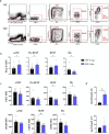
Fig. 1
Fig. 1
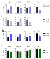
FACS sorting and characterization of osteochondral SSC (ocSSCs) and downstream progenitors in WT and Rankl−/− mice. a Representative contour plots of the gating strategy for both genotypes. b Abundance of the different ocSSPC populations, expressed as frequency of CD51+ cells, in 5-week-old mice of both genotypes. c Expression level of the positive markers in the indicated cell populations, expressed as mean fluorescence intensity (MFI). d Gene expression level of the RANKL receptors Rank and Lgr4 in FACS-sorted ocSSCs, normalized on Gapdh and expressed as Arbitrary Units (A.U.). All the data are expressed as mean ± SEM. * p < 0.05, ** p < 0.01; Mann-Whitney test. KO: knockout, Rankl−/−
Fig. 2
Fig. 2
Comparative analysis of ocSSCs and downstream progenitors in WT and Rankl−/− mice at different ages and in different skeletal compartments, and in oc/oc mice. a Abundance of the different ocSSPC populations, expressed as frequency of CD51+ cells, in 3-week-old WT and Rankl−/− mice. b Analysis of ocSSPCs in 1-week-old WT and Rankl−/− mice. c Analysis of ocSSPCs isolated from the skull of 5-week-old WT and Rankl−/− mice. d Abundance of the different ocSSPC populations in WT and oc/oc mice. All the data are expressed as mean ± SEM. * p < 0.05, *** p < 0.001; Mann-Whitney test. KO: knockout, Rankl−/−
Fig. 3
Fig. 3
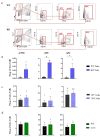
FACS sorting and characterization of perivascular SSC (pvSSC) and downstream progenitors. a Representative contour plots of the gating strategy for WT and Rankl−/− mice. b Abundance of the different pvSSPC populations, expressed as frequency of live cells, in 5-week-old WT and Rankl−/− mice. c Analysis of pvSSPCs in 3-week-old WT and Rankl−/− mice. d Abundance of the different pvSSPC populations in WT and oc/oc mice. All the data are expressed as mean ± SEM. * p < 0.05; Mann-Whitney test. KO: knockout, Rankl−/−
Fig. 4
Fig. 4
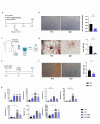
Characterization of the osteogenic potential of WT and Rankl−/− ocSSCs and primary osteoblasts. a Schematic representation of the experimental protocol for the assessment of the osteogenic potential of ocSSCs in clonogenic conditions. Briefly, Rankl−/− and WT ocSSCs were plated immediately after sorting at clonal density in basal medium. After 7 days, the cultures were exposed to OIM for 3 weeks. b Representative images of Alizarin Red Staining (ARS) at the end of osteogenic induction of the cultures described in a. The stain was chemically extracted, quantified by absorbance (Abs) reading at 405 nm and normalized on the number of colonies formed. c Schematic representation of the experimental protocol for the assessment of the osteogenic potential of WT and Rankl−/− ocSSC lines. Briefly, after in vitro expansion of sorted Rankl−/− and WT ocSSCs, the cell lines obtained were treated with OIM for 3 weeks. d Representative images of ARS at the end of osteogenic induction of the cultures described in c and quantization of the extracted stain by Abs reading at 405 nm. e Schematic representation of the experimental protocol for the assessment of the osteogenic potential of WT and Rankl−/− osteoblasts. Briefly, Rankl−/− and WT primary osteoblast cultures were established and induced to mineralize according to standard protocols. f Representative images of ARS at the end of osteogenic induction of the cultures described in e and quantization as in d. Scale bar in b, d and f: 500 μm. Higher magnifications of this panels are provided in Figure S2. g Gene expression analysis of representative osteogenic genes in WT and Rankl−/− pre- and mature osteoblasts, normalized on Gapdh and expressed as Arbitrary Units (A.U.). All the data are expressed as mean ± SEM. * p < 0.05; b, d, f: Mann-Whitney test; g: Kruskal-Wallis test. OIM: Osteogenic Induction Medium. pOBs: primary osteoblasts. KO: knockout, Rankl−/−
Fig. 5
Fig. 5
Expression profile of WT and Rankl−/− ocSSC lines. a Representative contour plots of the gating strategy for FACS analysis of WT and Rankl−/− ocSSC lines, using the same surface markers exploited for their isolation from bone cell suspensions. Cell populations were gated on CD45/Ter119; as expected, all cells were negative for these markers. b Frequency of cells displaying the immunophenotype of SSCs (as defined by Chan et al., 2015 and adopted in this work) after the establishment of WT and Rankl−/− ocSSC lines. c Gene expression analysis of representative stemness markers in WT and Rankl−/− ocSSC lines, normalized on Gapdh and expressed as Arbitrary Units (A.U.). All the data are expressed as mean ± SEM. * p < 0.05; Kruskal-Wallis test. KO: knockout, Rankl−/−
Fig. 6
Fig. 6
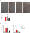
Impact of Denosumab treatment on the mineralization capacity of human BMSCs. a Representative image of ARS-stained cultures of human BMSCs from healthy donors after 2 weeks of culture either in basal medium (CTR) or in OIM alone or in OIM + isotype control (OIM + ISO) or in OIM + Denosumab (OIM + Dmab); in all the conditions, the culture medium was changed twice a week. At the end, the mineral deposition was quantified and indicated by Abs reading at 405 nm. Scale bar: 500 μm. Higher magnifications are provided in Figure S2. b Gene expression analysis of representative osteogenic genes in human BMSCs in the different treatment conditions indicated; normalization on 18 S. Results are expressed as Arbitrary Units (A.U.). All the data are represented as mean ± SEM. * p < 0.05; a: Friedman test; b: Kruskal-Wallis test. CTR: control. OIM: Osteogenic Induction Medium. ISO: isotype control. Dmab: Denosumab

Similar articles

Cited by

References

    1. Bianco P, Robey PG. Skeletal stem cells. Development. 2015;142(6):1023–7. doi: 10.1242/dev.102210. - DOI - PMC - PubMed
    1. Ortinau LC, Wang H, Lei K, Deveza L, Jeong Y, Hara Y, Grafe I, Rosenfeld SB, Lee D, Lee B, et al. Identification of functionally distinct Mx1 + αSMA + periosteal skeletal stem cells. Cell Stem Cell. 2019;25(6):784–e7965. doi: 10.1016/j.stem.2019.11.003. - DOI - PMC - PubMed
    1. Matsushita Y, Ono W, Ono N. Skeletal stem cells for Bone Development and Repair: diversity matters. Curr Osteoporos Rep. 2020;18(3):189–98. doi: 10.1007/s11914-020-00572-9. - DOI - PMC - PubMed
    1. Menon S, Salhotra A, Shailendra S, Tevlin R, Ransom RC, Januszyk M, Chan CKF, Behr B, Wan DC, Longaker MT, et al. Skeletal stem and progenitor cells maintain cranial suture patency and prevent craniosynostosis. Nat Commun. 2021;12(1):4640. doi: 10.1038/s41467-021-24801-6. - DOI - PMC - PubMed
    1. Xu J, Wang Y, Li Z, Tian Y, Li Z, Lu A, Hsu CY, Negri S, Tang C, Tower RJ, et al. PDGFRα reporter activity identifies periosteal progenitor cells critical for bone formation and fracture repair. Bone Res. 2022;10(1):7. doi: 10.1038/s41413-021-00176-8. - DOI - PMC - PubMed

LinkOut - more resources