. 2024 Sep;131(5):918-930.
doi: 10.1038/s41416-024-02774-9. Epub 2024 Jul 5.

HIV-protease inhibitors potentiate the activity of carfilzomib in triple-negative breast cancer

Affiliations

HIV-protease inhibitors potentiate the activity of carfilzomib in triple-negative breast cancer

Andrej Besse et al. Br J Cancer. 2024 Sep.

Abstract

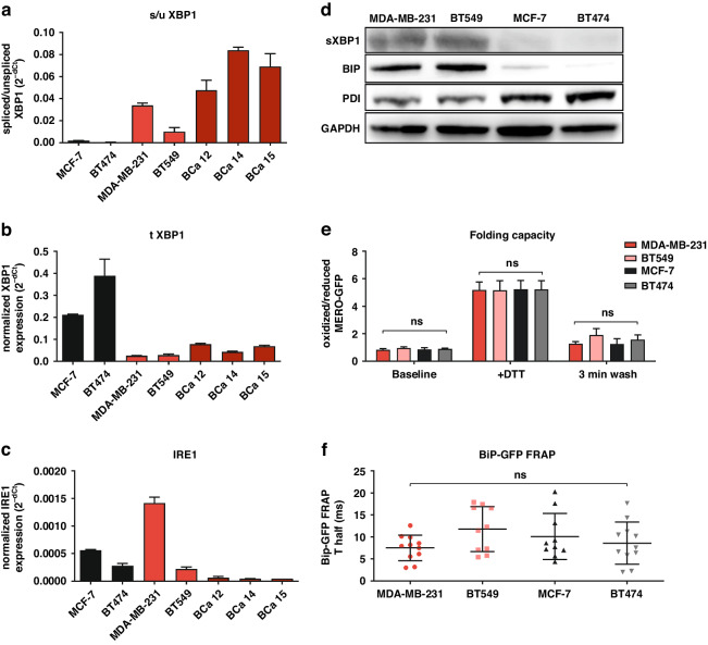
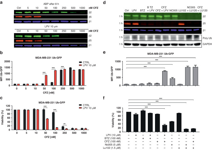
Background: Resistance to chemotherapy is a major problem in the treatment of patients with triple-negative breast cancer (TNBC). Preclinical data suggest that TNBC is dependent on proteasomes; however, clinical observations indicate that the efficacy of proteasome inhibitors in TNBC may be limited, suggesting the need for combination therapies.

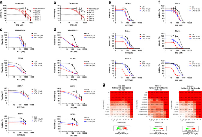
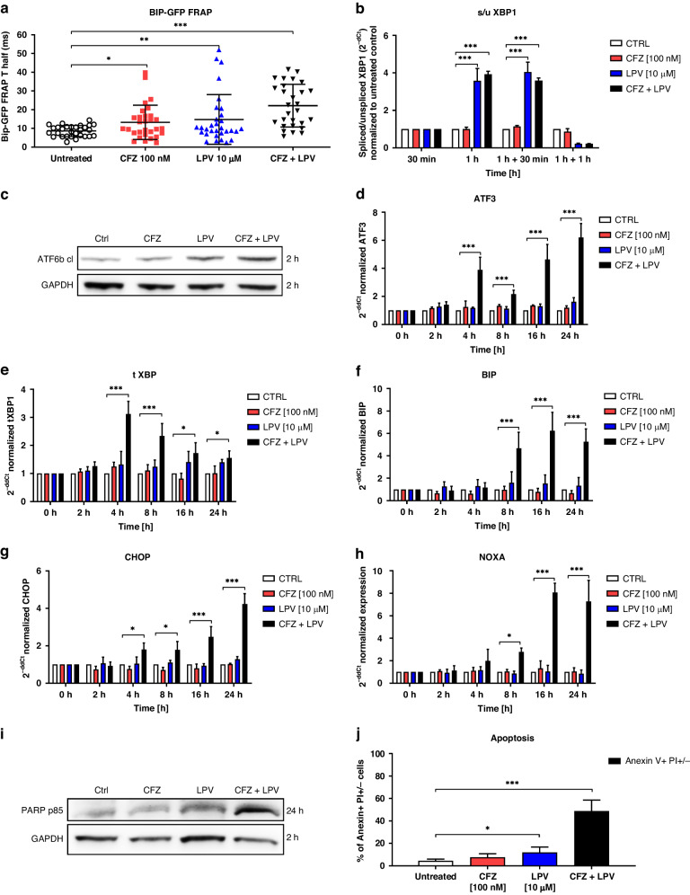
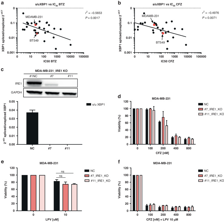
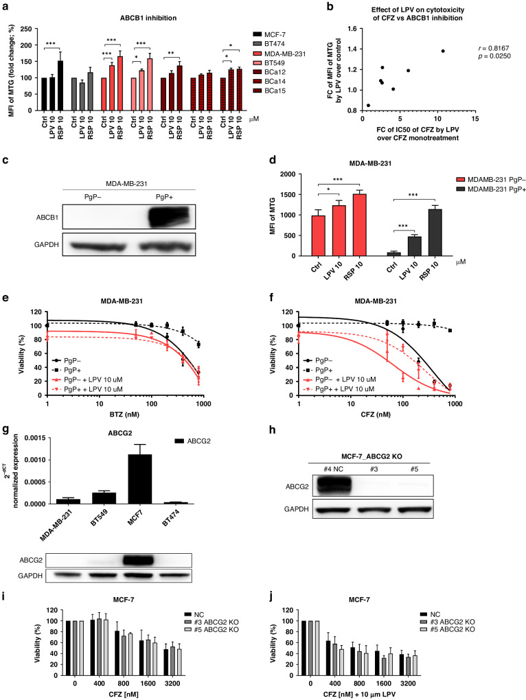
Methods: We compared bortezomib and carfilzomib and their combinations with nelfinavir and lopinavir in TNBC cell lines and primary cells with regard to their cytotoxic activity, functional proteasome inhibition, and induction of the unfolded protein response (UPR). Furthermore, we evaluated the involvement of sXBP1, ABCB1, and ABCG2 in the cytotoxic activity of drug combinations.

Results: Carfilzomib, via proteasome β5 + β2 inhibition, is more cytotoxic in TNBC than bortezomib, which inhibits β5 + β1 proteasome subunits. The cytotoxicity of carfilzomib was significantly potentiated by nelfinavir or lopinavir. Carfilzomib with lopinavir induced endoplasmic reticulum stress and pro-apoptotic UPR through the accumulation of excess proteasomal substrate protein in TNBC in vitro. Moreover, lopinavir increased the intracellular availability of carfilzomib by inhibiting carfilzomib export from cells that express high levels and activity of ABCB1, but not ABCG2.

Conclusion: Proteasome inhibition by carfilzomib combined with nelfinavir/lopinavir represents a potential treatment option for TNBC, warranting further investigation.


Conflict of interest statement

The authors declare no competing interests.

Figures

Fig. 1
Fig. 1. UPR activation status and folding capacity in breast cancer.
a Basal state of spliced XBP1 (sXBP1) in cell lines and patient-derived primary cells evaluated by qPCR, assessed as a ratio of spliced vs unspliced XBP1 and normalized to GAPDH, which served as a housekeeping gene. The data represent the mean ± SD from 3 independent experiments. b Basal state of total XBP1 (tXBP1) in cell lines and patient-derived primary cells evaluated by qPCR and normalized to GAPDH, which served as a housekeeping gene. The data represent the mean ± SD from 3 independent experiments. c Basal state of IRE1 in cell lines and patient-derived primary cells evaluated by qPCR and normalized to GAPDH, which served as a housekeeping gene. The data represent the mean ± SD from 3 independent experiments. d Basal state of proteins involved in the UPR and protein folding. Western blot analysis was performed with TNBC (MDA-MB-231 and BT549) and non-TNBC (MCF-7 and BT474) cells. Representative images of three independent experiments are shown. GAPDH served as an internal loading control. e Ratio of oxidized vs reduced MERO-GFP in the cell lines evaluated by flow cytometry at baseline, immediately after the exposure to DTT and 3 min after washing DTT away. The data represent the mean ± SD from 3 independent experiments. f BiP-GFP FRAP analysis of the cell lines at the basal state. The data represent the mean ± SD of T half (in ms) recovery of BiP-GFP fluorescence, evaluated in the individual cells in two independent experiments. BiP-GFP Binding immunoglobulin Protein green-fluorescent protein, DTT dithiothreitol, GAPDH Glyceraldehyde-3-Phosphate Dehydrogenase, IRE1 Inositol-Requiring Enzyme 1, MERO-GFP Mammalian endoplasmic reticulum-localized redox-sensitive green-fluorescent protein, PDI protein disulfide isomerase, XBP1 X-Box-Binding Protein 1.
Fig. 2
Fig. 2. Response of breast cancer cell lines, primary cells and PDX-organoids to bortezomib/carfilzomib alone or in combination with nelfinavir/lopinavir.
TNBC cell lines MDA-MB-231 and BT549, non-TNBC cell lines MCF-7 and BT474, and TNBC-primary cells were treated for 1 h with increasing doses of bortezomib (a) or carfilzomib (b). Subsequently, the cells were placed in a drug-free medium for the next 48 h. The data represent the mean ± SD of 3 independent experiments. Cell lines were treated for 1 h with increasing doses of bortezomib (c) or carfilzomib (d). Subsequently, the cells were placed in a drug-free medium or incubated with 10 µM nelfinavir or lopinavir for the next 48 h. The data represent the mean ± SD of 3 independent experiments. Viability at the selected time point at which CDI was calculated is depicted in Fig. S1, S2. TNBC patient-derived cells were treated for 1 h with increasing doses of bortezomib (e) or carfilzomib (f). Subsequently, the cells were placed in a drug-free medium or incubated with 10 µM nelfinavir or lopinavir for the next 48 h. Cell viability was measured 48 h after treatment, and the data represent the mean ± SD of 2 independent experiments. Viability at a selected time point at which CDI was calculated is depicted in Fig. S3, S4. In all experiments, the corresponding IC50 values were determined from the dose-response curves and are presented in Supplementary Table S3. g Cytotoxicity of carfilzomib and nelfinavir in the three PDxOs. Numbers represent the percentage of growth inhibition. Drug synergy was modelled using SynergyFinder+, CDI of the most synergistic drug combination is presented. BTZ bortezomib, CFZ carfilzomib, LPV lopinavir, NFV nelfinavir.
Fig. 3
Fig. 3. Induction of proteotoxic stress, UPR, and apoptosis in MDA-MB-231 cell line treated with carfilzomib and lopinavir.
a BiP-GFP FRAP analysis in MDA-MB-231 cells treated with carfilzomib, lopinavir, or their combination. FRAP images were acquired 1 h after the 1 h pulse treatment with carfilzomib or continuous treatment with lopinavir. The data represent the mean ± SD of T half (in ms) recovery of BiP-GFP fluorescence in the individual cells in three independent experiments. Statistical significance was determined with one-way ANOVA with Tukey post-test. * represents p < 0.05; ** represents p < 0.01; *** represents p < 0.001. b Induction of spliced XBP1 (sXBP1) presented as a ratio of spliced versus unspliced XBP1 RNA variants normalized to GAPDH and a time-point 30 min prior to the treatment. c Representative western blot image of the cleavage of ATF6 protein, represented by a cleaved form of ATF6 and obtained 2 h after the 1 h pulse treatment with carfilzomib or continuous treatment with lopinavir. GAPDH served as an internal loading control. d Induction of ATF3 expression normalized to GAPDH and a time-point 0 h after the 1 h pulse treatment with carfilzomib or continuous treatment with lopinavir. e Induction of total XBP1 (tXBP1) expression, normalized to GAPDH and a time-point 0 h after the 1 h pulse treatment with carfilzomib or continuous treatment with lopinavir. f Induction of BIP expression normalized to GAPDH and a time-point 0 h after the 1 h pulse treatment with carfilzomib or continuous treatment with lopinavir. g Induction of CHOP expression normalized to GAPDH and a time-point 0 h after the 1 h pulse treatment with carfilzomib or continuous treatment with lopinavir. h Induction of NOXA expression normalized to GAPDH and a time-point 0 h after the 1 h pulse treatment with carfilzomib or continuous treatment with lopinavir. i Representative western blot image of the induction of PARP on a protein level, represented by a cleaved form of PARP p85 and obtained 24 h after the 1 h pulse treatment with carfilzomib or continuous treatment with lopinavir. GAPDH served as an internal loading control. j Induction of early and late apoptosis represented by Anexin V + and PI− + PI+ positivity, evaluated by flow cytometry 48 h after the 1 h pulse treatment with carfilzomib or continuous treatment with lopinavir. The data represent the mean ± SD from 3 independent experiments. Statistical significance was determined with an unpaired two-sided t-test. * represents p < 0.05, *** represents p < 0.001. In all qPCR experiments, the data represent the mean ± SD from 3 independent experiments. Statistical significance was determined with one-way ANOVA with Tukey post-test. * represents p < 0.05, *** represents p < 0.001. ATF3 Activating Transcription Factor 6, ATF6 Activating Transcription Factor 6, BiP-GFP Binding immunoglobulin Protein green-fluorescent protein, BTZ bortezomib, CFZ carfilzomib, CHOP DNA Damage-Inducible Transcript 3, GAPDH Glyceraldehyde-3-Phosphate Dehydrogenase, LPV lopinavir, mero-GFP Mammalian Endoplasmic Reticulum-localized redox-sensitive Green-Fluorescent Protein, NOXA Phorbol-12-Myristate-13-Acetate-Induced Protein 1, PARP Poly(ADP-Ribose) Polymerase 1, XBP1 X-Box Binding Protein 1.
Fig. 4
Fig. 4. Dissection of the role of sXBP1 in monotherapy with carfilzomib and lopinavir or in the drug combination in MDA-MB-231.
a Correlation between spliced XBP1 expression level, represented as a ratio of spliced / unspliced XBP1 and normalized to GAPDH, and the sensitivity to bortezomib, represented by the IC50 values. b Correlation between spliced XBP1 expression level, represented as a ratio of spliced/unspliced XBP1 and normalized to GAPDH, and the sensitivity to carfilzomib, represented by the IC50 values. In both a and b, TNBC are marked in red. The data represent the mean of two independent experiments. c Representative western blot image of IRE1α knock-out in control or in two single-cell derived clones #7 and #11 of MDA-MB-231 cell line (upper part), leading to a functional decrease of sXBP1 evaluated by qPCR, assessed as a ratio of spliced vs unspliced XBP1 and normalized to GAPDH, which served as a housekeeping gene (bottom part). d Cytotoxicity of carfilzomib in MDA-MB-231 cells with a normal level of IRE1α (NC) or in single-cell derived clones (#7 and #11) with knocked-out IRE1α. Viability was assessed after 1 h pulse treatment and continuous 48 h incubation in drug-free medium. The data represent the mean ± SD from 3 independent experiments. Statistical significance was determined with an unpaired t-test. e Cytotoxicity of lopinavir in MDA-MB-231 cells with a normal level of IRE1α (NC) or in single-cell derived clones (#7 and #11) with knocked-out IRE1α. Viability was assessed after 48 h continuous treatment. The data represent the mean ± SD from 3 independent experiments. Statistical significance was determined with unpaired t-test. f Cytotoxicity of carfilzomib and lopinavir combination in MDA-MB-231 cells with a normal level of IRE1α (NC) or in single-cell derived clones (#7 and #11) with knocked-out IRE1α. Viability was assessed after 1 h pulse treatment with carfilzomib and continuous 48 h treatment with lopinavir. The data represent the mean ± SD from 3 independent experiments. Statistical significance was determined with an unpaired t-test. BTZ bortezomib, CFZ carfilzomib, GAPDH Glyceraldehyde-3-Phosphate Dehydrogenase, IRE1α Inositol-Requiring Enzyme 1, LPV lopinavir, XBP1 X-Box-Binding Protein 1.
Fig. 5
Fig. 5. Effect of the drugs and their combination on proteasome activity in MDA-MB-231 cells stably expressing UbG76V-GFP.
a Representative gel image of residual activity of the proteasome β2, β5, and β1 subunits, visualized by ABP labelling 8 h after the treatment. MDA-MB-231_UbG76VGFP cells were treated with carfilzomib alone for 1 h and subsequently placed into drug-free medium or into medium containing 10 µM lopinavir for 8 h. b Median fluorescence intensity (MFI) of UbG76V-GFP, corresponding to functional proteasome inhibition in cells treated with carfilzomib alone or co-treated with lopinavir for 8 h. The data represent the mean ± SD from 3 independent experiments. Statistical significance was determined with two-way ANOVA and Sidak’s post-test, *** represents p < 0.001. c Viability corresponding to a and b. The cells were treated with carfilzomib for 1 h and subsequently placed into a drug-free medium or into the medium with lopinavir for 48 h. The data represent the mean ± SD from 3 independent experiments. Statistical significance was determined with two-way ANOVA and Sidak’s post-test, ** represents p < 0.01; *** represents p < 0.001. d Representative gel image of residual activity of the proteasome β2, β5, and β1 subunits, visualized by ABP labelling and corresponding poly-Ub accumulation 1 h after the treatment. MDA-MB-231_UbG76VGFP cells were treated for 1 h with 10 µM lopinavir, 100 nM bortezomib, or 100 nM carfilzomib in monotherapy or in combination, as well as with β5 specific inhibitor NC005, β2 specific inhibitor LU102 or their combination. e MFI of UbG76V-GFP, representing functional proteasome inhibition 8 h after the treatment. The cells were treated with PIs, as shown in d for 1 h and subsequently placed into a drug-free medium or into the medium with lopinavir for 8 h. The data represent the mean ± SD from 3 independent experiments. Statistical significance was determined with one-way ANOVA with Dunnet post-test, *** represents p < 0.001. f Viability corresponding to d and e. The cells were treated with PIs for 1 h and subsequently placed into a drug-free medium or into a medium containing lopinavir for 48 h. The data represent the mean ± SD from 3 independent experiments. Statistical significance was determined with one-way ANOVA with Dunnet post-test, *** represents p < 0.001. BTZ bortezomib, CFZ carfilzomib, GAPDH Glyceraldehyde-3-Phosphate Dehydrogenase, LPV lopinavir, MFI median fluorescence intensity, UbG76V-GFP mutated uncleavable ubiquitin moiety-Green Fluorescent Protein.
Fig. 6
Fig. 6. Involvement of multi-drug resistance transporters in CFZ sensitivity.
a Functional inhibition of ABCB1 (PgP) multi-drug transporters by lopinavir and reserpine in 4 studied cell lines and patient-derived primary cells. The data represent the mean MFI of MTG ± SD from 3 independent experiments for cell lines and the single value for patient-derived primary samples. Statistical significance was determined with unpaired t-test, ** represents p < 0.01; *** represents p < 0.001. b Correlation between fold change of MFI of MTG by lopinavir and fold change of IC50 of carfilzomib by lopinavir. The data represent the correlation between the mean values from three independent experiments from cell lines and single values from patient-derived primary cells. The p-value and correlation coefficient (r) were determined by Pearson correlation. c Representative western blot image of ABCB1 level in MDA-MB-231 cells stably transduced with ABCB1 protein (PgP+). GAPDH serves as a protein loading control. d Functional inhibition of ABCB1 by lopinavir and reserpine in PgP+ and PgP- MDA-MB-231 cells. The data represent the mean MFI of MTG ± SD from 3 independent experiments. Statistical significance was determined with unpaired t-test, ** represents p < 0.01; *** represents p < 0.001. Dose-response curves of MDA-MB-231 cells without PgP (PgP-) or with introduced PgP (PgP + ) to bortezomib (e) and carfilzomib (f) in monotherapy or in combination with lopinavir. The cells were treated with PIs for 1 h and subsequently placed into a drug-free medium or into the medium with lopinavir for 48 h. The data represent the mean ± SD from 3 independent experiments. g Estimation of ABCG2 in 4 studied cell lines on the mRNA level by qPCR (upper part) and on the protein level (lower part) by western blot. In both experiments, the levels were normalized to GAPDH, which served as a control. h Representative western blot image of ABCG2 knock-out in control or in two single-cell derived clones #3 and #5 of MCF-7 cell line. i Cytotoxicity of carfilzomib in MCF-7 cells with a normal level of ABCG2 (NC) or in single-cell derived clones (#3 and #5) with knocked-out ABCG2. Viability was assessed after 1 h pulse treatment with carfilzomib and continuous 48 h incubation in a drug-free medium. The data represent the mean ± SD from 3 independent experiments. Statistical significance was determined with an unpaired t-test. j Cytotoxicity of carfilzomib and lopinavir combination in MCF-7 cells with a normal level of ABCG2 (NC) or in single-cell derived clones (#3 and #5) with knocked-out ABCG2. Viability was assessed after 1 h pulse treatment with carfilzomib and continuous 48 h treatment with lopinavir. The data represent the mean ± SD from 3 independent experiments. Statistical significance was determined with an unpaired t-test. ABCB1 ATP Binding Cassette Subfamily B Member 1, ABCG2 ATP Binding Cassette Subfamily G Member 2, BTZ bortezomib, CFZ carfilzomib, GAPDH Glyceraldehyde-3-Phosphate Dehydrogenase, LPV lopinavir, MFI median fluorescence intensity, MTG MitoTracker Green FM, PgP P-glycoprotein (ABCB1).

Similar articles

References

    1. Mendillo ML, Santagata S, Koeva M, Bell GW, Hu R, Tamimi RM, et al. HSF1 drives a transcriptional program distinct from heat shock to support highly malignant human cancers. Cell. 2012;150:549–62. 10.1016/j.cell.2012.06.031 - DOI - PMC - PubMed
    1. Chen L, Brewer MD, Guo L, Wang R, Jiang P, Yang X. Enhanced degradation of misfolded proteins promotes Tumorigenesis. Cell Rep. 2017;18:3143–54. 10.1016/j.celrep.2017.03.010 - DOI - PMC - PubMed
    1. Petrocca F, Altschuler G, Tan SM, Mendillo ML, Yan H, Jerry DJ, et al. A genome-wide siRNA screen identifies proteasome addiction as a vulnerability of basal-like triple-negative breast cancer cells. Cancer Cell. 2013;24:182–96. 10.1016/j.ccr.2013.07.008 - DOI - PMC - PubMed
    1. Ciechanover A. Proteolysis: from the lysosome to ubiquitin and the proteasome. Nat Rev Mol Cell Biol. 2005;6:79–87. 10.1038/nrm1552 - DOI - PubMed
    1. Heinemeyer W, Fischer M, Krimmer T, Stachon U, Wolf DH. The active sites of the eukaryotic 20 S proteasome and their involvement in subunit precursor processing. J Biol Chem. 1997;272:25200–9. 10.1074/jbc.272.40.25200 - DOI - PubMed

MeSH terms

LinkOut - more resources