. 2024 Jun 13:2024:3282679.
doi: 10.1155/2024/3282679. eCollection 2024.

Upregulation of TCPTP in Macrophages Is Involved in IL-35 Mediated Attenuation of Experimental Colitis

Affiliations

Upregulation of TCPTP in Macrophages Is Involved in IL-35 Mediated Attenuation of Experimental Colitis

Baoren Zhang et al. Mediators Inflamm. .

Abstract

Ulcerative colitis (UC) is a chronic intestinal inflammatory disease with complex etiology. Interleukin-35 (IL-35), as a cytokine with immunomodulatory function, has been shown to have therapeutic effects on UC, but its mechanism is not yet clear. Therefore, we constructed Pichia pastoris stably expressing IL-35 which enables the cytokines to reach the diseased mucosa, and explored whether upregulation of T-cell protein tyrosine phosphatase (TCPTP) in macrophages is involved in the mechanisms of IL-35-mediated attenuation of UC. After the successful construction of engineered bacteria expressing IL-35, a colitis model was successfully induced by giving BALB/c mice a solution containing 3% dextran sulfate sodium (DSS). Mice were treated with Pichia/IL-35, empty plasmid-transformed Pichia (Pichia/0), or PBS by gavage, respectively. The expression of TCPTP in macrophages (RAW264.7, BMDMs) and intestinal tissues after IL-35 treatment was detected. After administration of Pichia/IL-35, the mice showed significant improvement in weight loss, bloody stools, and shortened colon. Colon pathology also showed that the inflammatory condition of mice in the Pichia/IL-35 treatment group was alleviated. Notably, Pichia/IL-35 treatment not only increases local M2 macrophages but also decreases the expression of inflammatory cytokine IL-6 in the colon. With Pichia/IL-35 treatment, the proportion of M1 macrophages, Th17, and Th1 cells in mouse MLNs were markedly decreased, while Tregs were significantly increased. In vitro experiments, IL-35 significantly promoted the expression of TCPTP in macrophages stimulated with LPS. Similarly, the mice in the Pichia/IL-35 group also expressed more TCPTP than that of the untreated group and the Pichia/0 group.


Conflict of interest statement

The authors have no relevant financial or nonfinancial interests to disclose.

Figures
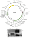
Figure 1
Figure 1
Identification of the successful construction of Pichia/IL-35. (a) Plasmid construction diagram. (b) To ensure the successful construction of Pichia/IL-35, the total cell lysate proteins from the strain Pichia/0 or Pichia/IL-35 were obtained, and a western blot analysis was performed.
Figure 2
Figure 2
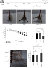
Pichia/IL-35 ameliorated symptoms of experimental colitis. (a) Details of the dextran sodium sulfate (DSS) used for the induction of experimental colitis and treatment of Pichia/0 or Pichia/IL-35. (b) Representative photos showing bloody stool at day 10 after DSS administration. Body weight loss (c) and disease activity index (DAI) score (d) of mice from untreated, Pichia/0 and Pichia/IL-35 groups (n = 6 per group). (e) Representative image of the colon after DSS administration and Pichia/0 or Pichia/IL-35 treatment. (f) Colon length of mice from untreated, Pichia/0 and Pichia/IL-35 groups was measured. All data are presented as mean ± SD. One-way ANOVA was used to perform statistical analysis. ns = no significant,  ∗∗∗∗p < 0.0001.
Figure 3
Figure 3
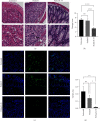
Pichia/IL-35 alleviated colon histological damage and apoptosis in colitis mice. (a) Representative H&E-stained sections of the distal colon after DSS administration and Pichia/0 or Pichia/IL-35 treatment (magnification: 200x). (b) Histology scores of mice colon in the untreated, Pichia/0 and Pichia/IL-35 groups at day 10 after DSS administration (n = 6 per group). (c) TUNEL images acquired by fluorescence microscope (magnification: 200x). The green part in the picture represents apoptotic cells in colon tissue (d) Percentage of apoptotic cells. All data are presented as mean ± SD. Inside the box is a 400x magnification image. One-way ANOVA was used to perform statistical analysis. ns = no significant,  p < 0.05,  ∗∗p < 0.01, and  ∗∗∗∗p < 0.0001.
Figure 4
Figure 4
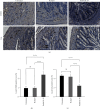
Pichia/IL-35 increased the proportion of M2 macrophages and reduced the level of pro-inflammatory cytokines in DSS-induced colitis mice. To evaluate the effect of Pichia/IL-35 on local immune microenvironment, CD206 and IL-6 (a) immunohistochemistry of colon sections in untreated, Pichia/0 and Pichia/IL-35 treated mice were performed. Quantitative data of cell positive area for each group are shown in (b) and (c) respectively (n = 6 per group). All data are presented as mean ± SD. The arrow shows positive cells. Statistics analyzed by One-way ANOVA. ns = no significant,  ∗∗∗∗p < 0.0001.
Figure 5
Figure 5
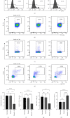
Pichia/IL-35 decreased the percentages of Th1, Th17, and M1 macrophages but increased the percentage of Tregs in MLNs of DSS-induced colitis. In addition to evaluation of the colon tissue, cell suspensions of MLNs were also prepared and stained for flow cytometry analysis. The representative histogram of (a) M1 (F4/80+CD86+) macrophages, the pseudocolor of (b) Th17 (CD4+IL-17A+), (c) Th1 (CD4+IFN-γ+) cells and (d) Tregs (CD4+CD25+Foxp3+) were depicted. In addition, the percentage of (e) M1 macrophages, (f) Th17 cells, (g) Th1 cells, (h) Tregs were calculated and graphed (n = 6 per group). All data are presented as mean ± SD. Statistics analyzed by One-way ANOVA. ns = no significant,  ∗∗p < 0.01,  ∗∗∗p < 0.001, and  ∗∗∗∗p < 0.0001.
Figure 6
Figure 6
IL-35 promoted TCPTP expression in vivo and in vitro. To access the effect of IL-35 on the expression of TCPTP in macrophages in vitro, RAW264.7 cell line and bone-marrow derived macrophages (BMDMs) were used. Representative western blots image of the TCPTP proteins from RAW264.7 (a) and BMDMs (b) were shown. The western blots image of SiRNA knockdown was shown as (e). The percentages of M1 macrophages and the western blots image in different groups was shown as (g, i). In vivo, we chose to detect the expression of TCPTP in colon tissue, and the representative western blots image was shown as (k). GAPDH was used as a loading control. (c, d, f, h, j, and l) shows the statistical graphs, respectively. All data are presented as mean ± SD. Statistics analyzed by one-way ANOVA. ns = no significant,  p < 0.05,  ∗∗p < 0.01,  ∗∗∗p < 0.001, and  ∗∗∗∗p < 0.0001.
Figure 7
Figure 7
Upregulation of TCPTP in Macrophages is Involved in IL-35 Mediated Attenuation of Experimental Colitis. The Pichia pastoris expressing IL-35 was intragastrically injected into colitis mice, and the results showed that Pichia/IL-35 treatment induced T cells and macrophages to play an anti-inflammatory role in promoting intestinal epithelial repair in vivo (a). In addition, IL-35 significantly increased TCPTP expression in LPS-stimulated macrophages in vitro (b). (Created with BioRender.com).

Similar articles

References

    1. Yamamoto-Furusho J. K., Harbi O. A., Armuzzi A., et al. Incidence of suboptimal response to tumor necrosis factor antagonist therapy in inflammatory bowel disease in newly industrialised countries: the EXPLORE study. Digestive and Liver Disease . 2020;52(8):869–877. doi: 10.1016/j.dld.2020.05.031. - DOI - PubMed
    1. The International IBD Genetics Consortium (IIBDGC), Jostins L., Ripke S., et al. Host-microbe interactions have shaped the genetic architecture of inflammatory bowel disease. Nature . 2012;491(7422):119–124. doi: 10.1038/nature11582. - DOI - PMC - PubMed
    1. Moller F. T., Andersen V., Wohlfahrt J., Jess T. Familial risk of inflammatory bowel disease: a population-based cohort study 1977–2011. American Journal of Gastroenterology . 2015;110(4):564–571. doi: 10.1038/ajg.2015.50. - DOI - PubMed
    1. Du L., Ha C. Epidemiology and pathogenesis of ulcerative colitis. Gastroenterology Clinics of North America . 2020;49(4):643–654. doi: 10.1016/j.gtc.2020.07.005. - DOI - PubMed
    1. Laass M. W., Roggenbuck D., Conrad K. Diagnosis and classification of Crohn’s disease. Autoimmunity Reviews . 2014;13(4-5):467–471. doi: 10.1016/j.autrev.2014.01.029. - DOI - PubMed

Substances

Supplementary concepts