Review
. 2024 Jul 24;124(14):8740-8786.
doi: 10.1021/acs.chemrev.4c00120. Epub 2024 Jul 3.

Noncanonical Amino Acids in Biocatalysis

Affiliations
Review

Noncanonical Amino Acids in Biocatalysis

Zachary Birch-Price et al. Chem Rev. .

Abstract

In recent years, powerful genetic code reprogramming methods have emerged that allow new functional components to be embedded into proteins as noncanonical amino acid (ncAA) side chains. In this review, we will illustrate how the availability of an expanded set of amino acid building blocks has opened a wealth of new opportunities in enzymology and biocatalysis research. Genetic code reprogramming has provided new insights into enzyme mechanisms by allowing introduction of new spectroscopic probes and the targeted replacement of individual atoms or functional groups. NcAAs have also been used to develop engineered biocatalysts with improved activity, selectivity, and stability, as well as enzymes with artificial regulatory elements that are responsive to external stimuli. Perhaps most ambitiously, the combination of genetic code reprogramming and laboratory evolution has given rise to new classes of enzymes that use ncAAs as key catalytic elements. With the framework for developing ncAA-containing biocatalysts now firmly established, we are optimistic that genetic code reprogramming will become a progressively more powerful tool in the armory of enzyme designers and engineers in the coming years.


Conflict of interest statement

The authors declare no competing financial interest.

Figures

Figure 1
Figure 1
NcAAs discussed in this review. (A) NcAAs incorporated via selective pressure incorporation (SPI), expressed protein ligation (EPL), or solid-phase peptide synthesis (SPPS). (B) NcAAs incorporated by GCE. The orthogonal translation system(s) used to incorporate each ncAA are listed. For several ncAAs, multiple incorporation techniques are discussed in this review, and these are also listed. DAP is incorporated as a precursor featuring a photocleavable group, which matures to DAP upon irradiation at 365 nm. 4-NH2Phe is incorporated as 4-AzPhe, which is then chemically reduced in situ to form 4-NH2Phe.
Figure 2
Figure 2
SPI of ncAAs. SPI employs an auxotrophic expression system to globally replace a target canonical amino acid (cAA) with a close structural analogue. An endogenous aaRS loads its cognate tRNA with the ncAA which is incorporated into proteins. Created with BioRender.com.
Figure 3
Figure 3
Strategies for the generation of ncAA-loaded tRNAs employ either chemoenzymatic methods (top left, PDB: 2C5U(102)) or Flexizymes (bottom left, PDB: 3CUN(103)). These ncAA-tRNAs can then be incorporated into a polypeptide chain using cell-free expression (CFE) systems (right). Created with BioRender.com.
Figure 4
Figure 4
Positive and negative selection processes can be used to engineer orthogonal aaRS-tRNA pairs to improve incorporation efficiency and/or specificity. The engineered aaRS catalyzes an aminoacylation reaction between its cognate tRNA and ncAA, with the ncAA added to the growing polypeptide chain during translation in response to a repurposed codon (e.g., the amber stop codon, UAG). Created with BioRender.com.
Figure 5
Figure 5
DAP incorporation into Valinomycin synthetase. (A) Genetically encoded (2S)-2-amino-3-([(2-[1-(6-nitrobenzo[d][1,3]dioxol-5-yl)ethyl]thio)ethoxy)carbonyl] ncAA is photodeprotected by irradiation at 365 nm to give DAP, which forms stable acyl-enzyme intermediates with an amide bond that is resistant to hydrolysis. (B) The active site of Valinomycin synthetase (protein shown as a gray cartoon, PDB: 6ECE(213)) with a noncanonical DAP nucleophile in position 2463 (atom-colored sticks, brown carbons) bound to a dodecadepsipeptide substrate (atom colored sticks, blue carbons). (C) Large structural differences are observed in the lid region of Valinomycin synthetase when bound to a dodecadepsipeptidyl intermediate (gray cartoon, PDB: 6ECE(213)) in comparison to a tetradepsipeptidyl intermediate (blue cartoon, PDB: 6ECD(213)).
Figure 6
Figure 6
Mechanistic studies on RNRs using ncAAs have shed light on the electron transfer pathway and enabled structural characterization of the active form of the multimer. (A) A cryogenic electron microscopy structure of RNR (PDB: 6W4X(231)) in its active α2β2 form was captured using a 2,3,5-F3Tyr122 mutation. The protein chains are shown as cartoons, and GDP and TPP are shown as red and gray spheres, respectively. (B) The mechanism of RNRs, which catalyze the conversion of nucleoside di- and triphosphates to deoxynucleotides. TR = thioredoxin. (C) DEER experiments provided information on the relative distances between the Tyr122 radical in the unreacted α/β pair and radicals on an N3NDP mechanistic inhibitor or radicals trapped on 3-NH2Tyr.
Figure 7
Figure 7
Mechanistic proposal for the FtmOx1-catalyzed hydrogen atom transfer from Tyr68 to C26•.
Figure 8
Figure 8
3-ClTyr incorporation into Ketosteroid Isomerase (KSI) to tune the active site electric field. (A) The mechanism of KSI. (B) The active site of KSI (PDB: 5KP1(254)) with the ncAA 3-ClTyr in the active site, shown with orange carbons. The protein backbone is shown as a gray cartoon. Active site residues and the substrate and transition state analogue equilenin are shown as atom-colored sticks, with gray and blue carbons, respectively. (C) The product analogue 19-nortestosterone used for VSE experiments.
Figure 9
Figure 9
Active site of WT NiSOD (left) and a variant with a secondary amine backbone substitution (right).
Figure 10
Figure 10
Electron donation to the iron center affects ferryl reactivity. (top) Cytochrome P450s are capable of hydrogen atom abstraction by the intermediate Compound I. Increased electron donation through an ncAA selenolate ligand increases the rate compared to WT P450. (bottom) Heme peroxidase compound II is reduced through proton coupled electron transfer. His to MeHis substitution decreases the electron donation to the ferryl intermediate and reduces its proton affinity, slowing the rate of compound II reduction.
Figure 11
Figure 11
Anaerobic X-ray crystal structures of the active sites of Human Cysteine Dioxygenase (CDO, PDB: 6N43(306)) and CDO Tyr157F2-Tyr (PDB: 6BPR(306)) in complex with the substrate cysteine and NO. CDO and CDO Tyr157F2-Tyr are shown as cartoons in blue and gray, respectively, with key active site residues and the substrate cysteine shown as atom-colored sticks with blue and gray carbon atoms. The noncanonical F2-Tyr157 is shown with orange carbon atoms.
Figure 12
Figure 12
NcAA-mediated noncovalent interactions influence enzyme stability. (A) SPI of 4-R-FPro in KlenTaq DNA polymerase switches many Pro puckers from endo to exo, as illustrated by the substitution of Pro555 (left, gray carbons) to 4-R-FPro555 (right, orange carbons) (PDB: 4DLG, 4DLE(335)). (B) Evolutionary trajectory of TFLeu-incorporating CAT (orange bars) starting from WT CAT (gray bar) against the half-life of enzyme inactivation at 60 °C. (C) Structures of T4 lysozyme with canonical Tyr18 (left, gray carbons) and noncanonical 3-ClTyr18 (right, orange carbons). Glu11 and Gly28 backbone atoms shown (white carbons). Halogen bond between Gly28 backbone oxygen and 3-ClTyr18 chlorine atom indicated with a dashed line (PDB: 1L63,5V7E).
Figure 13
Figure 13
Covalent cross-links mediated by ncAAs. (A) Cross-links generated between cAAs (black) and ncAAs (orange). Cross-linking bonds shown in gray. Top left: canonical Cys-Cys cross-link. Top right: Cys-SbuTyr cross-link. Middle left: Cys-BpAla cross-link. Middle right: amino group-4-NCSPhe cross-link. Bottom left: Cys-O-2-BeTyr cross-link. Bottom right: Cys-4-CaaPhe cross-link. (B) Structures of Cys-O-2-BeTyr cross-link (left) and Cys-4-CaaPhe cross-link (right) in Mb(H64V,V68A), with Tm increases given by one and two cross-links indicated. ncAAs shown with orange carbons and Cys with white carbons (PDB: 7SPE, 7SPH(351)).
Figure 14
Figure 14
NcAA-mediated enzyme immobilization. (A) Schematic representation of nonspecific enzyme immobilization, mediated by cross-linking at multiple reactive surface residues (gray circles), resulting in multiple enzyme orientations relative to the solid support, as well as enzyme–enzyme cross-linking leading to multilayer immobilization. (B) Schematic representation of site-specific enzyme immobilization, mediated by a ncAA (orange circles) incorporated site specifically, resulting in a monolayer with a single defined enzyme orientation. (C) Immobilization chemistries utilizing ncAAs (orange). From top to bottom: CuAAC, SPAAC, DOPhe–amine coupling, tetrazine-sTCO Diels–Alder cycloaddition, 3-NH2Tyr-acryloyl Diels–Alder cycloaddition, Glaser–Hay alkynyl coupling, and 4-SHPhe-BODIPY coupling.
Figure 15
Figure 15
Introduction of 4-AcPhe into PikC, a CYP450 enzyme, enabled biosynthetic reprogramming through allowing C(sp3)–H oxidation to occur in the absence of an amino-sugar moiety (brown).
Figure 16
Figure 16
Incorporation of ncAAs at various positions within P450BM3 alters the oxidation product distributions for (S)-ibuprofen-OMe and (+)-nootkatone substrates.
Figure 17
Figure 17
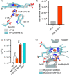
Peroxidases with MeHis proximal ligands. (A) An overlay of the crystal structures of APX2 (PDB: 1OAG(382)) and APX2 MeHis163 (PDB: 5L86(381)). Key active site residues and the heme are shown as atom-colored sticks with gray and blue carbons, respectively. MeHis is shown with brown carbons. (B) TTN achieved by APX2 and APX2 MeHis. (C) The catalytic efficiency toward guaiacol (2-methoxyphenol) oxidation for Mb variants and horseradish peroxidase (HRP). (D) An overlay of the crystal structures of Mb (PDB: 1A6K(383)) and Mb MeHis93 (PDB: 5OJ9(384)). The protein backbones are shown as cartoons, and key active site residues and the heme are shown as atom-colored sticks with gray and blue carbons, respectively. MeHis93 is shown with brown carbon atoms.
Figure 18
Figure 18
Biocatalytic cyclopropanations by Mb* MeHis93. (A) The bridged ion carbenoid intermediate observed by X-ray crystallography (PDB: 6F17(386)). A 2FO–FC map contoured at 1.5 σ is shown around the bridged carbenoid intermediate and the iron atom. (B) The cyclopropanation reaction catalyzed by engineered Mbs. (C) The non-native cofactor and MeHis ligand used to expand the scope of biocatalytic cyclopropanations.
Figure 19
Figure 19
Introduction of 4-AzPhe into selected sites of formate dehydrogenase (FDH) and mannitol dehydrogenase (MNDH) created bioorthogonal handles for SPAAC conjugation to either a heterobifunctional linker harboring a tetrazine handle or an alternative linker with a cyclooctene handle (PDB: 3WR5,1LJ8). FDH and MNDH are shown as gray and blue cartoons, respectively. The sites of 4-AzPhe incorporation are shown as red spheres.
Figure 20
Figure 20
Introduction of a photocaged ncAA into a DNA polymerase through GCE occludes the active site, preventing the diffusion of nucleotides for extension. Brief irradiation with UV light cleaves the O-NB moiety to reveal the catalytic Tyr and restore polymerase activity. Created with BioRender.com.
Figure 21
Figure 21
Photoresponsive ncAAs used in the allosteric light regulation of ImGPS. AzoPhe undergoes light induced reversible E/Z isomerizations enabling on–off switching of HisH activity.
Figure 22
Figure 22
Introduction of a pair of BpyAlas into Pfu POP (PDB: 5T88(458)) enabled inhibition of protease activity when incubated in divalent metal salts. Metal binding of the noncanonical ligands holds POP in a closed inactive conformation, which can be released through chelation of metal ions with EDTA addition, thereby allowing reversible allosteric control of biocatalyst activity. Created with BioRender.com.
Figure 23
Figure 23
Catalytic metal-coordinating ncAAs. (A) Crystal structure of dimeric LmrR, with the positions Val15, Met89, and Trp96 in the binding pocket shown with blue carbons (PDB: 3F8B(474)). (B) BpyAla-coordinated Cu(II) complex which activates 1-(1-methyl-1H-imidazol-2-yl)but-2-en-1-one toward nucleophilic attack. (C) Schemes of vinylogous Friedel–Crafts alkylations (top) and α,β-unsaturated 2-acyl pyridine hydrations (bottom) catalyzed by BpyAla-Cu(II) or 3-HqAla-Cu(II) metalloenzymes.
Figure 24
Figure 24
4-AzPhe-anchored metalloenzymes. (A) BCN-Derivatised dirhodium complex. OAc = acetate anion. (B) Crystal structure of POP, with positions of 4-AzPhe incorporation (orange spheres) and pore-opening alanine mutations (blue spheres) shown (PDB: 5T88(479)). (C) Schemes of styrene cyclopropanations (top) and the diazo cross-coupling cascade (bottom) catalyzed by POP variants containing 4-AzPhe-tethered dirhodium complexes.
Figure 25
Figure 25
Nucleophilic catalysis utilizing MeHis. (A) Scheme of ester hydrolysis, showing the reactive covalent intermediate formed between the substrate and MeHis23 (orange). (B) Structure of OE1.3, with MeHis23 (orange carbons) and sites of mutations installed during evolution (blue spheres) shown (PDB: 6Q7Q(486)). (C) Scheme highlighting the proton transfer role of Glu26 (gray) in the evolved MBHase BHMeHis1.8. Intermediates 2 (left) and 3 (right) are shown, covalently bound to MeHis23 (orange).
Figure 26
Figure 26
Nucleophilic catalysis utilizing 4-NH2Phe. (A) Scheme of hydrazone (X = N) and oxime (X = O) formations catalyzed by 4-NH2Phe (orange) incorporated into LmrR, with the covalent adduct formed by the carbonyl substrate and 4-NH2Phe15 shown. (B) Scheme of vinylogous Friedel–Crafts alkylations catalyzed by LmrR_V15_4-NH2Phe_RGN, with the activated imine intermediate formed between 4-NH2Phe15 (orange) and the aldehyde substrate shown. At the end of the reaction time NaBH4 is added to reduce the enzymatic product to the corresponding alcohol (right).
Figure 27
Figure 27
[2 + 2] Photocycloadditions catalyzed by BpAla. (A) Schemes of intramolecular [2 + 2] photocycloadditions of derivatized quinolones (top) and indoles (bottom). X = O or C, n = 1 or 2. (B) Crystal structure of EnT1.3 with product (green carbons) bound between BpAla (orange carbons), Trp244, and His287 (blue carbons) (PDB: 7ZP7(28)).
Figure 28
Figure 28
Metal-dependent ncAA-incorporating photoenzymes. (A) Chromophore autocatalytically generated in sfYFP and in PSP2, which incorporates BpAla (orange side chain) at position 66. (B) Structure of PSP2, with a chromophore shown (backbone indicated with gray carbons, BpAla side chain with orange carbons). The Cys95 site of nickel–terpyridine complex ligation is shown in dark gray (PDB: 5YR3(506)). (C) Scheme of dehalogenation reactions catalyzed by BpAla-incorporating PSP2T2 or by BpyAla-incorporating Mb. X = Cl, Br, or I. (D) Structure of Mb incorporating BpyAla (orange carbons) and with an iridium photocatalyst (green carbons) ligated to Cys45 (gray carbons) (PDB: 7YLK(516)).

Similar articles

References

    1. Punekar N. S.ENZYMES: Catalysis, Kinetics and Mechanisms; Springer Nature Singapore, 2018.10.1007/978-981-13-0785-0 - DOI
    1. Richter M. Functional Diversity of Organic Molecule Enzyme Cofactors. Nat. Prod. Rep. 2013, 30 (10), 1324–1345. 10.1039/c3np70045c. - DOI - PubMed
    1. Wagner C. R.; Benkovic S. J. Site Directed Mutagenesis: a Tool for Enzyme Mechanism Dissection. Trends Biotechnol. 1990, 8, 263–270. 10.1016/0167-7799(90)90189-5. - DOI - PubMed
    1. Arnold F. H. Directed Evolution: Bringing New Chemistry to Life. Angew. Chem. Int. Ed. 2018, 57 (16), 4143–4148. 10.1002/anie.201708408. - DOI - PMC - PubMed
    1. Turner N. J. Directed Evolution Drives the Next Generation of Biocatalysts. Nat. Chem. Biol. 2009, 5, 567–573. 10.1038/nchembio.203. - DOI - PubMed

LinkOut - more resources