. 2024 Aug 14;15(8):e0105324.
doi: 10.1128/mbio.01053-24. Epub 2024 Jul 2.

Colletotrichum fructicola co-opts cytotoxic ribonucleases that antagonize host competitive microorganisms to promote infection

Affiliations

Colletotrichum fructicola co-opts cytotoxic ribonucleases that antagonize host competitive microorganisms to promote infection

Chunhao Wang et al. mBio. .

Abstract

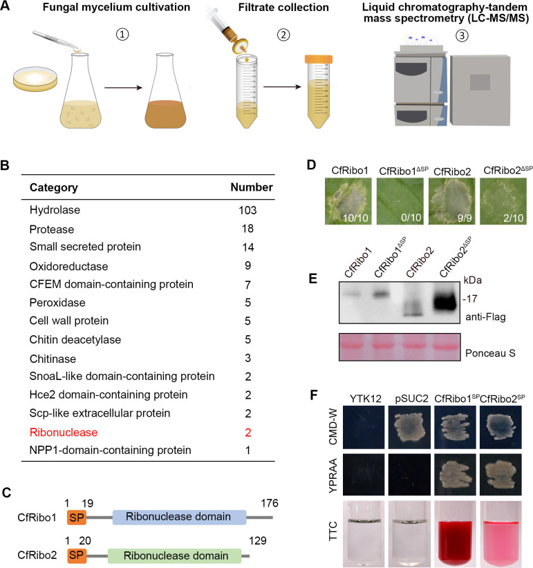
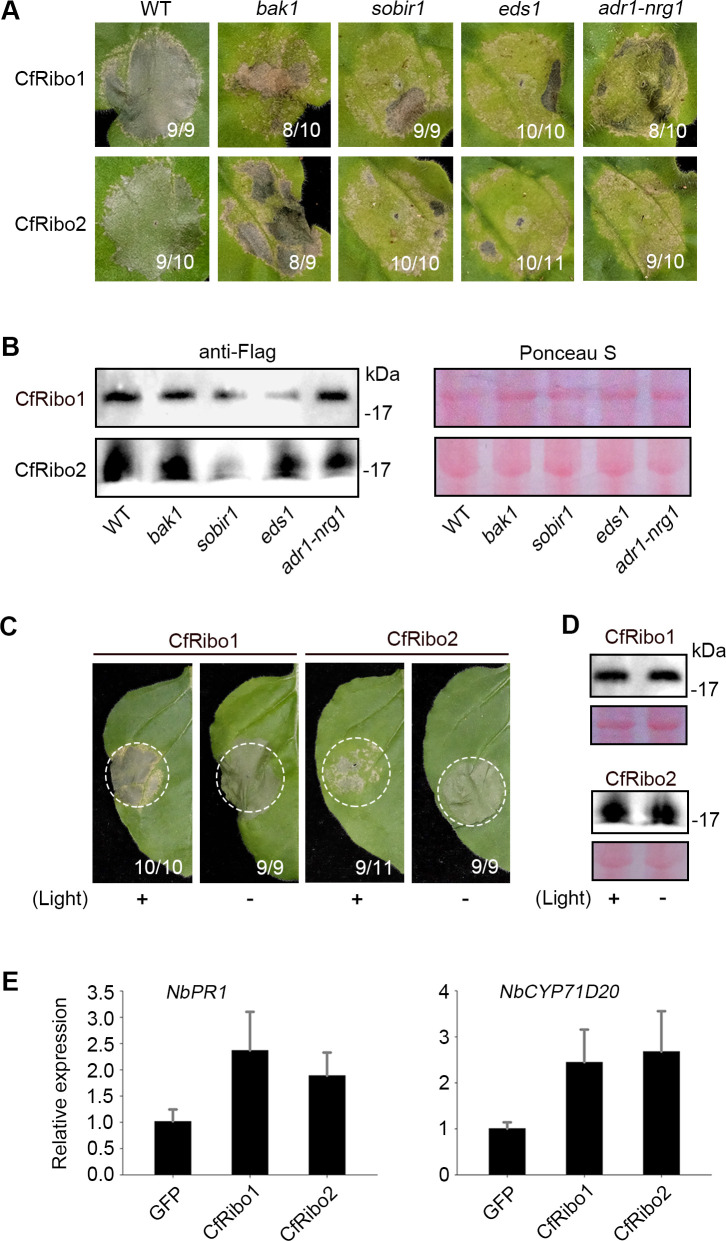
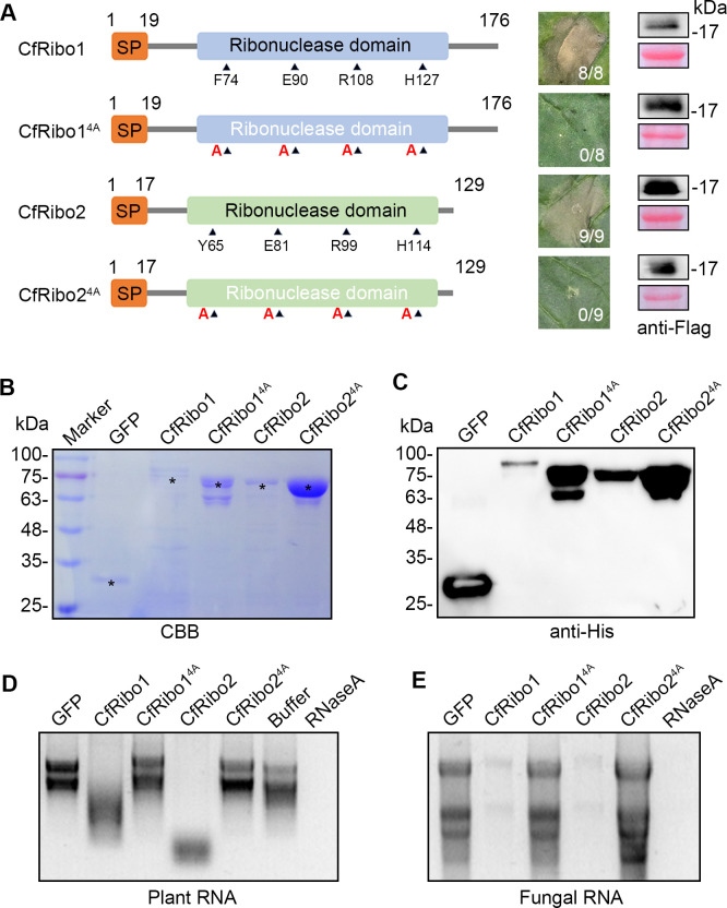
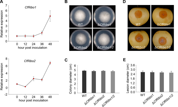
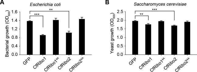
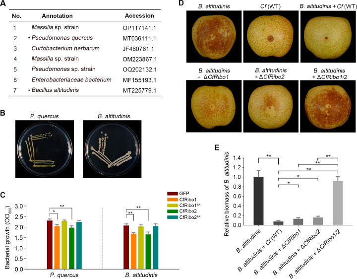
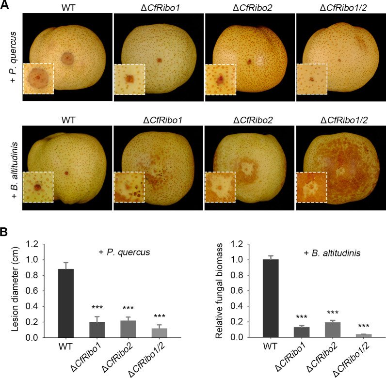
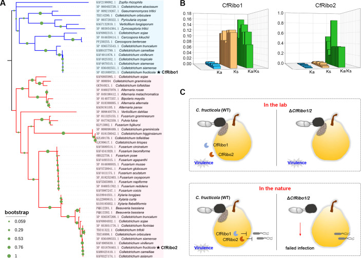
Phytopathogens secrete numerous molecules into the environment to establish a microbial niche and facilitate host infection. The phytopathogenic fungus Colletotrichum fructicola, which causes pear anthracnose, can colonize different plant tissues like leaves and fruits, which are occupied by a diversity of microbes. We speculate that this fungus produces antimicrobial effectors to outcompete host-associated competitive microorganisms. Herein, we identified two secreted ribonucleases, CfRibo1 and CfRibo2, from the C. fructicola secretome. The two ribonucleases both possess ribonuclease activity and showed cytotoxicity in Nicotianan benthamiana without triggering immunity in an enzymatic activity-dependent manner. CfRibo1 and CfRibo2 recombinant proteins exhibited toxicity against Escherichia coli, Saccharomyces cerevisiae, and, importantly, the phyllosphere microorganisms isolated from the pear host. Among these isolated microbial strains, Bacillus altitudinis is a pathogenic bacterium causing pear soft rot. Strikingly, CfRibo1 and CfRibo2 were found to directly antagonize B. altitudinis to facilitate C. fructicola infection. More importantly, CfRibo1 and CfRibo2 functioned as essential virulence factors of C. fructicola in the presence of host-associated microorganisms. Further analysis revealed these two ribonucleases are widely distributed in fungi and are undergoing purifying selection. Our results provide the first evidence of antimicrobial effectors in Colletotrichum fungi and extend the functional diversity of fungal ribonucleases in plant-pest-environment interactions.

Importance: Colletotrichum fructicola is emerging as a devastating pathogenic fungus causing anthracnose in various crops in agriculture, and understanding how this fungus establishes successful infection is of great significance for anthracnose disease management. Fungi are known to produce secreted effectors as weapons to promote virulence. Considerable progress has been made in elucidating how effectors manipulate plant immunity; however, their importance in modulating environmental microbes is frequently neglected. The present study identified two secreted ribonucleases, CfRibo1 and CfRibo2, as antimicrobial effectors of C. fructicola. These two proteins both possess toxicity to pear phyllosphere microorganisms, and they efficiently antagonize competitive microbes to facilitate the infection of pear hosts. This study represents the first evidence of antimicrobial effectors in Colletotrichum fungi, and we consider that CfRibo1 and CfRibo2 could be targeted for anthracnose disease management in diverse crops in the future.

Keywords: Colletotrichum; antimicrobial activity; cell death; host-associated microorganisms; ribonuclease.


Conflict of interest statement

The authors declare no conflict of interest.

Figures

Fig 1
Fig 1
CfRibo1 and CfRibo2 are two cytotoxic ribonucleases identified in Colletotrichum fructicola secretome. (A) Schematic representation of C. fructicola secretome analysis. (B) Classification of proteins identified in C. fructicola secretome. The numbers of each protein family are shown. (C) Schematic diagram illustration of the sequence structure of CfRibo1 and CfRibo2. (D) Nicotiana benthamiana leaves showing cell death triggered by CfRibo1, CfRibo2, and their signal peptide-deletion versions (CfRibo1ΔSP and CfRibo2ΔSP). The proteins were transiently expressed in N. benthamiana leaves via agroinfiltration. Cell death was observed 3–5 days post-agroinfiltration, with representative leaves photographed. The number of leaves showing cell death phenotype (numerator) and the number of total surveyed leaves (denominator) are indicated at the bottom of each photographed leaf. (E) Immunoblotting analysis of the transiently expressed proteins with anti-Flag antibody. Total proteins were stained with Ponceau S to serve as loading controls. (F) Functional validation of the SP of CfRibo1 and CfRibo2 using a yeast secretion system. The empty pSUC2 vector was used as a negative control.
Fig 2
Fig 2
CfRibo1 and CfRibo2 do not activate immunity in N. benthamiana. (A) Phenotype of cell death triggered by CfRibo1 and CfRibo2 in the wild-type N. benthamiana and mutants of central immunity regulators. CfRibo1 and CfRibo2 were agroinfiltrated in the wild-type N. benthamiana as well as the CRISPR/Cas9-edited bak1, sobir1, eds1, and adr1-nrg1 mutants. Cell death was visualized and photographed at 3–5 days post-agroinfiltration (dpa). The number of leaves showing cell death phenotype (numerator) and the number of total surveyed leaves (denominator) are indicated at the bottom of the photographed leaves. (B) Immunoblotting analysis of CfRibo1 and CfRibo2 transiently expressed in N. benthamiana with anti-Flag antibody. Ponceau S-stained total proteins were shown as loading controls. (C) Light independence of CfRibo1- and CfRibo2-triggered cell death. CfRibo1 and CfRibo2 were transiently expressed in the wild-type N. benthamiana, followed by incubation under light or dark conditions. Representative leaves showing the phenotypes were photographed at 3–5 dpa. The number of leaves showing cell death phenotype (numerator) and the number of total surveyed leaves (denominator) are indicated at the bottom of the photographed leaves. (D) Immunoblotting analysis of transiently expressed CfRibo1 and CfRibo2 with anti-Flag antibody. Total proteins were stained with Ponceau S to serve as loading controls. (E) Relative expression analysis of NbPR1 and NbCYP71D20 after the transient expression of CfRibo1 and CfRibo2. CfRibo1, CfRibo2, and the GFP control were agroinfiltrated into N. benthamiana Leaves. Leaves expressing indicated proteins were sampled at 2 days post-agroinfiltration, and transcripts of NbPR1 and NbCYP71D20 were evaluated by reverse transcription-quantitative polymerase chain reaction.
Fig 3
Fig 3
Ribonucleolytic activity of CfRibo1 and CfRibo2 determines their cytotoxicity. (A) Cell death triggered by CfRibo1, CfRibo2, and their catalytic site-mutated versions CfRibo14A and CfRibo24A in N. benthamiana. Schematic diagram illustration of these proteins was indicated. Each protein was transiently expressed in N. benthamiana via agroinfiltration. Cell death was observed at 3–5 days post-agroinfiltration, with representative leaves photographed. The number of leaves showing cell death phenotype (numerator) and the number of total surveyed leaves (denominator) are indicated at the bottom of the photographed leaves. The expression of these proteins was confirmed by immunoblotting with anti-Flag antibody, and the Ponceau S-stained total proteins were shown as loading controls. (B) Detection of recombinant proteins on gels by Coomassie brilliant blue staining. GFP, CfRibo1, CfRibo2, CfRibo14A, and CfRibo24A recombinant proteins produced in Escherichia coli were subjected to sodium dodecyl sulfate-polyacrylamide gel electrophoresis. The protein bands of expected size were indicated with black asterisks. (C) Immunoblotting detection of the recombinant proteins with anti-His antibody. (D and E) Ribonucleolytic activity tests using the recombinant proteins. The proteins were incubated with either plant total RNA or fungal total RNA at 37°C for 30 min. GFP recombinant protein and buffer were used as negative controls, and RNase A was used as a positive control.
Fig 4
Fig 4
Deletion of CfRibo1 and CfRibo2 does not affect C. fructicola virulence. (A) Expression profiles of CfRibo1 and CRibo2 during C. fructicola infection of the pear host. Pear fruits inoculated with C. fructicola were sampled at 0, 12, 24, 36, and 48 h post-inoculation, respectively. Relative expression of CfRibo1 and CfRibo2 was determined by reverse transcription-quantitative polymerase chain reaction. CfActin was used as an internal reference to normalize gene expression. (B and C) Filamentous growth phenotype of C. fructicola and the gene deletion transformants. The wild-type (WT) C. fructicola DSCF-02, ΔCfRibo1, ΔCfRibo2, and the double deletion mutant ΔCfRibo1/2 were cultured on potato dextrose agar plates at 28°C. Photographs were taken, and colony diameter was calculated 3 days later. (D and E) Virulence phenotype of C. fructicola and the gene deletion transformants. Conidia of C. fructicola strains and the gene deletion transformants were inoculated in pear fruits. Disease lesions were photographed, and lesion diameters were calculated 3 days post-inoculation.
Fig 5
Fig 5
CfRibo1 and CfRibo2 exhibit antimicrobial activity against E. coli and Saccharomyces cerevisiae. GFP, CfRibo1, CfRibo2, CfRibo14A, and CfRibo24A recombinant proteins were incubated with E. coli and S. cerevisiae at 1 µM concentration, followed by culturing at 37°C and 30°C, respectively. Both bacterial growth (A) and yeast growth (B) were monitored 24 h later. Error bars indicate the mean ± SD (Student’s t-test, **P < 0.01 and ***P < 0.001).
Fig 6
Fig 6
CfRibo1 and CfRibo2 synergistically antagonize a pathogenic bacterium during the infection of pear fruits. (A) Seven bacterial strains were identified from pear leaves. Pseudomonas quercus and Bacillus altitudinis chosen for further analysis were marked with black asterisks. (B) Colony phenotype of P. quercus and B. altitudinis on lysogeny broth plates. (C) Antimicrobial activity of CfRibo1 and CfRibo2 against P. quercus and B. altitudinis. GFP, CfRibo1, CfRibo2, CfRibo14A, and CfRibo24A recombinant proteins were incubated with P. quercus and B. altitudinis at 1 µM concentration, followed by culturing at 37°C. The growth of each strain was monitored 24 h post-incubation. Error bars indicate the mean ± SD (Student’s t-test, **P < 0.01). (D and E) CfRibo1 and CfRibo2 synergistically antagonize B. altitudinis during infection. C. fructicola strains including the wild type [Cf (WT)] and the gene deletion transformants (ΔCfRibo1, ΔCfRibo2, and ΔCfRibo1/2) were co-inoculated with B. altitudinis in pear fruits. As controls, Cf (WT) and B. altitudinis were individually inoculated at the same time. Disease symptoms were photographed at 40 hpi. Infection of B. altitudinis was quantified by relative biomass using reverse transcription-quantitative polymerase chain reaction. CfActin from C. fructicola and PbrTubulin from pear were used as reference genes. Error bars indicate the mean ± SD (Student’s t-test, *P < 0.05 and **P < 0.01).
Fig 7
Fig 7
CfRibo1 and CfRibo2 play essential roles in fungal virulence in the presence of P. quercus and B. altitudinis. (A) Virulence phenotype of the wild-type C. fructicola and gene deletion transformants in the presence of P. quercus and B. altitudinis. Conidia of the wild-type DSCF-02 strain and the gene deletion transformants (ΔCfRibo1, ΔCfRibo2, and ΔCfRibo1/2) were co-inoculated with P. quercus or B. altitudinis in pear fruits. Disease lesions were photographed at 48 hpi. (B) Quantification of C. fructicola and B. altitudinis infections. Disease development of C. fructicola was calculated with lesion diameters. B. altitudinis infection was determined by reverse transcription-quantitative polymerase chain reaction, with CfActin and PbrTubulin used as reference genes. Error bars indicate the mean ± SD (Student’s t-test, ***P < 0.001).
Fig 8
Fig 8
CfRibo1 and CfRibo2 are widely distributed across fungal taxa. (A) Phylogeny distribution of CfRibo1 and CfRibo2 homologs from different fungal species. A maximum-likelihood method was adopted to construct the tree. Bootstrap percentage support for each branch is indicated by circle symbols. CfRibo1 and CrRibo2 are marked with black asterisks. (B) Selective pressure determination of CfRibo1 and CfRibo2 by Ka/Ks ratios. The nonsynonymous rates (Ka), synonymous rates (Ks), and Ka/Ks ratios of CfRibo1 and CfRibo2 are shown. (C) A proposed model for the function of CfRibo1 and CfRibo2 during C. fructicola-pear interactions. In the lab, when inoculation assays were performed with C. fructicola only, CfRibo1 and CfRibo2 were dispensable for fungal virulence. In nature where diverse microorganisms exist, CfRibo1 and CfRibo2 function as antimicrobial effectors that synergistically antagonize host-associated competitive microorganism(s) and execute essential virulence roles, thereby promoting host infection.

Similar articles

References

    1. Göhre V, Robatzek S. 2008. Breaking the barriers: microbial effector molecules subvert plant immunity. Annu Rev Phytopathol 46:189–215. doi:10.1146/annurev.phyto.46.120407.110050 - DOI - PubMed
    1. Tariqjaveed M, Mateen A, Wang S, Qiu S, Zheng X, Zhang J, Bhadauria V, Sun W. 2021. Versatile effectors of phytopathogenic fungi target host immunity. J Integr Plant Biol 63:1856–1873. doi:10.1111/jipb.13162 - DOI - PubMed
    1. Rovenich H, Boshoven JC, Thomma B. 2014. Filamentous pathogen effector functions: of pathogens, hosts and microbiomes. Curr Opin Plant Biol 20:96–103. doi:10.1016/j.pbi.2014.05.001 - DOI - PubMed
    1. Snelders NC, Kettles GJ, Rudd JJ, Thomma B. 2018. Plant pathogen effector proteins as manipulators of host microbiomes? Mol Plant Pathol 19:257–259. doi:10.1111/mpp.12628 - DOI - PMC - PubMed
    1. Snelders NC, Boshoven JC, Song Y, Schmitz N, Fiorin GL, Rovenich H, van den Berg GCM, Torres DE, Petti GC, Prockl Z, Faino L, Seidl MF, Thomma B. 2023. A highly polymorphic effector protein promotes fungal virulence through suppression of plant‐associated Actinobacteria. New Phytol 237:944–958. doi:10.1111/nph.18576 - DOI - PubMed

MeSH terms

LinkOut - more resources