Advances in Noninvasive Molecular Imaging Probes for Liver Fibrosis Diagnosis
- PMID: 38952717
- PMCID: PMC11214848
- DOI: 10.34133/bmr.0042
Advances in Noninvasive Molecular Imaging Probes for Liver Fibrosis Diagnosis
Abstract
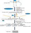
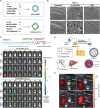
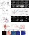
Liver fibrosis is a wound-healing response to chronic liver injury, which may lead to cirrhosis and cancer. Early-stage fibrosis is reversible, and it is difficult to precisely diagnose with conventional imaging modalities such as magnetic resonance imaging, positron emission tomography, single-photon emission computed tomography, and ultrasound imaging. In contrast, probe-assisted molecular imaging offers a promising noninvasive approach to visualize early fibrosis changes in vivo, thus facilitating early diagnosis and staging liver fibrosis, and even monitoring of the treatment response. Here, the most recent progress in molecular imaging technologies for liver fibrosis is updated. We start by illustrating pathogenesis for liver fibrosis, which includes capillarization of liver sinusoidal endothelial cells, cellular and molecular processes involved in inflammation and fibrogenesis, as well as processes of collagen synthesis, oxidation, and cross-linking. Furthermore, the biological targets used in molecular imaging of liver fibrosis are summarized, which are composed of receptors on hepatic stellate cells, macrophages, and even liver collagen. Notably, the focus is on insights into the advances in imaging modalities developed for liver fibrosis diagnosis and the update in the corresponding contrast agents. In addition, challenges and opportunities for future research and clinical translation of the molecular imaging modalities and the contrast agents are pointed out. We hope that this review would serve as a guide for scientists and students who are interested in liver fibrosis imaging and treatment, and as well expedite the translation of molecular imaging technologies from bench to bedside.
Copyright © 2024 Shaofang Chen et al.
Conflict of interest statement
Competing interests: The authors declare that they have no competing interests.
Figures


Similar articles
-
Liver Fibrosis Conventional and Molecular Imaging Diagnosis Update.J Liver. 2019;8(1):236. Epub 2019 Jan 22. J Liver. 2019. PMID: 31341723 Free PMC article.
-
Liver Fibrosis Leading to Cirrhosis: Basic Mechanisms and Clinical Perspectives.Biomedicines. 2024 Sep 30;12(10):2229. doi: 10.3390/biomedicines12102229. Biomedicines. 2024. PMID: 39457542 Free PMC article. Review.
-
Molecular Probes for Imaging Fibrosis and Fibrogenesis.Chemistry. 2019 Jan 24;25(5):1128-1141. doi: 10.1002/chem.201801578. Epub 2018 Nov 21. Chemistry. 2019. PMID: 30014529 Free PMC article. Review.
-
Imaging Fibrogenesis in a Diet-Induced Model of Nonalcoholic Steatohepatitis (NASH).Contrast Media Mol Imaging. 2019 Dec 1;2019:6298128. doi: 10.1155/2019/6298128. eCollection 2019. Contrast Media Mol Imaging. 2019. PMID: 31866798 Free PMC article.
-
Molecular MRI quantification of extracellular aldehyde pairs for early detection of liver fibrogenesis and response to treatment.Sci Transl Med. 2022 Sep 21;14(663):eabq6297. doi: 10.1126/scitranslmed.abq6297. Epub 2022 Sep 21. Sci Transl Med. 2022. PMID: 36130015 Free PMC article.
References
Publication types
LinkOut - more resources
Full Text Sources

