. 2024 Jul 1;26(1):108.
doi: 10.1186/s13058-024-01865-y.

Utilizing human cerebral organoids to model breast cancer brain metastasis in culture

Affiliations

Utilizing human cerebral organoids to model breast cancer brain metastasis in culture

Chenran Wang et al. Breast Cancer Res. .

Abstract

Background: Metastasis, the spread, and growth of malignant cells at secondary sites within a patient's body, accounts for over 90% of cancer-related mortality. Breast cancer is the most common tumor type diagnosed and the leading cause of cancer lethality in women in the United States. It is estimated that 10-16% breast cancer patients will have brain metastasis. Current therapies to treat patients with breast cancer brain metastasis (BCBM) remain palliative. This is largely due to our limited understanding of the fundamental molecular and cellular mechanisms through which BCBM progresses, which represents a critical barrier for the development of efficient therapies for affected breast cancer patients.

Methods: Previous research in BCBM relied on co-culture assays of tumor cells with rodent neural cells or rodent brain slice ex vivo. Given the need to overcome the obstacle for human-relevant host to study cell-cell communication in BCBM, we generated human embryonic stem cell-derived cerebral organoids to co-culture with human breast cancer cell lines. We used MDA-MB-231 and its brain metastatic derivate MDA-MB-231 Br-EGFP, other cell lines of MCF-7, HCC-1806, and SUM159PT. We leveraged this novel 3D co-culture platform to investigate the crosstalk of human breast cancer cells with neural cells in cerebral organoid.

Results: We found that MDA-MB-231 and SUM159PT breast cancer cells formed tumor colonies in human cerebral organoids. Moreover, MDA-MB-231 Br-EGFP cells showed increased capacity to invade and expand in human cerebral organoids.

Conclusions: Our co-culture model has demonstrated a remarkable capacity to discern the brain metastatic ability of human breast cancer cells in cerebral organoids. The generation of BCBM-like structures in organoid will facilitate the study of human tumor microenvironment in culture.

Keywords: Brain metastasis; Breast cancer; Cell-cell communication; Cerebral organoids; Neural cells; Tumor microenvironment.


Conflict of interest statement

The authors declare no competing interests.

Figures

Fig. 1
Fig. 1
2D co-culture of human breast cancer cells with human astrocytes. (A) Phase contrast and GFP fluorescence of MDA-MB-231, MCF-7, and HCC-1806 breast cancer cells on primary human astrocyte monolayer for 10 days. Arrows indicated GFP+ breast cancer cell colonies. Dashed lines circled the borders of breast cancer colonies based on GFP expression. (B) Mean ± SE of the number of breast cancer cell colonies formed from 1,000 cells seeded on human astrocytes. (C) Mean ± SE of the size of breast cancer cell colonies cultured on human astrocytes. One-way ANOVA was used for statistical analysis. n = 6 independent experiments. ns: no significance; **: p < 0.01, ***: p < 0.001. Bar = 100 μm
Fig. 2
Fig. 2
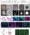
Generation of cerebral organoids and their co-culture with breast cancer cells in Eppendorf tube. (A) The schematic flow for generation of cerebral organoids from hESC. The phase contrast and RFP fluorescent images of H9-RFP derived hESC colony, EBs, and organoids at different developmental stages were shown. (B) H&E staining and immunofluorescent (IF) staining of DCX, SOX2, NEUN, and DAPI for mature organoids from H9 hESC. (C) IF staining of GFAP, OLIG2, IBA1, NG2, and DAPI for mature organoids from H9 hESC. Arrows indicated GFAP+, OLIG2+ and NG2+ cells. (D) A schematic depiction for generating organoid-breast cancer cells co-culture using Eppendorf tube. (E) GFP fluorescence of MCF-7 cells, MDA-MB-231cells, and HCC-1806 cells co-cultured with H9 organoids in Eppendorf tube. Representative images were from more than 10 cerebral organoids in 2–3 independent experiments. VZ/SVZ: ventricular zone/subventricular zone, CP: cortical plate. Bar = 500 μm in A and E, 100 μm in B and C
Fig. 3
Fig. 3
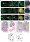
Co-culture of human breast cancer cells with hESC-derived cerebral organoids allowed growth and colonization of MDA-MB-231 and SUM159PT cell lines. (A) Phase contrast, GFP fluorescent, and RFP fluorescent images of co-cultured breast cancer cell lines in cerebral organoids from day 2 to day 14. Arrows indicated GFP+ breast cancer cells. (B) H&E staining of MCF-7 cells, MDA-MB-231 cells, SUM159PT cells, and HCC-1806 cells co-cultured with organoids. The arrow indicated apoptotic MDA-MB-231 cells and arrowheads indicated breast cancer cell colonies in organoids. Stars marked visible colonies from MCF-7 cells and HCC-1806 cells. (C and D) Mean ± SE of the number (C) of breast cancer cells and the percentage (D) of GFP+ breast cancer cells in organoid-breast cancer co-cultures. Representative images and quantification were from 13–19 cerebral organoids in 4–5 independent experiments. One-way ANOVA was used for statistical analysis. *: p < 0.05, **: p < 0.01, ***: p < 0.001. Bar = 200 μm
Fig. 4
Fig. 4
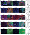
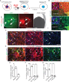
Growth of breast cancer cells in cerebral organoids. (A) IF of PCNA and GFP of breast cancer cells co-cultured with organoids at day 14. (B) Mean ± SE of the percentage of PCNA+ proliferative GFP+ breast cancer cells in cerebral organoids. (C) IF of EpCAM and GFP of breast cancer cells co-cultured with organoids. The arrow indicated a dead GFP-HCC-1806 cells in cerebral organoid. (D) Mean ± SE of the areas occupied by EpCAM+ breast cancer cells in cerebral organoids. (E) IF of TUJ1 and GFP of breast cancer cells co-cultured with organoids. Boxed area shown in detail as inset. Arrows indicated apoptotic HCC-1806 cells. (F) Mean ± SE of the percentage of the areas occupied by TUJ1+ cells in co-cultured cerebral organoids. (G) IF of GFAP and GFP of breast cancer cells co-cultured with organoids. (H) Mean ± SE of the number of GFAP+ cells in cerebral organoids around tumor colonies. (I) Fluorescence of TUNEL and DAPI of co-cultured breast cancer cells with organoids. Boxed area shown in detail as inset. (J) Mean ± SE of the percentage of TUNEL+ apoptotic breast cancer cells co-cultured with cerebral organoids. Dashed line indicated the border of breast cancer cells within organoid. Selected areas were shown in detail as inset. Representative images and quantification were from 13–19 cerebral organoids in 4–5 independent experiments. One-way ANOVA was used for statistical analysis. ND: not determined, ns: no significance, *: p < 0.05, **: p < 0.01, ***: p < 0.001. Bar = 100 μm
Fig. 5
Fig. 5
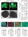
Increased tumor colony formation in cerebral organoid from MDA-MB-231 Br-EGFP cells. (A) Phase contrast and GFP fluorescence of co-cultured MDA-MB-231 Br-EGFP breast cancer cells in cerebral organoids at 10 cells/organoid and 100 cells/organoid for 14 days. (B) Mean ± SE of the number of GFP+ tumor colonies from parental MDA-MB-231 and MDA-MB-231 Br-EGFP cells in cerebral organoids. (C) Mean ± SE of the size of GFP+ tumor colonies from parental MDA-MB-231 and MDA-MB-231 Br-EGFP cells in cerebral organoids. (D, F, H, and J) IF of PCNA (D), TUJ1 (F), GFAP (H), TUNEL (J) with GFP and DAPI of Br-EGFP breast cancer cells co-cultured with cerebral organoids. (E, G, I, K) Mean ± SE of the relative percentage of PCNA+ cells (E), TUJ1+ areas (G), number of GFAP+ signaling around tumor colonies (I), and TUNEL+ apoptotic breast cancer cells (K) from parental MDA-MB-231 (set as 100%) and 100-1,000 MDA-MB-231 Br-EGFP cells in cerebral organoids. Representative images and quantification were from 12 cerebral organoids in 4 independent experiments. Two-way ANOVA and Student’s t-test were used for statistical analysis. ND: not determined, ns: no difference, *: p < 0.05, **: p < 0.01. Bar = 200 μm for A, 100 μm for D, F, H, and J
Fig. 6
Fig. 6
The transfer of sLP–mCherry from MDA-MB-231 breast cancer cells to neurons and astrocytes in cerebral organoids. (A) A schematic description for sLP-mCherry system to label neighbor recipient cells. Phase contrast and fluorescent images of GFP and mCherry of MDA-MB-231 cells in naïve WIBR3 organoids for 7 days. Arrows indicated mCherry+ organoid cells. Arrowhead indicated mCherry+ GFP+ breast cancer cells. Boxed area shown in detail as inset. (B) Fluorescence of GFP, mCherry, and DAPI of MDA-MB-231 cells in WIBR3 derived cerebral organoids. Arrows indicated mCherry+ cells in organoids. The dotted line indicated the boundary of breast cancer cells and organoids. Boxes shown in detail as inset. (C) IF of NEUN, mCherry, and DAPI of cerebral organoid cells and MDA-MB-231 cells expressing sLP–mCherry. Arrows indicated mCherry+ neurons while arrowheads indicated mCherry neurons. (D) IF of GFAP, mCherry, and DAPI of cerebral organoid cells and MDA-MB-231 cells expressing sLP–mCherry. Arrows indicated mCherry+ astrocytes while arrowheads indicated mCherry astrocytes. (E) Mean ± SE of percentage of NEUN+ mCherry+ cells and GFAP+ mCherry+ cells of total mCherry+ cells in the organoids. (F and G) Mean ± SE of percentage of NEUN+ mCherry+ cells of total NeuN+ cells (F) and GFAP+ mCherry+ cells of total GFAP+ cells (G) within different radius to tumor colony in organoid. Representative images and quantification were from 10 cerebral organoids in 3 independent experiments. One-way ANOVA and Student’s t-test were used for statistical analysis. ND: not determined, **: p < 0.01, ***: p < 0.001. Bar = 200 μm for A, 50 μm for B, C, and D

Similar articles

References

    1. Seyfried TN, Huysentruyt LC. On the origin of cancer metastasis. Crit Rev Oncog. 2013;18(1–2):43–73. doi: 10.1615/CritRevOncog.v18.i1-2.40. - DOI - PMC - PubMed
    1. Giaquinto AN, Sung H, Miller KD, Kramer JL, Newman LA, Minihan A, Jemal A, Siegel RL. Breast Cancer statistics, 2022. CA Cancer J Clin. 2022;72(6):524–41. doi: 10.3322/caac.21754. - DOI - PubMed
    1. Bailleux C, Eberst L, Bachelot T. Treatment strategies for breast cancer brain metastases. Br J Cancer. 2021;124(1):142–55. doi: 10.1038/s41416-020-01175-y. - DOI - PMC - PubMed
    1. Arslan C, Dizdar O, Altundag K. Systemic treatment in breast-cancer patients with brain metastasis. Expert Opin Pharmacother. 2010;11(7):1089–100. doi: 10.1517/14656561003702412. - DOI - PubMed
    1. Leone JP, Leone BA. Breast cancer brain metastases: the last frontier. Exp Hematol Oncol. 2015;4:33. doi: 10.1186/s40164-015-0028-8. - DOI - PMC - PubMed