Review
. 2024 Oct;1869(7):159529.
doi: 10.1016/j.bbalip.2024.159529. Epub 2024 Jun 28.

Mammalian START-like phosphatidylinositol transfer proteins - Physiological perspectives and roles in cancer biology

Affiliations
Review

Mammalian START-like phosphatidylinositol transfer proteins - Physiological perspectives and roles in cancer biology

Adrija Pathak et al. Biochim Biophys Acta Mol Cell Biol Lipids. 2024 Oct.

Abstract

PtdIns and its phosphorylated derivatives, the phosphoinositides, are the biochemical components of a major pathway of intracellular signaling in all eukaryotic cells. These lipids are few in terms of cohort of unique positional isomers, and are quantitatively minor species of the bulk cellular lipidome. Nevertheless, phosphoinositides regulate an impressively diverse set of biological processes. It is from that perspective that perturbations in phosphoinositide-dependent signaling pathways are increasingly being recognized as causal foundations of many human diseases - including cancer. Although phosphatidylinositol transfer proteins (PITPs) are not enzymes, these proteins are physiologically significant regulators of phosphoinositide signaling. As such, PITPs are conserved throughout the eukaryotic kingdom. Their biological importance notwithstanding, PITPs remain understudied. Herein, we review current information regarding PITP biology primarily focusing on how derangements in PITP function disrupt key signaling/developmental pathways and are associated with a growing list of pathologies in mammals.

Keywords: Lipid signaling; Mammalian disease; Phosphatidylinositol transfer proteins; Phosphoinositides.


Conflict of interest statement

Declaration of competing interest Vytas A. Bankaitis reports financial support was provided by National Institutes of Health. Vytas A. Bankaitis reports financial support was provided by Robert A. Welch Foundation. If there are other authors, they declare that they have no known competing financial interests or personal relationships that could have appeared to influence the work reported in this paper.

Figures

Fig. 1.
Fig. 1.
The phosphatidylinositol cycle and its synthesis. Phosphatidylinositol (PtdIns) is synthesized at the endoplasmic reticulum (ER) via the sequential action of cytidine diphosphate diacylglycerol (CDP-DAG) synthase 1/2, which converts phosphatidic acid (PtdOH) to the CPD-DAG intermediate, and PtdIns synthase which produces PtdIns. Michell’s PtdIns cycle hypothesis envisions mobilization of PtdIns from ER to the plasma membrane (PM) via soluble PtdIns transfer protein (PITP). PtdIns is converted to PtdIns4P by Type III PtdIns 4-kinase (PI4K) while Type I PtdIns 4-phosphate 5-kinase (PI4P5K) generates PtdIns(4,5)P2 from PtdIns4P at the plasma membrane. PtdIns(4,5)P2 undergoes hormone (agonist)-stimulated hydrolysis via the action of phospholipase C (PLC) to produce 1,4,5-trisphosphate (IP3) and DAG as soluble and membrane second messengers, respectively. Subsequently, DAG is converted to PtdOH by DAG kinase, and PtdOH is retrieved back to the ER via a soluble lipid transfer protein such as a PITP.
Fig. 2.
Fig. 2.
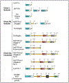
Mammalian START-like PITP structural architecture. Schematic representation of class I and -II PITPs with their specific domains and splice variants. PITPs are grouped into two major classes. Class I PITPs consists of soluble PtdIns/PtdCho exchange proteins while class II proteins are subdivided into two classes: class IIa consisting of insoluble multi-domain proteins and the class IIb soluble protein PITPnc1. The STAMP PITP of Toxoplama gondii is also included. Relevant amino acid numbers are indicated for each protein/domain and different domains are color coded. Three splice variants of PITPβ and two splice variants of human PITPnc1 are also shown. The proteins with different C-terminus splice variants are shown in separate colors. Domain abbreviations: FFAT, the diphenylalanine (FF) in an acidic tract motif that serves as binding site for VAP proteins [243]; DDHD, an ~180 residue domain that represents a potential metal binding unit [242]; LNS2, the PtdOH-binding Lipin/ Nde1 /Smp2 domain [208]; TM, putative membrane-spanning domain.
Fig. 3.
Fig. 3.
Heterotypic lipid exchange cycle of PITP (The “presentation” model): START-like PITPs exchange a second ligand (PtdCho for class I proteins and PtdOH for class II proteins) for PtdIns, and present PtdIns to an otherwise biologically insufficient PI4K enzyme, thereby potentiating PI4K activity and stimulating production of PtdIns4P pools dedicated to specific signaling reactions. The PtdIns4P pool available for signaling is depleted by PtdIns4P antagonists such as Osh proteins and the phosphoinositide phosphatase Sac1.
Fig. 4.
Fig. 4.
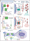
Role of Class I PITPs in human disease. (A) Class I PITPα, and PITPβ are identified as upstream regulators of the Hippo signaling pathway that enhance PtdIns4P synthesis in the plasma membrane. The absence of PITPα and PITPβ promotes YAP phosphorylation and cytoplasmic retention. PITPα and PITPβ are identified as the molecular targets of Microcolin B (MCB) and its derivative VT01454, which upregulate the Hippo pathway by preferentially depleting plasma membrane PtdIns4P pools [108]. (B) Class I START PITPα and PITPβ are involved in the polarized distribution of Golgi in neuronal stem cells. PITPα and PITPβ exchange PtdCho and PtdIns, stimulating PI4K activity to produce PtdIns4P on trans-Golgi network (TGN) membranes. That PtdIns4P pool recruits GOLPH3 and CERT and likely other effectors to TGN membranes where GOLPH3 interacts with the non-conventional actin protein myosin, Myo18A. The recruitment promotes apical enrichment of the Golgi system in the apical process of neural stem cells [110]. (C) PITPα is downregulated in human T2D pancreatic beta cells and its ablation in both murine and human beta cells results in impaired glucose stimulated insulin secretion and granule maturation leading to chronic ER stress. PITPα executes heterotypic PtdCho/PtdIns exchange on beta cell TGN membranes and thereby stimulates PI4K activity and PtdIns4P synthesis. PITPα re-expression in islets donated by type 2 diabetic (T2D) patients rescue pancreatic beta cell dysfunction by restoring insulin granule biogenesis and glucose stimulated insulin secretion to those islets [111]. (D) PITPα/PITPβ regulate non-canonical Planar Cell Polarity (ncPCP) signaling in neural stem cells (NSCs) by potentiating the trafficking of specific cohorts of receptors of the ncPCP pathway such as Vangl2 from the TGN to the plasma membrane [109]. This in turn controls actomyosin dynamics at the nuclear periphery thereby regulating interkinetic nuclear migration (IKNM). PITPs promote PtdIns4P signaling in the NSC TGN/endosomal system to support efficient membrane trafficking of a subset of ncPCP receptors (e,g, VANGL 2) to the plasma membrane. ncPCP receptor signaling activates the cytoplasmic actomyosin system in the nuclear periphery which, in turn, provides the intracellular force for IKNM in proliferating NSCs. IKNM is a distinctive feature of the NSC cell cycle and facilitates asymmetric expansion of newborn NSCs in the lateral, as opposed to the radial, dimension. Actomyosin dynamics at the apical tip of the NSCs might also contribute to IKNM by altering fluidity/plasticity of the apical plasma membrane. IKNM shapes morphogenesis of the neocortex as a thin tissue with an expansive surface area [109]. Defects in tangential expansion of the neocortex are associated with diseases of intellectual disability and autism.
Fig. 5.
Fig. 5.
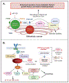
Role of class IIb PITPnc1 in human diseases. (A) PITPnc1 is proposed to enhance secretion of metastasis factors in breast cancer through recruitment of GOLPH3 and Rab1b to trans Golgi network (TGN). PITPnc1 expression is silenced by ‘anti-metastatic’ micro-RNA miR-126 resulting in angiogenesis and tumor metastasis. PITPnc1 exchanges PtdIns for PtdOH and stimulates PI4K activity to produce an expanded TGN PtdIns4P pool. PtdIns4P mediates GOLPH3 and Rab1b recruitment to the cancer cell TGN, resulting in enhanced secretion of pro-metastatic factors. These include growth factors and matrix metalloproteases [156]. (B) PITPnc1 is identified as a downstream effector of KRAS in lung and pancreatic ductal adenocarcinoma (LUAD and PDAC). A PITPnc1 regulated signature links KRAS to MYC stability thereby regulating autophagy. PITPnc1 depletion inhibits lung and pancreatic cancer cell proliferation in vitro and tumor proliferation in vivo (i.e. lung colonization and liver metastasis) [158]. PITPnc1 inhibition results in Myc downregulation which in turn perturbs mTORC1 localization and promotes autophagy. JAK2 and KRASG12C inhibitors identified as surrogate PITPnc1 inhibitors show anti-tumor effects in LUAD and PDAC.
Fig. 6.
Fig. 6.
The role of RdgBα protein in Drosophila melanogaster. Schematic representation of the phospholipase C (PLC) signaling pathway and the PtdIns(4,5)P2 cycle between microvillar plasma membrane and submicrovillar cisternae (SMC) in fly photoreceptor cells. RdgBα is posited to function in transfer of PtdIns from SMC to plasma membrane and PtdOH from plasma membrane back to the SMC. The lipid transfer cycle is proposed to occur at ER-PM contact sites. The PITP domain is both necessary and sufficient to support the cycle.

Similar articles

References

    1. Balla T, Phosphoinositides: tiny lipids with giant impact on cell regulation, Physiol. Rev 93 (3) (2013) 1019–1137, 10.1152/physrev.00028.2012. - DOI - PMC - PubMed
    1. Wenk MR, Lucast L, Paolo GD, Romanelli AJ, Suchy SF, Nussbaum RL, Cline GW, Shulman GI, McMurray W, De Camilli P, Phosphoinositide profilingin complex lipid mixtures using electrospray ionization mass spectrometry, Nat. Biotechnol 21 (7) (2003) 813–817, 10.1038/nbt837. - DOI - PubMed
    1. Berridge MJ, Discovery of the second messenger inositol trisphosphate, Messenger 1 (1) (2012) 3–15, 10.1166/msr.2012.1006. - DOI
    1. Dittmer JC, Dawson RMC, The isolation of a new lipid, triphosphoinositide, and monophosphoinositide from ox brain, Biochem. J 81 (3) (1961) 535–540. - PMC - PubMed
    1. Hokin LE, Hokin MR, Acetylcholine and the exchange of phosphate in phosphatidic acid in brain microsomes, J. Biol. Chem 233 (4) (1958) 822–826. - PubMed

MeSH terms

Substances

LinkOut - more resources