

Plasmodium vivax serological exposure markers: PvMSP1-42-induced humoral and memory B-cell response generates long-lived antibodies
- PMID: 38941356
- PMCID: PMC11239109
- DOI: 10.1371/journal.ppat.1012334
Plasmodium vivax serological exposure markers: PvMSP1-42-induced humoral and memory B-cell response generates long-lived antibodies
Abstract
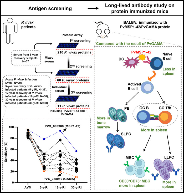
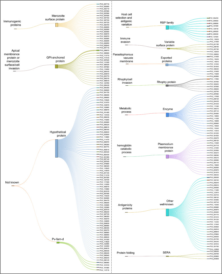
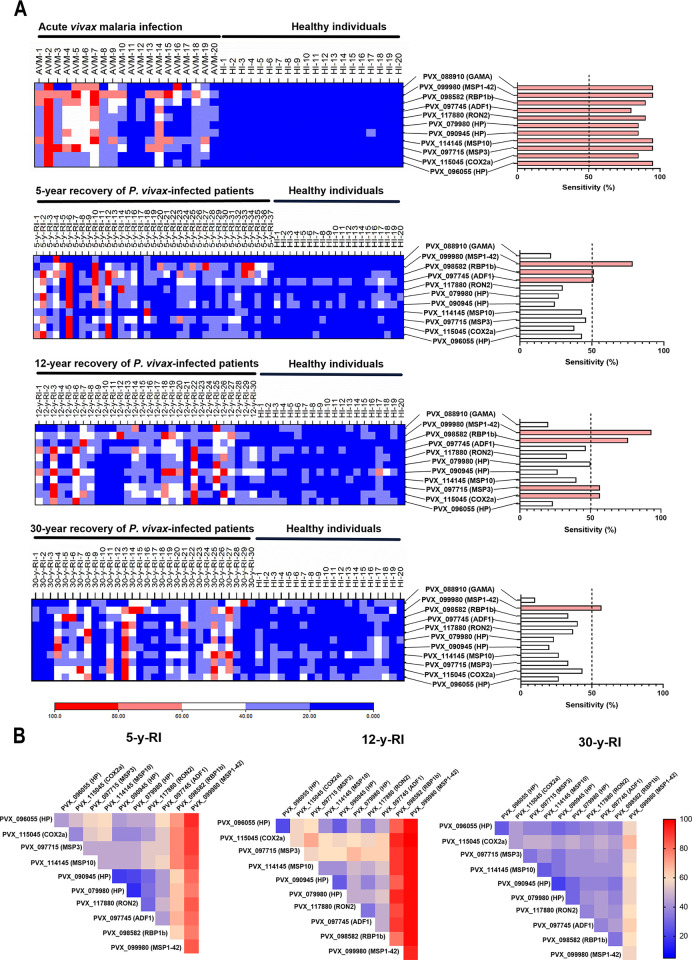
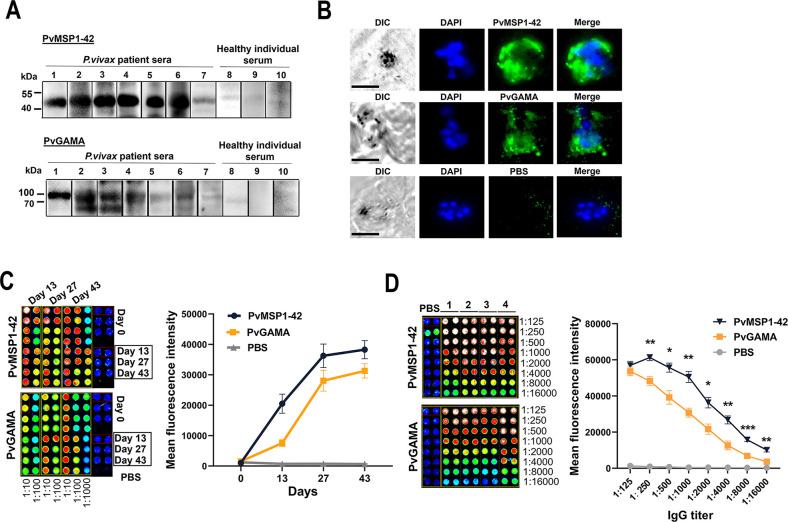
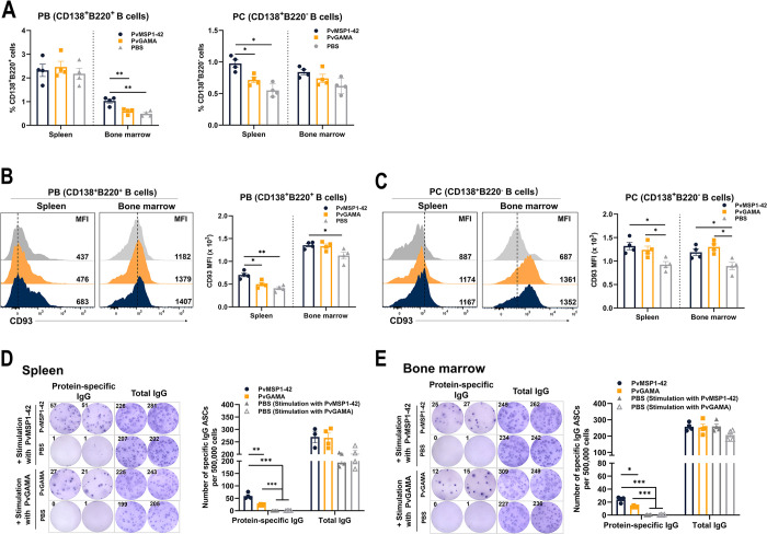
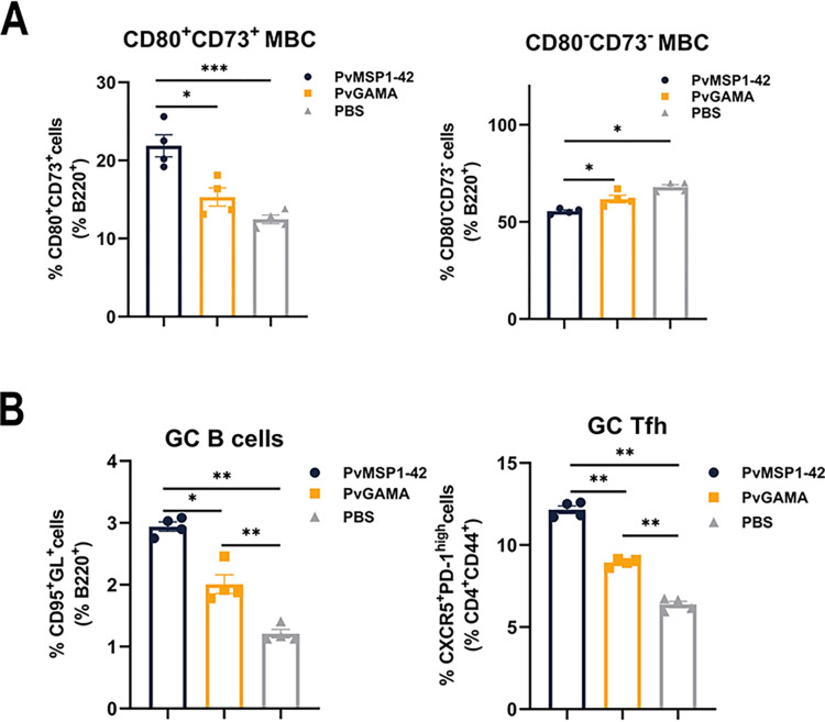
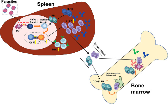
Plasmodium vivax serological exposure markers (SEMs) have emerged as promising tools for the actionable surveillance and implementation of targeted interventions to accelerate malaria elimination. To determine the dynamic profiles of SEMs in current and past P. vivax infections, we screened and selected 11 P. vivax proteins from 210 putative proteins using protein arrays, with a set of serum samples obtained from patients with acute P. vivax and documented past P. vivax infections. Then we used a murine protein immune model to initially investigate the humoral and memory B cell response involved in the generation of long-lived antibodies. We show that of the 11 proteins, especially C-terminal 42-kDa region of P. vivax merozoite surface protein 1 (PvMSP1-42) induced longer-lasting long-lived antibodies, as these antibodies were detected in individuals infected with P. vivax in the 1960-1970s who were not re-infected until 2012. In addition, we provide a potential mechanism for the maintenance of long-lived antibodies after the induction of PvMSP1-42. The results indicate that PvMSP1-42 induces more CD73+CD80+ memory B cells (MBCs) compared to P. vivax GPI-anchored micronemal antigen (PvGAMA), allowing IgG anti-PvMSP1-42 antibodies to be maintained for a long time.
Copyright: © 2024 Lu et al. This is an open access article distributed under the terms of the Creative Commons Attribution License, which permits unrestricted use, distribution, and reproduction in any medium, provided the original author and source are credited.
Conflict of interest statement
The authors have declared that no competing interests exist.
Figures
Similar articles
-
Immunogenicity of the Plasmodium vivax merozoite surface protein 1 paralog in the induction of naturally acquired antibody and memory B cell responses.Malar J. 2017 Aug 30;16(1):354. doi: 10.1186/s12936-017-2000-z. Malar J. 2017. PMID: 28854974 Free PMC article.
-
The acquisition of long-lived memory B cell responses to merozoite surface protein-8 in individuals with Plasmodium vivax infection.Malar J. 2019 May 31;18(1):188. doi: 10.1186/s12936-019-2821-z. Malar J. 2019. PMID: 31151441 Free PMC article.
-
Antibody response to the N and C-terminal regions of the Plasmodium vivax Merozoite Surface Protein 1 in individuals living in an area of exclusive transmission of P. vivax malaria in the north of Brazil.Acta Trop. 1999 Jan 15;72(1):13-24. doi: 10.1016/s0001-706x(98)00078-3. Acta Trop. 1999. PMID: 9924957
-
Development and longevity of naturally acquired antibody and memory B cell responses against Plasmodium vivax infection.PLoS Negl Trop Dis. 2024 Oct 24;18(10):e0012600. doi: 10.1371/journal.pntd.0012600. eCollection 2024 Oct. PLoS Negl Trop Dis. 2024. PMID: 39446698 Free PMC article. Review.
-
Natural acquisition of immunity to Plasmodium vivax: epidemiological observations and potential targets.Adv Parasitol. 2013;81:77-131. doi: 10.1016/B978-0-12-407826-0.00003-5. Adv Parasitol. 2013. PMID: 23384622 Review.
Cited by
-
Characterization of T and B cell epitopes in PvCyRPA by studying the naturally acquired immune response in Brazilian Amazon communities.Sci Rep. 2024 Nov 9;14(1):27343. doi: 10.1038/s41598-024-72671-x. Sci Rep. 2024. PMID: 39521783 Free PMC article.
References
-
- WHO. World Malaria Report 2023 2023. Available from: https://www.who.int/publications/i/item/9789240086173.
-
- Sutanto I, Kosasih A, Elyazar IRF, Simanjuntak DR, Larasati TA, Dahlan MS, et al.. Negligible Impact of Mass Screening and Treatment on Mesoendemic Malaria Transmission at West Timor in Eastern Indonesia: A Cluster-Randomized Trial. Clinical Infectious Diseases. 2018;67(9):1364–1372. doi: 10.1093/cid/ciy231 . - DOI - PMC - PubMed
MeSH terms
Substances
Grants and funding
LinkOut - more resources
Full Text Sources
Research Materials
Miscellaneous
