

The effects of field strength on stimulated echo and motion-compensated spin-echo diffusion tensor cardiovascular magnetic resonance sequences
- PMID: 38936803
- PMCID: PMC11283220
- DOI: 10.1016/j.jocmr.2024.101052
The effects of field strength on stimulated echo and motion-compensated spin-echo diffusion tensor cardiovascular magnetic resonance sequences
Abstract
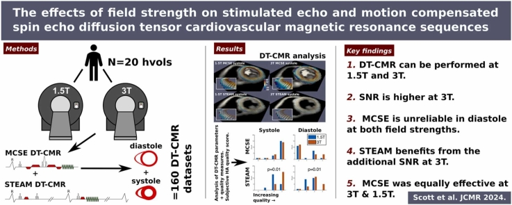
Background: In-vivo diffusion tensor cardiovascular magnetic resonance (DT-CMR) is an emerging technique for microstructural tissue characterization in the myocardium. Most studies are performed at 3T, where higher signal-to-noise ratio (SNR) should benefit this signal-starved method. However, a few studies have suggested that DT-CMR is possible at 1.5T, where echo planar imaging artifacts may be less severe and 1.5T hardware is more widely available.
Methods: We recruited 20 healthy volunteers and performed mid-ventricular short-axis DT-CMR at 1.5T and 3T. Acquisitions were performed at peak systole and end-diastole using both stimulated echo acquisition mode (STEAM) and motion-compensated spin-echo (MCSE) sequences at matched spatial resolutions. DT-CMR parameters were averaged over the left ventricle and compared between 1.5T and 3T sequences using both datasets with and without the blow reference data included.
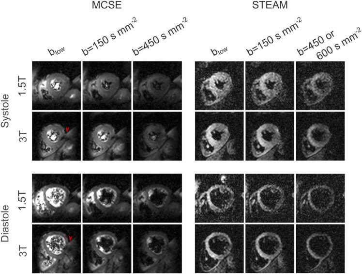
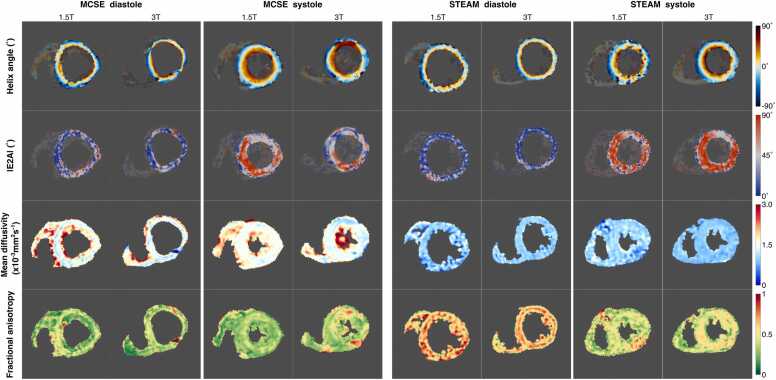
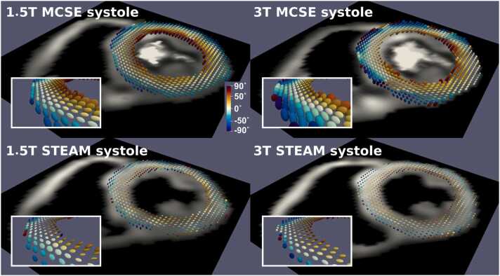
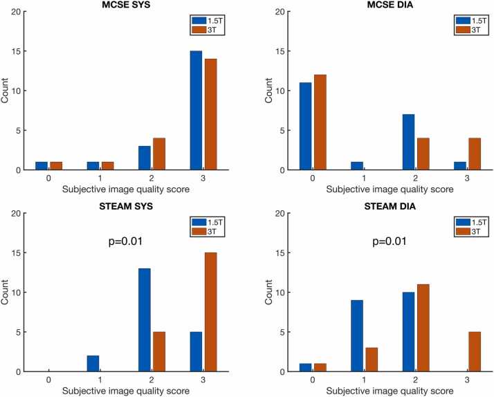
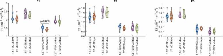
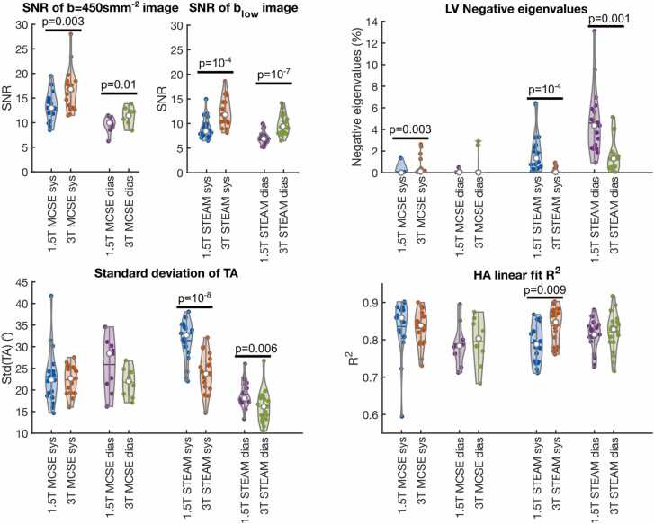
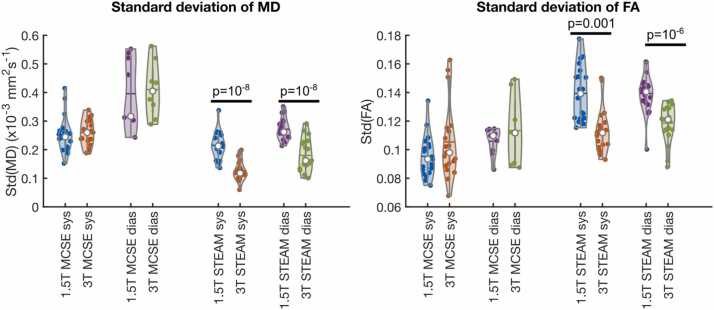
Results: Eleven (1.5T) and 12 (3T) diastolic MCSE acquisitions were rejected as the helix angle (HA) demonstrated <50% normal appearance circumferentially or the acquisition was abandoned due to poor image quality; a maximum of one acquisition was rejected for other datasets. Subjective HA map quality was significantly better at 3T than 1.5T for STEAM (p < 0.05), but not for MCSE and other DT-CMR quality measures were consistent with improvements in STEAM at 3T over 1.5T. When blow data were excluded, no significant differences in mean diffusivity were observed between field strengths, but fractional anisotropy was significantly higher at 1.5T than 3T for STEAM systole (p < 0.05). Absolute second eigenvector orientation (E2A, sheetlet angle) was significantly higher at 1.5T than 3T for MCSE systole and STEAM diastole, but significantly lower for STEAM systole (all p < 0.05). Transmural HA distribution was less steep at 1.5T than 3T for STEAM diastole data (p < 0.05). SNR was higher at 3T than 1.5T for all acquisitions (p < 0.05).
Conclusion: While 3T provides benefits in terms of SNR, both STEAM and MCSE can be performed at 1.5T. However, MCSE is unreliable in diastole at both field strengths and STEAM benefits from the improved SNR at 3T over 1.5T. Future clinical research studies may be able to leverage the wider availability of 1.5T CMR hardware where MCSE acquisitions are desirable.
Keywords: Cardiac microstructure; DTI; Diffusion tensor; Field strength; Healthy volunteers; Stimulated echo.
Copyright © 2024 The Author(s). Published by Elsevier Inc. All rights reserved.
Conflict of interest statement
Declaration of competing interests The authors declare the following financial interests/personal relationships which may be considered as potential competing interests: The Cardiovascular Magnetic Resonance Unit at the Royal Brompton Hospital receives research support from Siemens. Ke Wen and Yaqing Luo are partly funded by Siemens.
Figures

Similar articles
-
Defining the optimum strategy for identifying adults and children with coeliac disease: systematic review and economic modelling.Health Technol Assess. 2022 Oct;26(44):1-310. doi: 10.3310/ZUCE8371. Health Technol Assess. 2022. PMID: 36321689 Free PMC article.
-
Depressing time: Waiting, melancholia, and the psychoanalytic practice of care.In: Kirtsoglou E, Simpson B, editors. The Time of Anthropology: Studies of Contemporary Chronopolitics. Abingdon: Routledge; 2020. Chapter 5. In: Kirtsoglou E, Simpson B, editors. The Time of Anthropology: Studies of Contemporary Chronopolitics. Abingdon: Routledge; 2020. Chapter 5. PMID: 36137063 Free Books & Documents. Review.
-
Lamotrigine versus levetiracetam or zonisamide for focal epilepsy and valproate versus levetiracetam for generalised and unclassified epilepsy: two SANAD II non-inferiority RCTs.Health Technol Assess. 2021 Dec;25(75):1-134. doi: 10.3310/hta25750. Health Technol Assess. 2021. PMID: 34931602 Clinical Trial.
-
Falls prevention interventions for community-dwelling older adults: systematic review and meta-analysis of benefits, harms, and patient values and preferences.Syst Rev. 2024 Nov 26;13(1):289. doi: 10.1186/s13643-024-02681-3. Syst Rev. 2024. PMID: 39593159 Free PMC article.
-
Conservative, physical and surgical interventions for managing faecal incontinence and constipation in adults with central neurological diseases.Cochrane Database Syst Rev. 2024 Oct 29;10(10):CD002115. doi: 10.1002/14651858.CD002115.pub6. Cochrane Database Syst Rev. 2024. PMID: 39470206
References
-
- Nielles-Vallespin S., Scott A., Ferreira P., Khalique Z., Pennell D., Firmin D. Cardiac diffusion: technique and practical applications. J Magn Reson Imaging. 2020;52:348–368. - PubMed
-
- Reese T.G., Weisskoff R.M., Smith R.N., Rosen B.R., Dinsmore R.E., Wedeen V.J. Imaging myocardial fiber architecture in vivo with magnetic resonance. Magn Reson Med. 1995;34:786–791. - PubMed
-
- Stoeck C.T., von Deuster C., Fleischmann T., Lipiski M., Cesarovic N., Kozerke S. Direct comparison of in vivo versus postmortem second-order motion-compensated cardiac diffusion tensor imaging. Magn Reson Med. 2018;79:2265–2276. - PubMed
-
- Stoeck C.T., Von Deuster C., GeneT M., Atkinson D., Kozerke S. Second-order motion-compensated spin echo diffusion tensor imaging of the human heart. Magn Reson Med. 2016;75:1669–1676. - PubMed
Publication types
MeSH terms
LinkOut - more resources
Full Text Sources
