. 2024 Jun 13;16(6):958.
doi: 10.3390/v16060958.

Epidemiological and Genetic Characteristics of Respiratory Viral Coinfections with Different Variants of Severe Acute Respiratory Syndrome Coronavirus 2 (SARS-CoV-2)

Affiliations

Epidemiological and Genetic Characteristics of Respiratory Viral Coinfections with Different Variants of Severe Acute Respiratory Syndrome Coronavirus 2 (SARS-CoV-2)

Ivelina Trifonova et al. Viruses. .

Abstract

This study aimed to determine the incidence and etiological, seasonal, and genetic characteristics of respiratory viral coinfections involving severe acute respiratory syndrome coronavirus 2 (SARS-CoV-2). Between October 2020 and January 2024, nasopharyngeal samples were collected from 2277 SARS-CoV-2-positive patients. Two multiplex approaches were used to detect and sequence SARS-CoV-2, influenza A/B viruses, and other seasonal respiratory viruses: multiplex real-time polymerase chain reaction (PCR) and multiplex next-generation sequencing. Coinfections of SARS-CoV-2 with other respiratory viruses were detected in 164 (7.2%) patients. The most common co-infecting virus was respiratory syncytial virus (RSV) (38 cases, 1.7%), followed by bocavirus (BoV) (1.2%) and rhinovirus (RV) (1.1%). Patients ≤ 16 years of age had the highest rate (15%) of mixed infections. Whole-genome sequencing produced 19 complete genomes of seasonal respiratory viral co-pathogens, which were subjected to phylogenetic and amino acid analyses. The detected influenza viruses were classified into the genetic groups 6B.1A.5a.2a and 6B.1A.5a.2a.1 for A(H1N1)pdm09, 3C.2a1b.2a.2a.1 and 3C.2a.2b for A(H3N2), and V1A.3a.2 for the B/Victoria lineage. The RSV-B sequences belonged to the genetic group GB5.0.5a, with HAdV-C belonging to type 1, BoV to genotype VP1, and PIV3 to lineage 1a(i). Multiple amino acid substitutions were identified, including at the antibody-binding sites. This study provides insights into respiratory viral coinfections involving SARS-CoV-2 and reinforces the importance of genetic characterization of co-pathogens in the development of therapeutic and preventive strategies.

Keywords: AdV-C; BoV; PIV3; RSV-B; SARS-CoV-2; coinfections; influenza viruses; multiplex NGS; respiratory viruses.


Conflict of interest statement

The authors declare no conflicts of interest.

Figures

Figure 1
Figure 1
Distribution of proven coinfections by month from October 2020 to January 2024 in Bulgaria.
Figure 2
Figure 2
Comparison of Ct values of different respiratory viruses involved in mixed infections: (A) Ct of SARS-CoV-2 versus other respiratory viruses in a mixed infection (RSV, RV, BoV, HMPV, PIV2,3, and AdV); (B) Ct of influenza A/B viruses compared to Ct of other respiratory viruses involved in co-infection with SARS-CoV-2; (C) Ct of SARS-CoV-2 versus that of influenza A/B viruses involved in a co-infection; (D) Ct of SARS-CoV-2 co-infected with influenza A/B viruses versus Ct of SARS-CoV-2 in a mixed infection with RSV, RV, BoV, HMPV, PIV2/3, and AdV. Mean and median Ct values, SD, and p values (Fisher’s test) are shown. Values were calculated using the Mann–Whitney U-test.
Figure 3
Figure 3
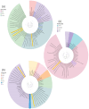
Phylogenetic analysis was performed to construct evolutionary trees based on a fragment of the HA gene of the influenza A(H1N1) virus (a). The Bulgarian sequences isolated in 2023 that belong to the influenza virus subtype A(H1N1) and that were detected in a co-infection with SARS-CoV-2 are marked in yellow. The tree was rooted based on the sequence deposited by a research team in Ghana in 2021 (A/Ghana/1894/2021-July). (b) Phylogenetic analysis of influenza virus A(H3N2). The Bulgarian sequences isolated in 2023 that belong to the influenza virus subtype A(H3N2) and that were detected in a mixed infection with SARS-CoV-2 are highlighted in yellow. The tree was rooted based on the sequence deposited by a research team in Brandenburg, Germany in 2022 (A/Brandenburg/15/2022). (c) Phylogenetic analysis of influenza virus B-Victoria. The sequences isolated in 2023 that belong to the B-Victoria influenza virus lineage and that were found in a mixed infection with SARS-CoV-2 are highlighted in yellow. The tree was rooted based on the sequence deposited by a research team in Colorado, USA in 2017 (B/Colorado/06/2 February 2017). Genetic distances were measured according to the Jukes–Cantor model. A phylogenetic tree was constructed using a neighbor-joining algorithm in Geneious Tree Builder. The sequences of the reference strains representative of the known genotypes were obtained from GenBank with the corresponding identification numbers.
Figure 4
Figure 4
Phylogenetic analysis of RSV-B in which a tree was constructed relative to the G protein and the entire genome of the virus. Genetic distances were measured according to the Jukes–Cantor model. The phylogenetic tree was constructed using a neighbor.
Figure 5
Figure 5
Phylogenetic analysis based on the hexon gene and the whole genome of HAdV. Genetic distances were measured according to the Jukes–Cantor model. The phylogenetic tree was constructed using a neighbor-joining algorithm in Geneious Tree Builder. The sequences of the reference strains representative of the known genotypes were obtained from GenBank with the corresponding identification numbers. The tree was rooted based on a 2010 strain (HAdV-A12 OQ518256.1). The sequence of HAdV-C1 isolated in mixed infections with SARS-CoV-2 is highlighted in yellow.
Figure 6
Figure 6
Amino acid analysis showing the sites of amino acid substitutions in the genetic sequence encoding the fiber and penton proteins of the Bulgarian strain of HAdV. Alignment was performed against the reference strain AC_000017.1.
Figure 7
Figure 7
Phylogenetic tree constructed based on the VP1/VP2 gene and the whole genome of hBov and other parvoviruses. Genetic distances were measured according to the Jukes–Cantor model. The phylogenetic tree was constructed using a neighbor-joining algorithm in Geneious Tree Builder. The sequences of the reference strains representative of the known genotypes were obtained from GenBank with the corresponding identification numbers. The tree was rooted based on the strain Human_parvovirus_4/3/NG-OR. Isolated sequences from confirmed co-infected patients with HBoV-V1 and SARS-CoV-2 are highlighted in yellow.
Figure 8
Figure 8
Amino acid analysis of Bulgarian strains isolated from cases with co-infection with SARS-CoV-2. Substitution sites in the genome of the Bulgarian HBoV strains encoding the VP1/VP2 protein are depicted. Alignment was performed against the prototype St1 strain.
Figure 9
Figure 9
Phylogenetic analysis based on the HN-coding region and the whole genome of human parainfluenza virus type 3 (HPIV3). Genetic distances were measured according to the Jukes–Cantor model. The phylogenetic tree was constructed using a neighbor-joining algorithm in Geneious Tree Builder. The sequences of the reference strains representative of the known genotypes were obtained from GenBank with the corresponding identification numbers. The tree was rooted based on the clone 2 strain HPIV3/AUS/3/2007 (KF530243.1). PIV3 sequences isolated from SARS-CoV-2 co-infected patients and belonging to subclade 1a(i) are shown in yellow.
Figure 10
Figure 10
Amino acid analysis showing the sites of substitutions in the genetic sequences encoding the fusion (F) protein and hemagglutinin–neuraminidase protein (HN) of the Bulgarian strain of PIV3. Alignment was performed against the reference strains and S82195.1 and JN089924.1.

Similar articles

Cited by

References

    1. Li P., Zhang Y., Shen W., Shi S., Zhao Z. dbGSRV: A manually curated database of genetic susceptibility to respiratory virus. PLoS ONE. 2022;17:e0262373. doi: 10.1371/journal.pone.0262373. - DOI - PMC - PubMed
    1. Wu X., Cai Y., Huang X., Yu X., Zhao L., Wang F., Li Q., Gu S., Xu T., Li Y., et al. Co-infection with SARS-CoV-2 and Influenza A Virus in Patient with Pneumonia, China. Emerg. Infect. Dis. 2020;26:1324–1326. doi: 10.3201/eid2606.200299. - DOI - PMC - PubMed
    1. Cuadrado-Payán E., Montagud-Marrahi E., Torres-Elorza M., Bodro M., Blasco M., Poch E., Soriano A., Piñeiro G.J. SARS-CoV-2 and influenza virus co-infection. Lancet. 2020;395:e84. doi: 10.1016/S0140-6736(20)31052-7. - DOI - PMC - PubMed
    1. Maltezou H.C., Papanikolopoulou A., Vassiliu S., Theodoridou K., Nikolopoulou G., Sipsas N.V. COVID-19 and Respiratory Virus Co-Infections: A Systematic Review of the Literature. Viruses. 2023;15:865. doi: 10.3390/v15040865. - DOI - PMC - PubMed
    1. Rubin R. Influenza’s Unprecedented Low Profile During COVID-19 Pandemic Leaves Experts Wondering What This Flu Season Has in Store. JAMA. 2021;326:899–900. doi: 10.1001/jama.2021.14131. - DOI - PubMed

MeSH terms

Supplementary concepts