. 2024 May 25;16(6):846.
doi: 10.3390/v16060846.

Genome-Wide Transcriptional Roles of KSHV Viral Interferon Regulatory Factors in Oral Epithelial Cells

Affiliations

Genome-Wide Transcriptional Roles of KSHV Viral Interferon Regulatory Factors in Oral Epithelial Cells

Seung Jin Jang et al. Viruses. .

Abstract

The viral interferon regulatory factors (vIRFs) of KSHV are known to dysregulate cell signaling pathways to promote viral oncogenesis and to block antiviral immune responses to facilitate infection. However, it remains unknown to what extent each vIRF plays a role in gene regulation. To address this, we performed a comparative analysis of the protein structures and gene regulation of the four vIRFs. Our structure prediction analysis revealed that despite their low amino acid sequence similarity, vIRFs exhibit high structural homology in both their DNA-binding domain (DBD) and IRF association domain. However, despite this shared structural homology, we demonstrate that each vIRF regulates a distinct set of KSHV gene promoters and human genes in epithelial cells. We also found that the DBD of vIRF1 is essential in regulating the expression of its target genes. We propose that the structurally similar vIRFs evolved to possess specialized transcriptional functions to regulate specific genes.

Keywords: EDC genes; IRF; KSHV; gammaherpesviruses; gene regulation; genomics; oral epithelial cells; vIRF.


Conflict of interest statement

The authors declare no conflict of interest.

Figures
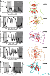
Figure 1
Figure 1
Structure prediction of KSHV vIRF1 (A), vIRF2 (B), vIRF3 (C), vIRF4 (D) and hIRF4 (E). Shown on the left are graphs of the predicted disorder disposition (Y-axis) for each of the vIRFs and hIRF4 along their amino acid sequences, calculated by PONDR_fit [26]. Regions with a disorder probability higher than 0.5 are predicted to be disordered. The light gray shaded area indicates the DNA-binding domain (DBD) and the dark gray shaded area displays the IRF association domain (IAD). On the right, the top-ranked AlphaFold structure predictions of the vIRFs and hIRF4 are shown as ribbon diagrams [27]. Beta strands are colored in gray and α-helices in red. The positions of the DBD and IAD are indicated.
Figure 2
Figure 2
Structural comparison of the DBD and the IAD of vIRFs and hIRF4. (A) Schematics of KSHV vIRFs and hIRF4 indicating the DBD and IAD domains and their amino acid positions. (B) The ribbon diagrams of the DBDs in the vIRF and hIRF4 proteins are displayed in the same orientation. Beta strands are colored in gray and α-helices in red. The positions of arginine (R) residues 163 and 172 in vIRF1 implicated in vIRF1’s DNA binding and the basic amino acids in the equivalent position in the other vIRFs and hIRF4 are indicated. (C) The ribbon diagrams of the IADs in the vIRF and hIRF4 proteins are shown. The structural superposition of the DBDs (D) and IADs (E) is displayed.
Figure 3
Figure 3
Identification of vIRF-regulated viral promoters in KSHV. (A) KSHV gene promoter screen with vIRFs using luciferase reporter assay. (B) Comparing the effects of vIRFs on the promoter activity of the KSHV ORF57 gene. The t tests were performed between the vector- and vIRF-transfected cells. (C) Measuring the induction of ORF57 promoter fragments by vIRF4. The t tests were calculated by comparing the promoter activity of the ORF57 promoter deletion mutants to the 450-bp full promoter of ORF57. (D) Testing the inducibility of the minimal promoter of the pGL4.27 reporter plasmid by vIRF4 to determine whether it can be increased via the 229–126-bp region of the ORF57 promoter. pGL4.15 containing the full ORF57 promoter was used as a positive control. The t tests were performed relative to the vector-transfected samples. (E) Luciferase reporter assay testing whether vIRF4 can induce the minimal promoter of pGL4.27 luciferase reporter plasmid via the RTA response element (RRE). RTA co-transfection was used a positive control. The t tests were performed relative to pGL4.27-RRE and vector co-transfection samples. The statistical significance is indicated as * p ≤ 0.05, and NS refers to statistically non-significant results (samples of n = 3).
Figure 4
Figure 4
Identification of host genes regulated by vIRFs in primary oral epithelial cells. (A) Immunoblot analysis of the expression of vIRFs in lenti-3xFLAG-vIRF-transduced HGEP cells. (B) Volcano plot of the differentially expressed host genes between HGEP cells expressing GFP (control) and each of the vIRFs. (C) Gene ontology analysis of vIRF-regulated host genes.
Figure 5
Figure 5
Analysis of host genes co-regulated by vIRFs in primary oral epithelial cells. (A) Venn diagrams showing the shared and unique upregulated and downregulated target genes of vIRFs. (B) Gene ontology analysis of the unique target genes of each vIRF. Note: vIRF4 is not included due to its small number of target genes.
Figure 6
Figure 6
RT-qPCR confirmation and measurement of the expression of vIRF-regulated host genes in HGEP cells. (A) vIRF1-regulated genes. (B) Epidermal differentiation complex-related genes induced by vIRF1 and vIRF3. (C) vIRF1- and vIRF2-induced genes. (D) Host genes inhibited by vIRF1. (E) Host genes that are inhibited by vIRF1 and induced by vIRF3. (F) Host genes that are induced by vIRF2 and vIRF4 and repressed by vIRF3. The t tests were performed between lenti-GFP and the indicated lenti-vIRF samples. The statistical significance is indicated as * p ≤ 0.05 (samples of n = 3).
Figure 7
Figure 7
Comparison of host genes regulated by vIRFs with genes dysregulated upon KSHV infection in oral epithelial cells. (A) Venn diagrams showing the common and unique host genes that are regulated by the lenti-vIRFs versus during KSHV infection of HGEP cells. (B) Gene ontology analysis of host genes whose expression is regulated by both vIRF expression and KSHV infection. (C) RT-qPCR analysis of select vIRF1-regulated host genes in HGEP cells following KSHV infection at 24 hpi. Student’s t tests were performed between the mock and KSHV-infected HGEP samples. The statistical significance is indicated as * p ≤ 0.05 (samples of n = 3). (D) Immunoblot detection of a viral protein (ORF45) and a vIRF1-regulated host factor in mock and KSHV-infected HGEP cells at 24 hpi.
Figure 8
Figure 8
Analyzing the effect of functional mutations in vIRF1 on vIRF1-mediated host gene regulation. (A) Schematic representation of the wild type (WT) and mutants of vIRF1. Red arrows at DBDm indicate the R163A and R172A mutations. Red box in the ΔEGPS mutant marks the internal deletion that has been shown to abrogate the binding of vIRF1 to USP7. (B) Immunoblot analysis of the expression of WT and mutants of 3xFLAG-vIRF1. (C) RT-qPCR analysis of the effect of vIRF1 mutations on the expression of vIRF1-inducible host gene LCE3D in HGEP cells. (D) RT-qPCR measurement of the effect of vIRF1 mutations on the expression of vIRF1-inhibited host gene CXCL3 in HGEP cells. Student’s t tests were performed between lenti-GFP and lenti-vIRF1 samples. The statistical significance is indicated as * p ≤ 0.05 (samples of n = 3).
Figure 9
Figure 9
DNA-binding activity of vIRF1 is needed to induce viral promoters by vIRF1. Luciferase reporter assay was performed with the indicated KSHV gene promoter luciferase reporter plasmids and WT or DBDm of vIRF1. Student’s t tests were performed between WT vIRF1- and vIRF1_DBDm-transfected cells. The statistical significance is indicated as * p ≤ 0.05 (samples of n = 3).
Figure 10
Figure 10
Comparing host gene expression changes between HGEP cells expressing WT vIRF1 and vIRF1 DBDm using RNA-seq analysis. (A) Immunoblot analysis of HGEP cells transduced with lenti-3xFLAG-vIRF1 WT or DBDm. (B) Volcano plot displaying the fold change in host gene expression between HGEP cells expressing GFP (control) or 3xFLAG-vIRF1 (WT or DBDm). (C) Venn diagram representation of the significantly upregulated or downregulated host genes that are differentially expressed between WT vIRF1 and DBDm vIRF1. Examples of genes for each category are shown. (D) RT-qPCR confirmation of differential expression of host genes by WT vs. DBDm vIRF1 in HGEP cells. Student’s t tests were performed between WT vIRF1- and vIRF1_DBDm-transduced cells. The statistical significance is indicated as * p ≤ 0.05 (samples of n = 3). (E) Immunoblot test of vIRF1 expression in lentivirus-transduced TIGK cells. (F) RT-qPCR confirmation of differential expression of host genes by WT vs. DBDm vIRF1 in TIGK cells. Student’s t tests were performed between WT vIRF1- and vIRF1_DBDm-transduced cells. The statistical significance is indicated as * p ≤ 0.05 (samples of n = 3).
Figure 11
Figure 11
vIRF1 increases the level of activating histone modifications on the TGM1 promoter. WT or DBDm of vIRF1 was expressed in HGEP cells for 2 days followed by ChIP analysis to test the accumulation of (A) H3K4me3 and (B) H3K27ac on the TGM1 promoter (0.7 kb upstream of the transcription start site of the TGM1 gene).

Similar articles

References

    1. Chang Y., Cesarman E., Pessin M.S., Lee F., Culpepper J., Knowles D.M., Moore P.S. Identification of herpesvirus-like DNA sequences in AIDS-associated Kaposi’s sarcoma. Science. 1994;266:1865–1869. doi: 10.1126/science.7997879. - DOI - PubMed
    1. Cesarman E., Chang Y., Moore P.S., Said J.W., Knowles D.M. Kaposi’s sarcoma-associated herpesvirus-like DNA sequences in AIDS-related body-cavity-based lymphomas. N. Engl. J. Med. 1995;332:1186–1191. doi: 10.1056/NEJM199505043321802. - DOI - PubMed
    1. Soulier J., Grollet L., Oksenhendler E., Cacoub P., Cazals-Hatem D., Babinet P., d’Agay M.F., Clauvel J.P., Raphael M., Degos L., et al. Kaposi’s sarcoma-associated herpesvirus-like DNA sequences in multicentric Castleman’s disease. Blood. 1995;86:1276–1280. doi: 10.1182/blood.V86.4.1276.bloodjournal8641276. - DOI - PubMed
    1. Atyeo N., Rodriguez M.D., Papp B., Toth Z. Clinical Manifestations and Epigenetic Regulation of Oral Herpesvirus Infections. Viruses. 2021;13:681. doi: 10.3390/v13040681. - DOI - PMC - PubMed
    1. Minhas V., Wood C. Epidemiology and transmission of Kaposi’s sarcoma-associated herpesvirus. Viruses. 2014;6:4178–4194. doi: 10.3390/v6114178. - DOI - PMC - PubMed

MeSH terms

Substances

LinkOut - more resources