. 2024 May 23;16(6):694.
doi: 10.3390/pharmaceutics16060694.

Archaeosomes for Oral Drug Delivery: From Continuous Microfluidics Production to Powdered Formulations

Affiliations

Archaeosomes for Oral Drug Delivery: From Continuous Microfluidics Production to Powdered Formulations

Ivan Vidakovic et al. Pharmaceutics. .

Abstract

Archaeosomes were manufactured from natural archaeal lipids by a microfluidics-assisted single-step production method utilizing a mixture of di- and tetraether lipids extracted from Sulfolobus acidocaldarius. The primary aim of this study was to investigate the exceptional stability of archaeosomes as potential carriers for oral drug delivery, with a focus on powdered formulations. The archaeosomes were negatively charged with a size of approximately 100 nm and a low polydispersity index. To assess their suitability for oral delivery, the archaeosomes were loaded with two model drugs: calcein, a fluorescent compound, and insulin, a peptide hormone. The archaeosomes demonstrated high stability in simulated intestinal fluids, with only 5% of the encapsulated compounds being released after 24 h, regardless of the presence of degrading enzymes or extremely acidic pH values such as those found in the stomach. In a co-culture cell model system mimicking the intestinal barrier, the archaeosomes showed strong adhesion to the cell membranes, facilitating a slow release of contents. The archaeosomes were loaded with insulin in a single-step procedure achieving an encapsulation efficiency of approximately 35%. These particles have been exposed to extreme manufacturing temperatures during freeze-drying and spray-drying processes, demonstrating remarkable resilience under these harsh conditions. The fabrication of stable dry powder formulations of archaeosomes represents a promising advancement toward the development of solid dosage forms for oral delivery of biological drugs.

Keywords: archaeal lipids; archaeosomes; dry powder formulation; insulin; oral drug delivery; solid dosage form.


Conflict of interest statement

The co-authors Christina Horn and Julian Quehenberger are currently employed by the company NovoArc GmbH, Vienna, Austria. The remaining authors declare that the research was conducted in the absence of any commercial or financial relationships that could be construed as a potential conflict of interest.

Figures

Figure 1
Figure 1
Visualization of Sulfolobus acidocaldarius cells using (a) TEM and (b) SEM. The scale bar represents 200 nm. The sample preparation for SEM includes water displacement through a series of ethanol treatments that can cause cell shrinkage as evident in (b). The molecular structures of two ether lipid species isolated from the archaeal membrane are presented in (c,d), highlighting structural differences. (c) Features archaeol—the principal representative of DEL containing two isoprenoid chains and (d) caldarchaeol—a prominent representative of TEL, which is almost double in length having two polar groups connected with two isoprene-like chains containing 6 cyclic structures.
Figure 2
Figure 2
(a) TEM image of negatively stained archaeosomes using 1% uranyl acetate. The image of the air-dried archaeosomes shows single particles with fairly uniform size distribution. The scale bar represents 200 nm. (b) Global fit of the SAXS pattern of archaeosomes indicating unilamellar structures. The inset gives the corresponding electron density profile, in which the distance between the two maxima of the Gaussian modeling of the electron dense headgroup regions gives a measure for the lipid layer thickness.
Figure 3
Figure 3
Archaeosome stability in different media. The presented values are given as percentages of released calcein compared to the maximum fluorescence signal obtained after detergent-induced membrane disruption (defined as 100% release).
Figure 4
Figure 4
CLSM z-scans of co-culture (Caco-2/HT_29MTX of 7/3) cell layers incubated with different archaeosome formulations. (a,b) Blank samples incubated with pure buffer only. The cytoskeleton was labeled with phalloidin (either red or green), the nuclei were stained with Hoechst Blue. (c) Free calcein. (d) Calcein-loaded archaeosomes. The green stain detected on top of the red cytoskeleton layer implies that the calcein is found outside on top of the cell membrane with low penetration of calcein into the cell layer. (e) Co-localization (yellow) of red-labeled empty archaeosomes and the cell layer (in this case colored with a green phalloidin dye) indicates good adhesion of archaeosomes to the cell membrane. (f) shows the co-localization in yellow of calcein (green) still encapsulated within archaeosomes (red). In this case the cytoskeleton was not stained.
Figure 5
Figure 5
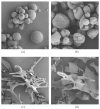
SEM images of dried powders. (a) Spray-dried empty archaeosomes. (b) Spray-dried archaeosomes loaded with insulin. (c) Lyophilized empty archaeosomes. (d) Lyophilized archaeosomes loaded with insulin.
Figure 6
Figure 6
SEM images of redissolved insulin-loaded archaeosomes after spray drying (a) and lyophilization (b).
Figure 7
Figure 7
Acrylamide gradient native gel (4–12%) of insulin-loaded archaeosomes. Lane 1 presents the low-molecular-weight marker panel. Lane 2, reference insulin (1 µg) used for the encapsulation process. Lane 3, archaeosome formulation with encapsulated insulin directly after the preparation using microfluidics. The majority of insulin is encapsulated within the archaeosomes, resulting in minimal Coomassie blue staining. Only a faint band is visible due to a portion of non-encapsulated insulin. Lane 4 shows the same formulation mixed with Triton X-100, which disrupts the archaeosomes, leading to the release of encapsulated insulin. Thus, lane 4 corresponds to the amount of initially encapsulated insulin. Lanes 5 and 7 contain insulin-loaded archaeosomes after spray-drying and lyophilization processes, respectively. The powdered formulations were dissolved in water and loaded onto gel. The absence of insulin bands in lanes 5 and 7 suggests negligible insulin release during the drying processes. Lanes 6 and 8 display the same formulations after disruption with Triton X-100, confirming the presence of insulin inside the spray-dried and lyophilized archaeosomes, respectively.

Similar articles

References

    1. De Rosa M., Gambacorta A., Gliozzi A. Structure, biosynthesis, and physicochemical properties of archaebacterial lipids. Microbiol. Rev. 1986;50:70–80. doi: 10.1128/mr.50.1.70-80.1986. - DOI - PMC - PubMed
    1. Charles-Orszag A., Lord S.J., Mullins R.D. High-Temperature Live-Cell Imaging of Cytokinesis, Cell Motility, and Cell-Cell Interactions in the Thermoacidophilic Crenarchaeon Sulfolobus acidocaldarius. Front. Microbiol. 2021;12:707124. doi: 10.3389/fmicb.2021.707124. - DOI - PMC - PubMed
    1. Quehenberger J., Shen L., Albers S.V., Siebers B., Spadiut O. Sulfolobus—A Potential Key Organism in Future Biotechnology. Front. Microbiol. 2017;8:2474. doi: 10.3389/fmicb.2017.02474. - DOI - PMC - PubMed
    1. Salvador-Castell M., Golub M., Erwin N., Deme B., Brooks N.J., Winter R., Peters J., Oger P.M. Characterisation of a synthetic Archeal membrane reveals a possible new adaptation route to extreme conditions. Commun. Biol. 2021;4:653. doi: 10.1038/s42003-021-02178-y. - DOI - PMC - PubMed
    1. Jacquemet A., Barbeau J., Lemiègre L., Benvegnu T. Archaeal tetraether bipolar lipids: Structures, functions and applications. Biochimie. 2009;91:711–717. doi: 10.1016/j.biochi.2009.01.006. - DOI - PubMed

LinkOut - more resources