. 2024 Jun 15;14(6):764.
doi: 10.3390/life14060764.

Acute Kaempferol Stimulation Induces AKT Phosphorylation in HepG2 Cells

Affiliations

Acute Kaempferol Stimulation Induces AKT Phosphorylation in HepG2 Cells

Beatriz Santana-Lima et al. Life (Basel). .

Abstract

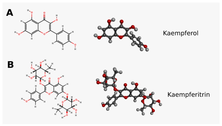
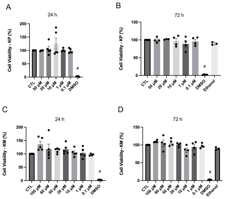
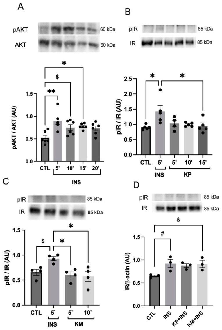
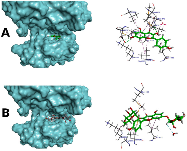
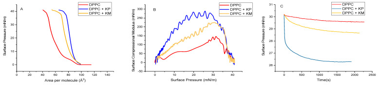
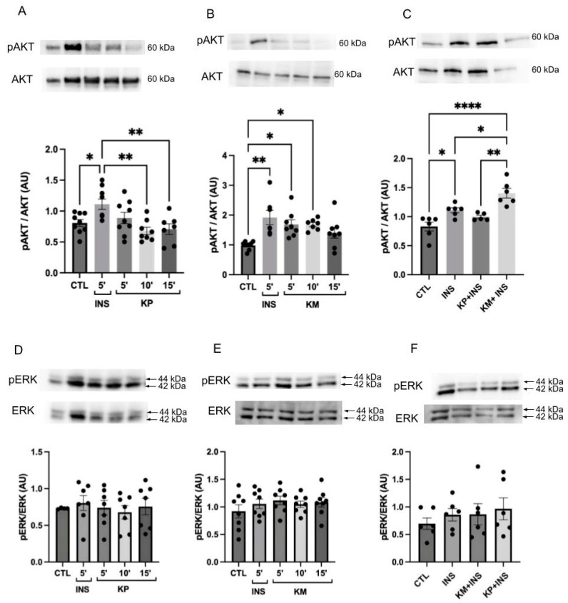
Type 2 diabetes mellitus (T2DM) stands as a prevalent global public health issue caused by deficiencies in the action of insulin and/or insulin production. In the liver, insulin plays an important role by inhibiting hepatic glucose production and stimulating glycogen storage, thereby contributing to blood glucose regulation. Kaempferitrin (KP) and kaempferol (KM), flavonoids found in Bauhinia forficata, exhibit insulin-mimetic properties, showing promise in managing T2DM. In this study, we aimed to assess the potential of these compounds in modulating the insulin signaling pathway and/or glucose metabolism. Cell viability assays confirmed the non-cytotoxic nature of both compounds toward HepG2 cells at the concentrations and times evaluated. Theoretical molecular docking studies revealed that KM had the best docking pose with the IR β subunit when compared to the KP. Moreover, Langmuir monolayer evaluation indicated molecular incorporation for both KM and KP. Specifically, KM exhibited the capability to increase AKT phosphorylation, a key kinase in insulin signaling, regardless of insulin receptor (IR) activation. Notably, KM showed an additional synergistic effect with insulin in activating AKT. In conclusion, our findings suggest the potential of KM as a promising compound for stimulating AKT activation, thereby influencing energy metabolism in T2DM.

Keywords: flavonoids; insulin signaling; liver cells; type 2 diabetes mellitus.


Conflict of interest statement

The authors report no declarations of interest. The graphical abstract was created with https://www.biorender.com/.

Figures

Figure 1
Figure 1
Chemical structure of kaempferol (KM) (A) and kaempferitrin (KP) (B). MarvinSketch20.11 and Biovia Discovery Studio Visualizer v.21.1.0.20298.
Figure 2
Figure 2
MTT assay. (A,B) Groups: control (CTL), administration of KP at concentrations 50, 20, 10, 1, and 0.1 μM, and 30% DMSO after incubation for 24 h and 72 h in HepG2 cells. n = 3–5. (C,D) Groups: control (CTL), administration of KM at concentrations 100, 80, 50, 20, 10, 1, and 0.1 μM, and 30% DMSO after incubation for 24 h and 72 h in HepG2 cells. n = 4–5. # DMSO 30% vs. all other groups; p < 0.0001. The dots are the representations of each n in the experiment.
Figure 3
Figure 3
AKT and insulin receptor phosphorylation were detected by Western blotting in the HepG2 cell line. (A) AKT phosphorylation. Groups: control (CTL), stimulation with insulin at a concentration of 100 nM for 5, 10, 15, and 20 min in HepG2 cells. n = 5–6 * CTL vs. 15 min; ** CTL vs. 5 min; $ CTL vs. 10 min (p = 0.0890). (B) Insulin receptor phosphorylation. Groups: control (CTL), stimulation with insulin at a concentration of 100 nM for 5 min (INS) and KP administration at a concentration of 10 μM during 5, 10, and 15 min. n = 4–5. * CTL vs. 5 min; * 5 vs. 15 min. (C) Insulin receptor phosphorylation. Groups: control (CTL), stimulation with insulin at a concentration of 100 nM for 5 min (INS) and KM administration at a concentration of 10 μM for 5 and 10 min. n = 4. $ CTL vs. INS (p = 0.0581); * INS vs. 5 min; * INS vs. 10 min. (D) Insulin receptor phosphorylation. Groups: control (CTL), stimulation with insulin at a concentration of 100 nM (INS); administration of KP at a concentration of 10 μM plus insulin at a concentration of 100 nM (KP + INS); administration of KM at a concentration of 10 μM plus insulin at a concentration of 100 nM (KM + INS); stimulation for 5 min. n = 3. # CTL vs. INS (p = 0.0984); and & CTL vs. KM + INS (p = 0.1005). The dots are the representations of each n in the experiment.
Figure 4
Figure 4
(A) General overview of kaempferol (KM) docked into the β subunit of the insulin receptor represented as a surface-covered structure and predicted binding mode of kaempferol (KM) highlighting the interaction with the cavity amino acids. Two hydrogen bonds were observed. (B) General overview of kaempferitrin (KP) docked into the insulin receptor kinase. Receptor represented as a surface-covered structure. Predicted binding mode of kaempferitrin (KP) highlighting the interaction with the cavity amino acids. One hydrogen bond was observed. Carbons in aquamarine, nitrogens in blue, oxygens in red, and hydrogens in white. Orange dotted lines—hydrogen bond-type interactions.
Figure 5
Figure 5
Tensiometric measurements of cell membrane models (DPPC monolayers) and their effect upon incorporating KP and KM. (A) surface pressure–area isotherms; (B) surface compressional modulus–surface pressure isotherms; and (C) surface pressure–time isotherms for previously compressed monolayers up to 30 mN/m, keeping the area constant.
Figure 6
Figure 6
BAM images (3600 × 2400 μm) of DPPC without (A,C) and with KP (B) and KC (D) at 30 mN/m. The arrow shows an example of a common aggregate formed when the monolayer is close to the collapse point.
Figure 7
Figure 7
AKT and ERK phosphorylation were detected by Western blotting in the HepG2 cell line. (A) AKT phosphorylation. Groups: control (CTL), stimulation with insulin at a concentration of 100 nM for 5 min (INS) and KP administration at a concentration of 10 μM during 5, 10, and 15 min in HepG2 cells. n = 7–9 * CTL vs. INS; ** INS vs. 10 and 15 min; (B) AKT phosphorylation. Groups: control (CTL), stimulation with insulin at a concentration of 100 nM for 5 min (INS) and KM administration at a concentration of 10 μM during 5, 10, and 15 min in HepG2 cells. n = 7–8 * CTL vs. 5 min; ** CTL vs. INS and 10 min; (C) AKT phosphorylation. Groups: control (CTL), stimulation with insulin at a concentration of 100 nM for 5 min (INS); administration of KP at a concentration of 10 μM plus insulin at a concentration of 100 nM (KP + INS); administration of KM at a concentration of 10 μM plus insulin at a concentration of 100 nM (KM + INS). Stimulation for 5 min. n = 5–6 * CTL vs. INS; * INS vs. KM + INS; ** KP + INS vs. KM + INS; **** CTL vs. KM + INS (D) ERK phosphorylation. Groups: control (CTL), stimulation with insulin at a concentration of 100 nM for 5 min (INS) and KP administration at a concentration of 10 μM during 5, 10, and 15 min in HepG2 cells. n = 4–7. No statistical difference between groups. (E) ERK phosphorylation. Groups: control (CTL), stimulation with insulin at a concentration of 100nM for 5 min (INS) and KM administration at a concentration of 10 μM during 5, 10, and 15 min in HepG2 cells. n = 7. No statistical difference between groups. (F) ERK phosphorylation. Groups: control (CTL), stimulation with insulin at a concentration of 100 nM for 5 min (INS); administration of KP at a concentration of 10 μM plus insulin at a concentration of 100 nM (KP + INS); administration of KM at a concentration of 10 μM plus insulin at a concentration of 100 nM (KM + INS). Stimulation for 5 min. n = 6. No statistical difference between groups. The representative images of total AKT (AC) are the same as those used in Figure 8 (Figure 8A–C). The representative images of total ERK (DF) are the same as those used in Figure 8 (Figure 8D–F). The dots are the representations of each n in the experiment.
Figure 8
Figure 8
AKT and ERK expression detected by Western blotting in HepG2 cell line. (A,D) Expression of AKT and ERK, respectively. Groups: control (CTL), stimulation with insulin at a concentration of 100 nM for 5 min (INS) and KP administration at a concentration of 10 μM during 5, 10, and 15 min in HepG2 cells. n = 6–9. No statistical difference between groups; (B,E) Expression of AKT and ERK, respectively. Groups: control (CTL), stimulation with insulin at a concentration of 100 nM for 5 min (INS) and KM administration at a concentration of 10 μM during 5, 10, and 15 min in HepG2 cells. n = 6–9. No statistical difference between groups; (C,F) Expression of AKT and ERK, respectively. Groups: control (CTL), stimulation with insulin at a concentration of 100 nM for 5 min (INS); administration of KM at a concentration of 10 μM plus insulin at a concentration of 100 nM (KM + INS); administration of KP at a concentration of 10 μM plus insulin at a concentration of 100 nM (KP + INS). Stimulation for 5 min. n = 4. The representative images of total AKT (Figure 7A–C) are the same as those used in Figure 8 (AC). The representative images of total ERK (Figure 7D–F) are the same as those used in Figure 8 (DF). The representative images of β-actin (A and D, B and E and C and F) are the same. The dots are the representations of each n in the experiment.

Similar articles

References

    1. IDF Diabetes Atlas. 10th ed. International Diabetes Federation; Brussels, Belgium: 2021.
    1. Souza B.V.C., Araújo R.S.R.M., Silva O.A., Faustino L.C., Gonçalves M.F.B., Santos M.L., Souza G.R., Rocha L.M., Cardoso M.L.S., Nunes L.C. Bauhinia forficata In Treatment Of Diabetes Mellitus: A Patent Review. Expert Opin. Ther. Pat. 2018;28:129–138. - PubMed
    1. Cechinel Filho V. Chemical composition and biological potential of plants from the genus Bauhinia. Phytother. Res. 2009;23:1347–1354. doi: 10.1002/ptr.2756. - DOI - PubMed
    1. Chandramohan G. Kaempferol, a flavonoid, ameliorates hyperglycemia by attenuating the key enzymes of carbohydrate metabolism in streptozotocin–induced experimental diabetic rats. Prog. Nutr. 2020;21:65–72. doi: 10.23751/pn.v21i2-S.6329. - DOI
    1. Luo C., Yang H., Tang C., Yao G., Kong L., He H., Zhou Y. Kaempferol alleviates insulin resistance via hepatic IKK/NF-κB signal in type 2 diabetic rats. Int. Immunopharmacol. 2015;28:744–750. doi: 10.1016/j.intimp.2015.07.018. - DOI - PubMed

LinkOut - more resources