. 2024 Jun 18;25(12):6695.
doi: 10.3390/ijms25126695.

Gut Microbiota Alleviates Intestinal Injury Induced by Extended Exposure to Light via Inhibiting the Activation of NLRP3 Inflammasome in Broiler Chickens

Affiliations

Gut Microbiota Alleviates Intestinal Injury Induced by Extended Exposure to Light via Inhibiting the Activation of NLRP3 Inflammasome in Broiler Chickens

Dandan Ma et al. Int J Mol Sci. .

Abstract

Light pollution is a potential risk for intestinal health in humans and animals. The gut microbiota is associated with the development of intestinal inflammation induced by extended exposure to light, but the underlying mechanism is not yet clear. The results of this study showed that extended exposure to light (18L:6D) damaged intestinal morphology, downregulated the expression of tight junction proteins, and upregulated the expression of the NLRP3 inflammasome and the concentration of pro-inflammatory cytokines. In addition, extended exposure to light significantly decreased the abundance of Lactobacillus, Butyricicoccus, and Sellimonas and increased the abundance of Bifidobacterium, unclassified Oscillospirales, Family_XIII_UCG-001, norank_f__norank_o__Clostridia_vadinBB60_group, and Defluviitaleaceae_UCG-01. Spearman correlation analysis indicated that gut microbiota dysbiosis positively correlated with the activation of the NLRP3 inflammasome. The above results indicated that extended exposure to light induced intestinal injury by NLRP3 inflammasome activation and gut microbiota dysbiosis. Antibiotic depletion intestinal microbiota treatment and cecal microbiota transplantation (CMT) from the 12L:12D group to 18L:6D group indicated that the gut microbiota alleviated intestinal inflammatory injury induced by extended exposure to light via inhibiting the activation of the NLRP3 inflammasome. In conclusion, our findings indicated that the gut microbiota can alleviate intestinal inflammation induced by extended exposure to light via inhibiting the activation of the NLRP3 inflammasome.

Keywords: NLRP3 inflammasome; broiler chickens; extended exposure to light; gut microbiota; intestinal inflammation.


Conflict of interest statement

The authors declare no conflict of interest.

Figures

Figure 1
Figure 1
Extended exposure to light caused intestinal injury and decreased the mRNA expression of tight junction protein. (A) representative histological images of intestinal tissue of 12L:12D group was captured by a microscope (NIKON DS-U3) at 200×; (B) representative histological images of intestinal tissue of 18L:6D group was captured by a microscope (NIKON DS-U3) at 200×. (CE) mRNA expression of Claudin-1, Occludin, and ZO-1 (n = 6). There were significant differences between different lower-letters (a and b) (p < 0.05), but no significant differences between the same lower-letters (p > 0.05).
Figure 2
Figure 2
Extended exposure to light activated NLRP3 inflammasome and increased pro-inflammatory cytokines (n = 6). (AC) mRNA expression of NLRP3, caspase1 and IL-1β in the duodenum of broiler chickens. (DG) the concentration of pro-inflammatory cytokines (IL-1β, IL-6, IL-18, and TNF-α) in the duodenum of broiler chickens. There were significant differences between different lower-letters (a and b) (p < 0.05), but no significant differences between the same lower-letters (p > 0.05).
Figure 2
Figure 2
Extended exposure to light activated NLRP3 inflammasome and increased pro-inflammatory cytokines (n = 6). (AC) mRNA expression of NLRP3, caspase1 and IL-1β in the duodenum of broiler chickens. (DG) the concentration of pro-inflammatory cytokines (IL-1β, IL-6, IL-18, and TNF-α) in the duodenum of broiler chickens. There were significant differences between different lower-letters (a and b) (p < 0.05), but no significant differences between the same lower-letters (p > 0.05).
Figure 3
Figure 3
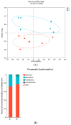
Extended exposure to light altered gut microbiota composition. (A) PCoA analysis of gut microbiota; (B) the abundance of the gut microbiota at the phylum level; (C) the abundance of the gut microbiota at the genus level; (D) differences in gut microbiota composition at the genus level between 12L:12D group and 18L:6D group. P1 refers to 12L:12D group; P2 refers to 18L:6D group. * and ** Indicate a significant correlation between two indicators (* indicates p < 0.05 and ** indicates p < 0.01, n = 6).
Figure 3
Figure 3
Extended exposure to light altered gut microbiota composition. (A) PCoA analysis of gut microbiota; (B) the abundance of the gut microbiota at the phylum level; (C) the abundance of the gut microbiota at the genus level; (D) differences in gut microbiota composition at the genus level between 12L:12D group and 18L:6D group. P1 refers to 12L:12D group; P2 refers to 18L:6D group. * and ** Indicate a significant correlation between two indicators (* indicates p < 0.05 and ** indicates p < 0.01, n = 6).
Figure 4
Figure 4
Correlation analysis between gut microbiota and NLRP3 inflammasome at the genus level. Note: the red and blue matrices represent positive and negative correlations, respectively, and the color depth represents the magnitude of the correlation coefficient. * and ** Indicate a significant correlation between two indicators (* indicates p < 0.05 and ** indicates p < 0.01, n = 6).
Figure 5
Figure 5
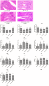
Cecal microbiota collected from broiler chickens of 12L:12D group transplanted to the broiler chickens of 18L:6D group alleviated intestinal inflammation (n = 6). (AD) representative histological images of intestinal tissue of 12L:12D, 18L:6D, 18L:6D+PBS, and 18L:6D+CMT groups were captured by a microscope (NIKON DS-U3) at 200×(the red arrows indicate the erosive intestinal villi and the shed epithelial cells of the mucosal layer; the yellow arrows indicate the infiltration of inflammatory cells). (EG) mRNA expression of Claudin-1, Occludin, and ZO-1. (HJ): mRNA expression of NLRP3, caspase1, and IL-1β. (KN) the concentration of IL-1β, IL-6, IL-18, and TNF-α. There were significant differences between different lower-letters (a, b and c) (p < 0.05), but no significant differences between the same lower-letters (p > 0.05).
Figure 6
Figure 6
Antibiotic treatment alleviated intestinal injury and inhibited the activation of NLRP3 inflammasome induced by extended exposure to light (n = 6). (AC) representative histological images of intestinal tissue of 12L:12D group, 18L:6D group, and 18L:6D+Antibiotic treatment group were captured by a microscope (NIKON DS-U3) at 200× (the red arrows indicate the erosive intestinal villi and the shed epithelial cells of the mucosal layer; the yellow arrows indicate the infiltration of inflammatory cells). (DF) mRNA expression of Claudin-1, Occludin, and ZO-1. (GI) mRNA expression of NLRP3, caspase1, and IL-1β. (JM) the concentration of IL-1β, IL-6, IL-18, and TNF-α. There were significant differences between different lower-letters (a, b and c) (p < 0.05), but no significant differences between the same lower-letters (p > 0.05).
Figure 6
Figure 6
Antibiotic treatment alleviated intestinal injury and inhibited the activation of NLRP3 inflammasome induced by extended exposure to light (n = 6). (AC) representative histological images of intestinal tissue of 12L:12D group, 18L:6D group, and 18L:6D+Antibiotic treatment group were captured by a microscope (NIKON DS-U3) at 200× (the red arrows indicate the erosive intestinal villi and the shed epithelial cells of the mucosal layer; the yellow arrows indicate the infiltration of inflammatory cells). (DF) mRNA expression of Claudin-1, Occludin, and ZO-1. (GI) mRNA expression of NLRP3, caspase1, and IL-1β. (JM) the concentration of IL-1β, IL-6, IL-18, and TNF-α. There were significant differences between different lower-letters (a, b and c) (p < 0.05), but no significant differences between the same lower-letters (p > 0.05).

Similar articles

References

    1. Zielinska-Dabkowska K.M., Schernhammer E.S., Hanifin J.P., Brainard G.C. Reducing nighttime light exposure in the urban environment to benefit human health and society. Science. 2023;380:1130–1135. doi: 10.1126/science.adg5277. - DOI - PubMed
    1. Kyba C.C.M., Altıntaş Y., Walker C.E., Newhouse M. Citizen scientists report global rapid reductions in the visibility of stars from 2011 to 2022. Science. 2023;379:265–268. doi: 10.1126/science.abq7781. - DOI - PubMed
    1. Deboer T., Détári L., Meijer J.H. Long term effects of sleep deprivation on the mammalian circadian pacemaker. Sleep. 2007;30:257–262. doi: 10.1093/sleep/30.3.257. - DOI - PubMed
    1. Dumont M., Beaulieu C. Light exposure in the natural environment: Relevance to mood and sleep disorders. Sleep Med. 2007;8:557–565. doi: 10.1016/j.sleep.2006.11.008. - DOI - PubMed
    1. Deaver J.A., Eum S.Y., Toborek M. Circadian Disruption Changes Gut Microbiome Taxa and Functional Gene Composition. Front. Microbiol. 2018;9:737. doi: 10.3389/fmicb.2018.00737. - DOI - PMC - PubMed

MeSH terms

LinkOut - more resources