Review
. 2024 Jun 26;22(1):376.
doi: 10.1186/s12951-024-02580-8.

Advanced gene nanocarriers/scaffolds in nonviral-mediated delivery system for tissue regeneration and repair

Affiliations
Review

Advanced gene nanocarriers/scaffolds in nonviral-mediated delivery system for tissue regeneration and repair

Wanheng Zhang et al. J Nanobiotechnology. .

Erratum in

Abstract

Tissue regeneration technology has been rapidly developed and widely applied in tissue engineering and repair. Compared with traditional approaches like surgical treatment, the rising gene therapy is able to have a durable effect on tissue regeneration, such as impaired bone regeneration, articular cartilage repair and cancer-resected tissue repair. Gene therapy can also facilitate the production of in situ therapeutic factors, thus minimizing the diffusion or loss of gene complexes and enabling spatiotemporally controlled release of gene products for tissue regeneration. Among different gene delivery vectors and supportive gene-activated matrices, advanced gene/drug nanocarriers attract exceptional attraction due to their tunable physiochemical properties, as well as excellent adaptive performance in gene therapy for tissue regeneration, such as bone, cartilage, blood vessel, nerve and cancer-resected tissue repair. This paper reviews the recent advances on nonviral-mediated gene delivery systems with an emphasis on the important role of advanced nanocarriers in gene therapy and tissue regeneration.

Keywords: Advanced gene/Drug nanocarriers; Cancer-resected tissue repair; Gene therapy; Nonviral-mediated delivery system; Tissue regeneration.


Conflict of interest statement

The authors declare no competing interests.

Figures
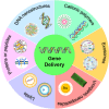
Fig. 1
Fig. 1
Nonviral-mediated advanced functional carriers for precise and targeting delivery of different nucleic acids, including dsDNA, ssDNA, siRNA, miRNA, mRNA and so on. Advanced biomaterials, such as lipid/lipid-like carriers, polymeric biomaterials, functional dendrimers, inorganic particles and exosomes, are intensively investigated for efficient gene delivery and therapy
Fig. 2
Fig. 2
Exosomes as gene delivery vectors and their biomedical applications in gene therapy. (A) The production of biomimetic vesicles derived from cells through extrusion/filtration method. (B) The biomedical application of exosomes. (C) Representative illustration of FA covalently attached to the exosome-polyethyleneimine matrix (EPM) as a transfer carrier for nucleic acid delivery. (D) The entrapment ability of EPM with siKRAS and siRNA. (E) Tissue distribution of bovine colostrum exosomes and EPM, with and without FA-functionalized exosomes. (C-E) Reproduced with permission [70]. Copyright 2021, Elsevier. (F) Schematic graph of HA and FA loading into mExo. (G) Scanning electron microscope (SEM) photos of mExo and HA-mExo-FA. (F, G) Reproduced with permission [71]. Copyright 2023, Wiley
Fig. 3
Fig. 3
Cationic polymers as gene delivery vectors and their biomedical potentials. (A) PEI-based nanoparticle formation is designed by the admixing of PEI polymer derivatives (linear or branched) with nucleic acid cargos to encode for any genes of interest. (B) Schematic overview of ternary complex formation by sequential addition of core and shell polymer to the nanovector DNA. (C) In vitro evaluation of toxicity and transfection efficiency in HuH-7 cells. (B, C) Reproduced with permission [107]. Copyright 2023, Wiley. (D) Structural illustration of selectively fluorinated PAMAM–Arg conjugates and transfected cell micrographs. Reproduced with permission [108]. Copyright 2023, American Chemical Society
Fig. 4
Fig. 4
ECM remodeling and skeleton-mediated cellular nanomechanics enhance gene delivery through activation of force-sensing-related signaling pathways. Interaction of tissue regeneration related cells, including progenitor cells, iPSCs, ESCs and MSCs with advanced biomaterials to promote the expression of some functional proteins related to cell anchoring junction, such as collagen, fibronectin and laminin. These proteins can connect a series of intracellular anchor proteins, such as talin, vinculin and β-catenin for cell spreading and adhesion to alter the cytoskeleton and nuclear transcription to promote cell migration, division and differentiation and functional cell fate. Gene editing (knock in and knock out of targeting genes) also participate in this process to enhance gene therapy ability in tissue regeneration
Fig. 5
Fig. 5
Biomaterials-based gene delivery vectors for bone regeneration. (A) Non-coding-RNA-activated core/chitosan shell nanounits coated with polyetheretherketone with the ability to promote bone regeneration. (B) Micro-CT images demonstrating bone regeneration surrounding implants, as well as reconstructed 3D images of samples and new bone. The color yellow denotes samples, whereas the color red represents fresh bone regeneration. Quantitative bone analysis 8 weeks after implantation. (A-B) Reproduced with permission [183]. Copyright 2023, American Chemical Society. (C) CRISPR activation of DANCR promotes calvarial bone formation. (D) µCT analysis of in vivo calvarial bone repair. 3D projection of osteomized sites of mock and Bac-Sa-VPR groups (left) and frontal views of osteomized sites (right). (C-D) Reproduced with permission [184]. Copyright 2021, Elsevier. (E) Schematic diagram of conjugating Asp8 to exosomal amine groups by click chemistry reaction. (F) H&E staining and Masson’s trichrome staining analysis of defect sites at 2 weeks, scale bar = 100 μm. (E-F) Reproduced with permission [185]. Copyright 2023, Elsevier
Fig. 6
Fig. 6
Biomaterials-based gene delivery vectors for cartilage regeneration. (A) Schematic illustration of the preparation of the Cu-based NPs. CuS NPs are reacted with ATPES to obtain CuS-NH2 NPs, and then electrostatically bonded with negatively charged TGF-β1 pDNA. Then PC is coated on the NPs for biomimetic modification. (B) Representative images of Safranin-O/Fast green staining and Toluidine blue staining of the knee joints after different treatments. (A-B) Reproduced with permission [162]. Copyright 2021, Elsevier. (C) The silencing of miR-221 in combination with TGF-β3 improved sGAG distribution. Representative histological images of cell-seeded scaffolds stained with (A) hematoxylin and eosin and (B) thionine after 28 days in culture +/− TGF-β3 supplementation. Scale bars represent 100 μm length. (D) Representation of GET peptide: a glycosaminoglycan (GAG)-binding peptide sequence (P21), fused to an amphiphilic region with an octa-arginine (8R) and a cell-penetrating peptide (CPP). (E) The incorporation of miR-221 inhibitor to CI/II-HyA scaffolds reduced the expression of miR-221 by hMSCs. (C-E) Reproduced with permission [200]. Copyright 2021, Wiley
Fig. 7
Fig. 7
Biomaterial-based gene delivery vectors for blood vessel regeneration. (A) Hypoxia-pretreated ADSC-derived exosome-embedded hydrogels promote angiogenesis and accelerate diabetic wound healing. Reproduced with permission [166]. Copyright 2023, Elsevier. (B) The gelation of the laponite hydrogels in vitro. (C) Co-staining of the injured rat common carotid arteries with eNOS and αSMA 14 days after the treatment with the miR-22 loading laponite hydrogels. (B, C) Reproduced with permission [211]. Copyright 2022, Elsevier. (D) Morphology of the prepared vascular scaffold. SEM micrograph of the cross section and larger magnification. Reproduced with permission [214]. Copyright 2016, Elsevier
Fig. 8
Fig. 8
Regulation of gene and protein expression using RNA/Nucleic acid in cancer therapy for tissue regeneration. Once delivered into the cells, RNA macromolecules can utilize diverse intracellular mechanism to control gene and protein expression in cancer cells for gene therapy. (I) Hybridization of antisense oligonucleotides (ASOs) to a targeting mRNA can result in specific inhibition of gene expression by the induction of RNase H endonuclease activity, which cleaves the mRNA-ASO heteroduplex. (II) Short interfering RNA (siRNA) is recognized by the RNA-induced silencing complex (RISC), guided by an antisense strand of the siRNA, specifically to bind and cleave targeting mRNA. (III) In vitro transcribed mRNA utilizes the protein synthesis machinery of host cells to translate the encoded genetic information into a protein. Ribosome subunits are recruited to mRNA with a cap and poly(A)-binding proteins, forming a translation initiation complex. (IV) In the clustered regularly interspaced short palindromic repeats (CRISPR-Cas9) system, co-delivery of a single guide RNA (sgRNA) together with the mRNA encoding the Cas9 DNA endonuclease allows site-specific cleavage of dsDNA, leading to the knockout of a target gene and its product. Then, this will realize cancer therapy by RNA/Nucleic acid delivery
Fig. 9
Fig. 9
Regulation of gene and protein expression using RNA/Nucleic acid in cancer therapy for tissue regeneration. (A) A schematic diagram of gene therapy in tissue repair after removing tumors. (B) A schematic diagram to depict the role of lnc030 in maintenance of BCSC stemness through regulating SQLE and cholesterol accumulation to stimulate PI3K/Akt signaling. (C) Intracellular cholesterol is measured in lnc030-knockdown (left) or SQLE-knockdown (right) Hs578T, BT549, MCF-7 derived spheres and their control spheres. (D) Tumor growth curve and tumor weight of each mouse group are measured. (B-D) Reproduced with permission [275]. Copyright 2020, Wiley. (E) Schematic illustration of therapeutic process of ADSC-loaded GO-GA-polymer scaffold with pH-triggered dual release of GO and GA. (F) The tumor growth curves after implantation of different formulations with and without NIR irradiation. (G) Histological analysis and histomorphometric measurements of adipose tissue regeneration in vivo. (E-G) Reproduced with permission [276]. Copyright 2019, Wiley. (H) A schematic diagram of nanocomposite multifunctional hydrogel for suppressing osteosarcoma recurrence and enhancing bone regeneration. Reproduced with permission [274]. Copyright 2022, Elsevier

Similar articles

Cited by

References

    1. Zhang Z, et al. Native tissue-based strategies for meniscus repair and regeneration. Cell Tissue Res. 2018;373:337–50. 10.1007/s00441-017-2778-6 - DOI - PubMed
    1. Salhotra A, et al. Mechanisms of bone development and repair. Nat Rev Mol Cell Biol. 2020;21:696–711. 10.1038/s41580-020-00279-w - DOI - PMC - PubMed
    1. Tzahor E, Poss KD. Cardiac regeneration strategies: staying young at heart. Science. 2017;356:1035–9. 10.1126/science.aam5894 - DOI - PMC - PubMed
    1. Rana D, et al. Development of decellularized scaffolds for stem cell-driven tissue engineering. J Tissue Eng Regen Med. 2017;11:942–65. 10.1002/term.2061 - DOI - PubMed
    1. Nerem RM, et al. Tissue engineering in the USA. Med Biol Eng Comput. 1992;30:CE8–12. 10.1007/BF02446171 - DOI - PubMed