Review
. 2024 Jun 26;33(172):240008.
doi: 10.1183/16000617.0008-2024. Print 2024 Apr.

Neuroimmune recognition and regulation in the respiratory system

Affiliations
Review

Neuroimmune recognition and regulation in the respiratory system

Jie Chen et al. Eur Respir Rev. .

Abstract

Neuroimmune recognition and regulation in the respiratory system is a complex and highly coordinated process involving interactions between the nervous and immune systems to detect and respond to pathogens, pollutants and other potential hazards in the respiratory tract. This interaction helps maintain the health and integrity of the respiratory system. Therefore, understanding the complex interactions between the respiratory nervous system and immune system is critical to maintaining lung health and developing treatments for respiratory diseases. In this review, we summarise the projection distribution of different types of neurons (trigeminal nerve, glossopharyngeal nerve, vagus nerve, spinal dorsal root nerve, sympathetic nerve) in the respiratory tract. We also introduce several types of cells in the respiratory epithelium that closely interact with nerves (pulmonary neuroendocrine cells, brush cells, solitary chemosensory cells and tastebuds). These cells are primarily located at key positions in the respiratory tract, where nerves project to them, forming neuroepithelial recognition units, thus enhancing the ability of neural recognition. Furthermore, we summarise the roles played by these different neurons in sensing or responding to specific pathogens (influenza, severe acute respiratory syndrome coronavirus 2, respiratory syncytial virus, human metapneumovirus, herpes viruses, Sendai parainfluenza virus, Mycobacterium tuberculosis, Pseudomonas aeruginosa, Staphylococcus aureus, amoebae), allergens, atmospheric pollutants (smoking, exhaust pollution), and their potential roles in regulating interactions among different pathogens. We also summarise the prospects of bioelectronic medicine as a third therapeutic approach following drugs and surgery, as well as the potential mechanisms of meditation breathing as an adjunct therapy.


Conflict of interest statement

Conflict of interest: All authors have nothing to disclose.

Figures

FIGURE 1
FIGURE 1
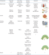
Innervation of the respiratory tract. Neurons projected to the respiratory tract are mainly from the olfactory nerve (cranial nerve I), trigeminal nerve (cranial nerve V), glossopharyngeal nerve (cranial nerve IX), vagus nerve (cranial nerve X), dorsal root ganglia (DRG), sympathetic ganglia and intrinsic neurons from peripheral organs such as the oesophagus and lungs. The sympathetic ganglia can be further divided into the superior cervical ganglion (SCG), the stellate ganglia (SG) and the thoracic sympathetic chain ganglion (TSG), each of which projects differently to the respiratory tract. The synaptic terminals of sensory neurons are hotspots distributed in the respiratory tract, including from pulmonary neuroendocrine cells, brush cells, solitary chemosensory cells and tastebuds. Neural recognition of pathogens is expanded through the expression of recognition receptors on these cells. TG: trigeminal ganglion; Pa5: paratrigeminal nucleus; PG: petrosal ganglion; JG: jugular ganglion; NG: nodose ganglion; NTS: solitary tractus nucleus; DMV: dorsal motor nucleus of the vagus nerve; Amb: nucleus ambiguus; IS: inferior salivatory nucleus.
FIGURE 2
FIGURE 2
Sensory neurons directly or indirectly perceive danger signals, with the vagus nerve as the example. Sensory neurons directly sense pathogens in a variety of ways. a) First, sensory neurons directly perceive pathogen-associated molecular patterns (PAMPs) or damage-associated molecular patterns (DAMPs) through the expression of pattern recognition receptors (PRRs). Sensory neurons utilise the expression of classical PRRs, such as toll-like receptors (TLRs), C-type lectin receptors (CLRs), RIG-I-like receptors (RLRs), nucleotide-binding and oligomerisation domain (NOD)-like receptors (NLRs), prostaglandin E receptor (EP)3 and FcεR1α to directly sense pathogens and immune mediators. As these receptors are activated, intracellular NF-κB and mitogen-activated protein kinase (MAPK) signalling pathways are initiated, leading to an increase in intracellular calcium (Ca2+) and sodium (Na+) flux, thereby activating neuronal signal transduction. These ion fluxes also activate ion channels within neurons, propelling neurons to further activation and enhancing sensitisation. b) Second, sensory neurons express a variety of ion channel receptors on their surface, including transient receptor potential (TRP) channels, purinergic P2X channels, and mechanosensitive ion channels. For example, transient receptor potential cation channel subfamily A member 1 (TRPA1) and transient receptor potential cation channel subfamily V member 1 (TRPV1) can directly detect lipopolysaccharide (LPS) in Gram-negative bacteria and respond to thermal stimuli. Recognition of these stimuli induces channel opening, influx of Ca2+ and Na+, and rapid changes in neuronal membrane potential, resulting in rapid activation of neurons. c) Third, sensory neurons can couple PRRs to ion channels to comediate recognition responses to pathogenic substances. Examples include the conjugation of TLR4 to TRPV1, TLR7 to TRPA1 and TLR3 to TRPV1. By coupling PRRs expressed on neurons with ion channels, the immune signals transmitted by immune-related receptors are rapidly converted into neuroelectrical signals, overcoming the slow transmission of immune signals. ss: single strand; ds: double strand; MDP: muramyl dipeptide.
FIGURE 3
FIGURE 3
Neuroepithelial interactions contribute to the recognition and regulation of pathogens. CGRP: calcitonin gene-related peptide; ACh: acetylcholine; NE: norepinephrine; 5-HT: 5-hydroxytryptamine; P2RY1: P2Y purinoceptor 1; SP: substance P; PNECs: pulmonary neuroendocrine cells; Pvalb: parvalbumin; Na: sodium; K: potassium; TRP: transient receptor potential; DRG: dorsal root ganglion; ATIII: alveolar type III cells; Npy2r: neuropeptide Y receptor type 2.
FIGURE 4
FIGURE 4
Examples of neuroimmune interactions in the respiratory tract. a) Bacterial infection: sensory neurons act as second-order neurons that receive input from respiratory epithelial cells, such as solitary chemosensory cells (SCCs). SCCs sense acyl-homoserine lactones (AHLs) [27, 87] produced by Gram-negative bacteria through bitter taste receptors (T2R). The release of acetylcholine (ACh) from SCCs activates trigeminal sensory neurons, triggering axonal reflexes, leading to the release of calcitonin gene-related peptide (CGRP) and substance P (SP) from peripheral nerve endings and neurogenic inflammation. Nerves can sense bacterial infections in a variety of ways. One way to do this is to identify pathogens directly through neurons. For example, transient receptor potential (TRP)V1+ vagus sensory neurons directly recognise Staphylococcus aureus, resulting in the release of CGRP from neurons, which acts on locally infected neutrophils and γδT-cells, inhibiting their inflammatory response. In addition, sensory neurons act as second-order neurons that receive input from respiratory epithelial cells, such as brush cells. Brush cells sense bitter-tasting quorum-sensing molecules (QSM) produced by Pseudomonas aeruginosa through TAS2R taste receptors. Similar to SCCs, the release of ACh from brush cells activates sensory neurons, triggering axonal reflexes, leading to the release of CGRP and SP from peripheral nerve endings and neurogenic inflammation. b) Viral infection: severe acute respiratory syndrome coronavirus 2 (SARS-CoV-2) can directly infect olfactory sensory neurons, resulting in olfactory epithelial destruction, leading to anosmia. In addition, SARS-CoV-2 can enter the brain through the olfactory nerve, triggering the activation of microglia and recruiting CD8 T-cells. This activation causes microglia and CD8 cells to aggregate, forming nodules that cause damage to brain tissue. In the case of influenza virus infection, γ-aminobutyric acid receptor subunit α1 (GABRA1+) petrosal neurons located in the posterior region of the nasopharynx expressing prostaglandin E2 (PGE2) receptor 3 (EP3) can sense pathogen-induced PGE2, thereby inducing disease behaviour. c) Allergic airway inflammation: multiple types of nerves are involved in regulating allergic airway inflammation. For the vagus nerve, TRPV1+ vagal neurons can sense IgE produced by the body's immune response through FcεR1 receptors. They release SP, promoting the inflammatory response of type 2 helper (Th2) cells and enhancing the secretion of mucus by goblet cells. Simultaneously, Mrgprc11+ jugular neurons of the vagus nerve activate parasympathetic neurons, leading to the release of ACh, which mediates bronchoconstriction, inducing airway hyperreactivity. The parasympathetic ganglia can also release neuromedin U (NMU), which acts on NMUR1 receptors on group 2 innate lymphoid cells (ILC2), activating them and amplifying allergic inflammation. The parasympathetic nervous system can also negatively regulate ILC2 cells by releasing ACh, which acts on α7 nicotinic acetylcholine receptors (a7nAChR) on ILC2 cells, inhibiting their transformation into inflammatory ILC2 cells. ILC2 cells play a crucial role in the neuroimmune interaction of airway allergic inflammation and can be regulated by various neurons. Regarding sensory neurons, they sense allergic inflammation and release vasoactive intestinal peptide (VIP), which acts on VPAC2 receptors on ILC2 cells, promoting the release of interleukin (IL)-5 and enhancing the inflammatory response of eosinophils. Activated eosinophils can increase airway epithelial nerve density, further leading to airway hyperreactivity. The sympathetic nervous system similarly regulates ILC2 cells by releasing dopamine and norepinephrine, which act on the D1 dopamine receptor (DRD1) and β2-adrenergic receptors (AR) on ILC2 cells, respectively, negatively modulating their response. The sympathetic nervous system can also release norepinephrine, which acts on β2-ARs on airway smooth muscle cells (ASMs), mediating airway dilation. Additionally, oesophagus intrinsic neurons can mediate airway dilation by releasing VIP and nitric oxide (NO). Not limited to neurons, ILC2 cells also receive regulation from pulmonary neuroendocrine cells (PNECs) that secrete CGRP and γ-aminobutyric acid (GABA), promoting the release of IL-5 and IL-3, leading to a robust inflammatory response. d) Acute lung injury. The parasympathetic nervous system plays a vital role in this process. ACh acts on the α7nAChR receptor of type 2 alveolar cells (AT2), promotes the differentiation of AT2 cells into AT1 cells, and mediates the repair of alveolar epithelial damage. This process depends on the vagus nerve. IAV: influenza virus; H: hydrogen; P2Y purinoceptor 1; OVA: ovalbumin; DC: dendritic cell; EET: eosinophil extracellular traps; NE: norepinephrine; CHAT: choline O-acetyltransferase; M3: M3 muscarinic ACh receptor; DRG: dorsal root ganglion; CNS: central nervous system.

Similar articles

References

    1. Chang RB, Strochlic DE, Williams EK,et al. . Vagal sensory neuron subtypes that differentially control breathing. Cell 2015; 161: 622–633. doi:10.1016/j.cell.2015.03.022 - DOI - PMC - PubMed
    1. Nonomura K, Woo SH, Chang RB, et al. . Piezo2 senses airway stretch and mediates lung inflation-induced apnoea. Nature 2017; 541: 176–181. doi:10.1038/nature20793 - DOI - PMC - PubMed
    1. Man WH, de Steenhuijsen Piters WA, Bogaert D. The microbiota of the respiratory tract: gatekeeper to respiratory health. Nat Rev Microbiol 2017; 15: 259–270. doi:10.1038/nrmicro.2017.14 - DOI - PMC - PubMed
    1. Prescott SL, Umans BD, Williams EK, et al. . An airway protection program revealed by sweeping genetic control of vagal afferents. Cell 2020; 181: 574–589. doi:10.1016/j.cell.2020.03.004 - DOI - PMC - PubMed
    1. Schmit D, Le DD, Heck S, et al. . Allergic airway inflammation induces migration of mast cell populations into the mouse airway. Cell Tissue Res 2017; 369: 331–340. doi:10.1007/s00441-017-2597-9 - DOI - PubMed

MeSH terms

LinkOut - more resources