. 2024 Aug;300(8):107492.
doi: 10.1016/j.jbc.2024.107492. Epub 2024 Jun 24.

Lack of mismatch repair enhances resistance to methylating agents for cells deficient in oxidative demethylation

Affiliations

Lack of mismatch repair enhances resistance to methylating agents for cells deficient in oxidative demethylation

Roberto Gutierrez et al. J Biol Chem. 2024 Aug.

Abstract

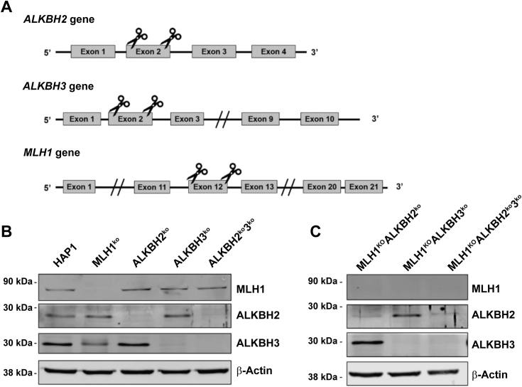
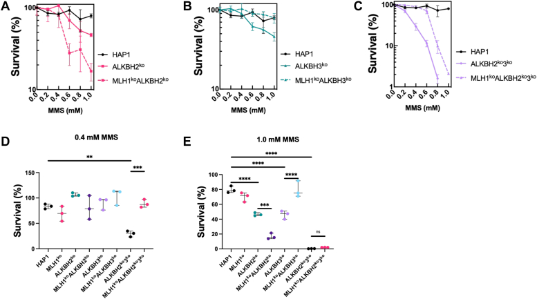
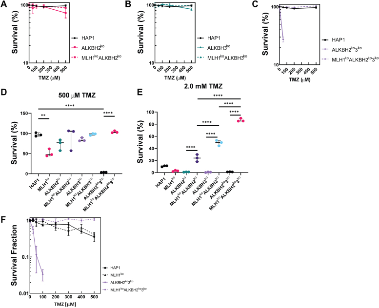
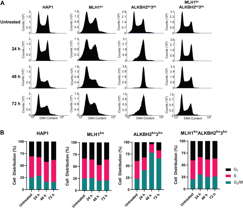
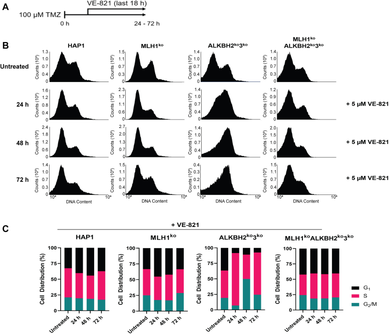
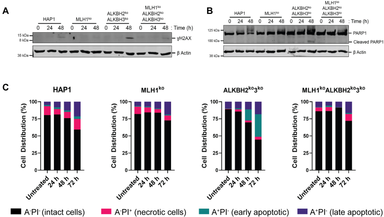
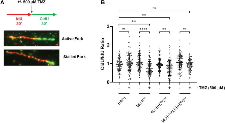
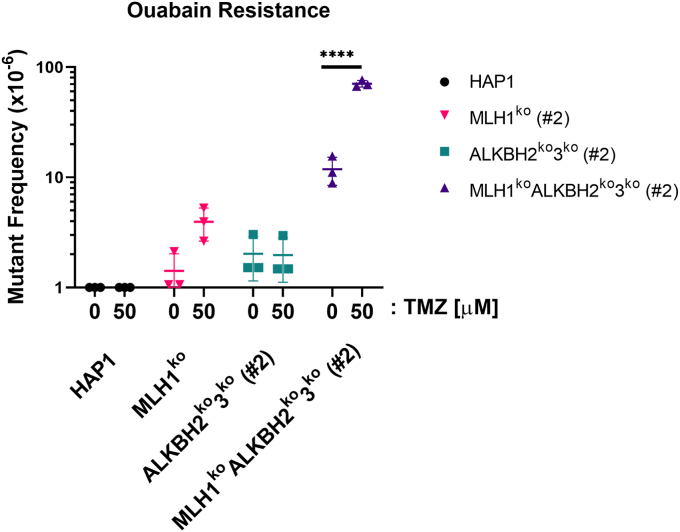
The human alkylation B (AlkB) homologs, ALKBH2 and ALKBH3, respond to methylation damage to maintain genomic integrity and cellular viability. Both ALKBH2 and ALKBH3 are direct reversal repair enzymes that remove 1-methyladenine (1meA) and 3-methylcytosine (3meC) lesions commonly generated by alkylating chemotherapeutic agents. Thus, the existence of deficiencies in ALKBH proteins can be exploited in synergy with chemotherapy. In this study, we investigated possible interactions between ALKBH2 and ALKBH3 with other proteins that could alter damage response and discovered an interaction with the mismatch repair (MMR) system. To test whether the lack of active MMR impacts ALKBH2 and/or ALKBH3 response to methylating agents, we generated cells deficient in ALKBH2, ALKBH3, or both in addition to Mlh homolog 1 (MLH1), another MMR protein. We found that MLH1koALKBH3ko cells showed enhanced resistance toward SN1- and SN2-type methylating agents, whereas MLH1koALKBH2ko cells were only resistant to SN1-type methylating agents. Concomitant loss of ALKBH2 and ALKBH3 (ALKBH2ko3ko) rendered cells sensitive to SN1- and SN2-agents, but the additional loss of MLH1 enhanced resistance to both types of damage. We also showed that ALKBH2ko3ko cells have an ATR-dependent arrest at the G2/M checkpoint, increased apoptotic signaling, and replication fork stress in response to methylation. However, these responses were not observed with the loss of functional MLH1 in MLH1koALKBH2ko3ko cells. Finally, in MLH1koALKBH2ko3ko cells, we observed elevated mutant frequency in untreated and temozolomide treated cells. These results suggest that obtaining a more accurate prognosis of chemotherapeutic outcome requires information on the functionality of ALKBH2, ALKBH3, and MLH1.

Keywords: AlkB; MLH1; demethylation; methylating agents; mismatch repair.


Conflict of interest statement

Conflict of interest The authors declare that they have no conflicts of interest with the contents of this article.

Figures

Figure 1
Figure 1
Targeting and KO validation of MLH1, ALKBH2, and ALKBH3. A, schematic illustrating the target regions ALKBH2, ALKBH3, and MLH1. Indicated regions were cleaved using a dual single-guide RNA CRISPR/Cas9 targeting scheme. B and C, confirmation of MLH1, ALKBH2, and ALKBH3 KOs through immunoblotting. Fifty micrograms of whole cell extracts was subjected to immunoblotting to detect MLH1, ALKBH2, and ALKBH3 expression. β-actin is the loading control. Molecular weights are indicated on the left of the panels. The same β-actin bands are found in Fig. S3. ALKBH, alkylation B homolog; MLH1, Mlh homolog 1.
Figure 2
Figure 2
Cell Survival in HAP1-knockout cells after treatment with MMS. Cell survival of ALKBH2ko, ALKBH3ko, MLH1koALKBH2ko, MLH1koALKBH3ko, and MLH1koALKBH2ko3ko cells was determined after treatment with SN2 methylating agent, MMS. Cells were treated with increasing concentrations of MMS for 1 h, then incubated in fresh-medium. Three days after treatment, cells were fixed with neutral buffered formalin, followed by staining with 0.1% crystal violet solution, and the dye was resolubilized with 10% acetic acid. Relative survival was determined by setting the untreated controls as the baseline. Survival of (A) ALKBH2koversus MLH1koALKBH2ko, (B) ALKBH3koversus MLH1koALKBH3ko, and (C) ALKBH2ko3koversus MLH1koALKBH2ko3ko cells was determined. Survival of all cells after treatment with (D) 0.4 mM or (E) 1.0 mM MMS was analyzed using crystal violet staining. Statistical analysis was carried out using an ordinary one-way ANOVA test. All experiments were performed in triplicate (n = 3). ∗p < 0.05, ∗∗p < 0.01, ∗∗∗p < 0.001, ∗∗∗∗p < 0.0001. ALKBH, alkylation B homolog; MMS, methyl methanesulfonate; MLH1, Mlh homolog 1.
Figure 3
Figure 3
Cell Survival and Colony Formation in HAP1-KO cells after treatment with TMZ. Cell survival of ALKBH2ko, ALKBH3ko, MLH1koALKBH2ko, MLH1koALKBH3ko, and MLH1koALKBH2ko3ko cells was determined after treatment with the SN1 methylating agent, TMZ, as previously stated. The survival of (A) ALKBH2ko versus MLH1koALKBH2ko, (B) ALKBH3koversus MLH1koALKBH3ko, and (C) ALKBH2ko3koversus MLH1koALKBH2ko3ko cells was determined. Survival of all cells after treatment with (D) 500 μM or (E) 2.0 mM TMZ was analyzed using crystal violet staining. F, a colony formation assay was carried out on ALKBH2ko3ko and MLH1koALKBH2ko3ko cell lines 7 days after treatment with TMZ. Statistical analysis was carried out using an ordinary one-way ANOVA test. All experiments were performed in triplicate (n = 3). ∗p < 0.05, ∗∗p < 0.01, ∗∗∗p < 0.001, ∗∗∗∗p < 0.0001. ALKBH, alkylation B homolog; MLH1, Mlh homolog 1; TMZ, temozolomide.
Figure 4
Figure 4
Replication kinetics of HAP1 cells after 100 μM TMZ treatment. FACS analysis of HAP1 cell cultures with 100 μM TMZ carried out at 24 to 72 h post treatment. The indicated KO cells were treated with 100 μM of TMZ for 1 h, then pulse labeled with 10 μM BrdU for 30 min before harvesting cells at the indicated time points. Cells were fixed with 70% ethanol and stained with PI before FACS analysis. A, FACS analysis carried out 24 to 72 post treatment in HAP1, MLH1ko, ALKBH2ko3ko, and MLH1koALKBH2ko3ko cells. B, cell distribution % of KO cells was collected by quantifying BrdU-labeling at 24, 48, and 72 h post TMZ treatment. Over 50,000 events were collected using the BD Accuri C6 Flow Cytometer. ALKBH, alkylation B homolog; FACS, fluorescent-activated cell sorting; MLH1, Mlh homolog 1; TMZ, temozolomide.
Figure 5
Figure 5
G2/M-arrest in ALKBH2ko3kocells is dependent on ATR activation, not ATM.A, schematic of protocol used to determine the role of ATR inhibition using VE-821. Parental HAP1, MLH1ko, ALKBH2ko3ko, and MLH1koALKBH2ko3ko cells received a treatment of 100 μM TMZ for 1 h. Cells were harvested at 24, 48, and 72 h post treatment in the presence of 5 μM VE-821 during the 18 h. Prior to harvest, cells were pulse labeled with 10 μM BrdU for 30 min. Cells were then fixed with 70% ethanol and stained with PI before FACS analysis. B, FACS analysis carried out 24 to 72 post treatment in HAP1, MLH1ko, ALKBH2ko3ko, and MLH1koALKBH2ko3ko cells. C, cell distribution % of KO cells was collected by quantifying BrdU-labeling at 24, 48, and 72 h post TMZ treatment. Over 50,000 events were collected using the BD Accuri C6 Flow Cytometer. ALKBH, alkylation B homolog; FACS, fluorescent-activated cell sorting; MLH1, Mlh homolog 1; TMZ, temozolomide.
Figure 6
Figure 6
ɣH2AX, and apoptotic signaling in HAP1 cells after TMZ treatment. HAP1 cells were treated with 100 μM TMZ for 1 h and collected at the indicated time point. A, Thirty micrograms of whole cell protein extracts were subjected to electrophoresis on a 10 to 15% SDS-PAGE followed by immunoblotting using anti-ɣH2AX antibodies. Apoptotic signaling was measured by subjecting 30 μg of whole cell protein extracts to SDS-PAGE followed by immunoblotting using (B) anti-PARP1. β-actin was used as the loading control. C, apoptotic cell death was analyzed in HAP1 cells after TMZ treatment using the Annexin-C/PI staining method. Data represent HAP1 cells stained with PI and Annexin-V conjugated with FITC at the indicated time point after treatment with TMZ. Data represented in this figure represents flow cytometry results of 10,000 events. ɣH2AX, phosphorylated histone variant H2AX; TMZ, temozolomide.
Figure 7
Figure 7
MMR, ALKBH2, and ALKBH3 protect against stalled replication forks from methylation damage. DNA fiber combing experiments showing replication fork stability after 500 μM TMZ in HAP1 cells. A, schematic representation of the assay conditions. HAP1 cells were pulse labeled with IdU for 30 min, followed by pulse labeling with CldU for 30 min, in combination with 500 μM TMZ. Fork images show an active fork with IdU and CldU labeling of equal lengths. Stalled forks are observed as reduced CldU length relative to IdU. B, DNA fiber combing experiment shows stalled replication forks in MLH1ko and ALKBH2ko3ko cell lines. CldU and IdU fiber tracts were measured using Zen 2.6 software (Zeiss). The ratio of CldU to IdU tracts lengths is presented. Statistical analysis was carried out using an ordinary one-way ANOVA test. Hundred fibers were counted per group (n = 100). ∗p < 0.05, ∗∗p < 0.01, ∗∗∗p < 0.001, ∗∗∗∗p < 0.0001. ALKBH, alkylation B homolog; MMR, mismatch repair; MLH1, Mlh homolog 1; TMZ, temozolomide.
Figure 8
Figure 8
Mutant frequency is elevated in MLH1koALKBH2ko3kocells after TMZ treatment. MLH1koALKBH2ko3ko cells show elevated mutation frequency after treatment with TMZ. HAP1 cells were treated with DMSO or 50 μM TMZ for 1 h. Treated cells were expanded for 5 days, followed by treatment of cells in 1 μM of ouabain for 10 days. Ouabain-resistant colonies were fixed and stained, and the mutation frequencies were calculated. Statistical analysis was carried out using a two-way ANOVA. All experiments were performed in triplicates (n = 3). ∗∗∗∗p < 0.0001. ALKBH, alkylation B homolog; DMSO, dimethylsulfoxide; MLH1, Mlh homolog 1; TMZ, temozolomide.

Similar articles

References

    1. Warwick G.P. The mechanism of action of alkylating agents. Cancer Res. 1963;23:1315–1333. - PubMed
    1. Fu D., Calvo J.A., Samson L.D. Balancing repair and tolerance of DNA damage caused by alkylating agents. Nat. Rev. Cancer. 2012;12:104–120. - PMC - PubMed
    1. Scharer O.D., Jiricny J. Recent progress in the biology, chemistry and structural biology of DNA glycosylases. Bioessays. 2001;23:270–281. - PubMed
    1. Seeberg E., Eide L., Bjoras M. The base excision repair pathway. Trends Biochem. Sci. 1995;20:391–397. - PubMed
    1. Falnes P.O., Johansen R.F., Seeberg E. AlkB-mediated oxidative demethylation reverses DNA damage in Escherichia coli. Nature. 2002;419:178–182. - PubMed

MeSH terms

Substances

LinkOut - more resources