. 2024 Aug 15;213(4):442-455.
doi: 10.4049/jimmunol.2300706.

A Broad-specificity Neutralizing Nanobody against Hepatitis E Virus Capsid Protein

Affiliations

A Broad-specificity Neutralizing Nanobody against Hepatitis E Virus Capsid Protein

Xueting Wang et al. J Immunol. .

Abstract

Hepatitis E virus (HEV) is a worldwide zoonotic and public health concern. The study of HEV biology is helpful for designing viral vaccines and drugs. Nanobodies have recently been considered appealing materials for viral biological research. In this study, a Bactrian camel was immunized with capsid proteins from different genotypes (1, 3, 4, and avian) of HEV. Then, a phage library (6.3 × 10⁸ individual clones) was constructed using peripheral blood lymphocytes from the immunized camel, and 12 nanobodies against the truncated capsid protein of genotype 3 HEV (g3-p239) were screened. g3-p239-Nb55 can cross-react with different genotypes of HEV and block Kernow-C1/P6 HEV from infecting HepG2/C3A cells. To our knowledge, the epitope recognized by g3-p239-Nb55 was determined to be a novel conformational epitope located on the surface of viral particles and highly conserved among different mammalian HEV isolates. Next, to increase the affinity and half-life of the nanobody, it was displayed on the surface of ferritin, which can self-assemble into a 24-subunit nanocage, namely, fenobody-55. The affinities of fenobody-55 to g3-p239 were ∼20 times greater than those of g3-p239-Nb55. In addition, the half-life of fenobody-55 was nine times greater than that of g3-p239-Nb55. G3-p239-Nb55 and fenobody-55 can block p239 attachment and Kernow-C1/P6 infection of HepG2/C3A cells. Fenobody-55 can completely neutralize HEV infection in rabbits when it is preincubated with nonenveloped HEV particles. Our study reported a case in which a nanobody neutralized HEV infection by preincubation, identified a (to our knowledge) novel and conserved conformational epitope of HEV, and provided new material for researching HEV biology.


Conflict of interest statement

The authors have no financial conflicts of interest.

Figures

None
Graphical abstract
FIGURE 1.
FIGURE 1.
Construction of the VHH library and screening of specific nanobodies against g3-p239 by phage display technology. (A) Titers of Abs against g3-p239 in the immunized camel after quintuple immunization with g1-p239, g3-p239, g4-p239, g3-rabbit p239, and ap237. (B) The percentage of positive clones among 48 randomly selected clones was determined by PCR. The expected size of the PCR products was ∼700 bp. (C) Identification of the periplasmic extracts from the 96 clones that specifically bind to g3-p239 via indirect ELISA. A total of 38 clones were identified as positive. (D) Alignments of amino acid sequences of 12 screened nanobodies. The sequences were grouped according to the variation in CDR3.
FIGURE 2.
FIGURE 2.
Production and characterization of 12 nanobodies against different genotypes of HEV p239. (A) SDS-PAGE analysis of 12 monovalent nanobodies against g3-p239 expressed by the bacterial system. (B) Western blot analysis of the 12 nanobodies using an anti-His mAb as the primary Ab. (C) The binding capacity of 12 nanobody-HRP fusion proteins expressed in HEK293T cells with g3-p239 by direct ELISA. The PPV-VP2 protein was used as a negative control. (D) Cross-reaction of 12 nanobody-HRP fusion proteins with different genotypes of HEV p239s and ap237 by direct ELISA. (E) IFA analysis of the blocking effect of 12 nanobodies on the attachment of g3-p239 to HepG2/C3A cells. The nuclei were stained by DAPI, and the g3-p239 protein was stained by Alexa Fluor 488_labeled goat anti-mouse Ab. The fluorescence intensity was scanned with ImageJ software. The data were analyzed by one-way ANOVA using GraphPad software. (F) Analysis of g3-p239-Nb1 and g3-p239-Nb55 neutralizing HEV Kernow-C1/P6 infection in HepG2/C3A cells by IFA. The nuclei were stained by DAPI, and the virus was stained by Alexa Fluor 488_labeled goat anti-mouse Ab. The fluorescence intensity was scanned with ImageJ software. The experiment was independently repeated three times. The error bars indicate the mean ± SD. **p < 0.01.
FIGURE 3.
FIGURE 3.
Expression, purification, and characterization of fenobody-55. (A) Analysis of the expression and purification of fenobody-55 by SDA-PAGE. The expected size of fenobody-55 was ∼34 kDa. (B) Western blot analysis of the expression and purification of fenobody-55 using an anti-His mAb as the primary Ab. (C) Observation of purified fenobody-55 by transmission electron microscopy. Fenobody-55 self-assembled into a 24-subunit nanocage. (D) Cross-reactivity of fenobody-55 with different genotypes of HEV p239s compared with g3-p239-Nb55. G3-p239-Nb55 (1 μg/ml) and fenbody-55 (1 μg/ml) can bind to HEV p239 proteins from mammals but do not react with ap237 from avians. The experiment was independently repeated three times. The error bars indicate the mean ± SD. ***p < 0.001.
FIGURE 4.
FIGURE 4.
Characterization of the in vivo affinity, capture ability, and half-life of g3-p239-Nb55 and fenobody-55. Comparisons of the affinities of g3-p239-Nb55 (A) and fenobody-55 (B) for binding to g3-p239 with a BLI assay. The purified g3-p239 (50 μg/ml) was fixed on the solid biosensor AR2G. Then, the nanobody (5 μg/ml) and fenobody (30 μg/ml) were serially diluted for union and disunion. The binding affinity of fenbody-55 for g3-p239 was ∼20 times greater than that for g3-p239-Nb55. The binding capacity of g3-p239-Nb55 and fenobody-55 with no pretreatment (C) and pretreatment with deoxycholate (DOC-Na) and 0.1% trypsin. (D) HEV Kernow-C1/P6 particles by immunocapture PCR. Pretreated HEV particles could be captured by 2.1 μM (3.2 μg) fenobody-55, but not by 2.1 μM (7.25 μg) g3-p239-Nb55. Neither of the unpretreated HEV particles could be captured by either of them. In addition, absolute qRT-PCR was performed to determine the number of HEV RNA copies in the captured particles. The experiment was independently repeated three times. The error bars indicate the mean ± SD. (E) Plasma concentrations of g3-p239-Nb55 and fenobody-55 at different time points after i.v. injection into SPF BALB/C mice. The half-life was represented by t1/2β, the time required for the drug concentration in plasma to decrease by half. The half-life of fenobody-55 was longer than that of g3-p239-Nb55. M, 2000 marker.
FIGURE 5.
FIGURE 5.
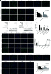
Neutralizing activities of g3-p239-Nb55 and fenobody-55 against the HEV ORF2 proteins and Kernow-C1/P6 virus in vitro. G3-p239-Nb55 and fenobody-55 can block g3-p239 attachment to HepG2/C3A cells, as determined by IFA. The nuclei were stained by DAPI, and the g3-p239 protein was stained by Alexa Fluor 488_labeled goat anti-mouse Ab. (A) and Western blot (B). (C) G3-p239-Nb55 and fenobody-55 can block different genotypes of HEV ORF2 proteins (g1-p239, g3-rabbit-p239, and g4-p239) from attaching to HepG2/C3A cells. The nuclei were stained by DAPI, and the p239 proteins were stained by Alexa Fluor 488_labeled goat anti-mouse Ab. Compared with those in the p239-only group, the majority of HEV ORF2 proteins were strongly inhibited by g3-p239-Nb55 and fenobody-55, especially g3-p239. Neutralizing analysis of g3-p239-Nb55 and fenobody-55, which inhibited the infection of Kernow-C1/P6 into HepG2/C3A cells, by IFA. The nuclei were stained by DAPI, and the virus was stained by Alexa Fluor 488_labeled goat anti-mouse Ab. (D) and real-time RT-PCR (E). The experiment was independently repeated three times. The error bars indicate the mean ± SD. *p <0.05, **p <0.01, ***p <0.001. ns, no significant difference.
FIGURE 6.
FIGURE 6.
In vivo neutralization analysis of g3-p239-Nb55 and fenobody-55 and optical micrographs of liver lesions from necropsied SPF rabbits. Fecal virus shedding and the level of anti-HEV Abs in the serum of SPF rabbits were detected. The plus (+) and minus (−) symbols indicate positive and negative results, respectively, for HEV RNA in the fecal samples from the different rabbits at 1–5 wpi according to nested RT-PCR. The lines show the levels of Abs against HEV in the sera of different rabbits at 1–5 wpi determined by indirect ELISA with the ORF2 protein as the coating Ag. The liver tissues were stained by hematoxylin-eosin. The arrows show locally intense lymphocytic venous phlebitis, lymphocytic inflammatory cell infiltration, and periphlebitis. SPF rabbits were inoculated with PBS and labeled No. 1–5 (A), with only rabbit HEV and labeled No. 6–10 (B). SPF rabbits were inoculated with rabbit HEV mixed with 133 μM NDV-Nb96 and labeled No. 11–15 (C), mixed with the same dosage of NDV-fenobody-96 and labeled No. 16–20 (D), mixed with g3-p239-Nb55 and labeled No. 21–25 (E), mixed with fenobody-55 and labeled No. 26–30 (F). SPF rabbits were first inoculated with rabbit HEV, and after 24 h, they were injected with 133 μM g3-p239-Nb55 and labeled No. 31–35 (G), and injected with fenobody-55 and labeled No. 36–40 (H). Three liver sections from each rabbit were stained with H&E to evaluate pathological and histopathological changes caused by HEV infection. The error bars indicate the mean ± SD. (I) The average number of inflammatory cells in three random 1-mm2 sections of each slice was determined via ImageJ software.
FIGURE 7.
FIGURE 7.
Identification and characterization of the epitope recognized by g3-p239-Nb55. (A) Schematic diagram of g3-p239 and various truncated proteins. The mutated amino acid residues of g3-p239 are marked in detail. (B) SDS-PAGE analysis of purified g3-p239 and various truncated proteins. (C) Identification of g3-p239-Nb55-HRP and Nb55D98A-M99A-D100A-D101A-M102A-HRP fusion proteins expressed in HEK293T cells by IFA. The nuclei were stained by DAPI, and the fusion proteins were stained by Alexa Fluor 488_labeled goat anti-mouse Ab. (D) Detection of the binding capacity between g3-p239 or truncated and mutated proteins and Nb55-HRP or Nb55 D98A-M99A-D100A-D101A-M102A-HRP by ELISA. Spatial locations on g3-p239 of the epitope recognized by g3-p239-Nb55. Overall structures of g3-p239 monomers (E) and pentamers (F) and a detailed assessment of the epitope. The spatial locations of the g3-p239-Nb55 (yellow) binding epitope (blue) protruding from g3-p239 (green) are marked. The experiment was independently repeated three times. The error bars indicate the mean ± SD.

Similar articles

References

    1. Pillot, J., Türkoglu S., Dubreuil P., Cosson A., Lemaigre G., Meng J., Lazizi Y.. 1995. Cross-reactive immunity against different strains of the hepatitis E virus transferable by simian and human sera. C. R. Acad. Sci III 318: 1059–1064. - PubMed
    1. Cuevas-Ferrando, E., Randazzo W., Pérez-Cataluña A., Sánchez G.. 2019. HEV occurrence in waste and drinking water treatment plants. Front. Microbiol. 10: 2937. - PMC - PubMed
    1. Smith., D. B., Simmonds P.; Members of the International Committee on the Taxonomy of Viruses Study Group ; Jameel S., Emerson S. U., Harrison T. J., Meng X.-J., Okamoto H., Van der Poel W. H. M., and Purdy M. A.. 2014. Consensus proposals for classification of the family Hepeviridae. J. Gen. Virol. 95: 2223–2232. - PMC - PubMed
    1. Purdy, M. A., Drexler J. F., Meng X.-J., Norder H., Okamoto H., Van der Poel W. H. M., Reuter G., de Souza W. M., Ulrich R. G., Smith D. B.. 2022. ICTV virus taxonomy profile: Hepeviridae 2022. J. Gen. Virol 103: jgv.0.001778. - PubMed
    1. Forni, D., Cagliani R., Clerici M., Sironi M.. 2018. Origin and dispersal of hepatitis E virus. Emerg. Microbes Infect. 7: 11–23. - PMC - PubMed