. 2024 May 23;13(11):897.
doi: 10.3390/cells13110897.

Reprogramming Glioblastoma Cells into Non-Cancerous Neuronal Cells as a Novel Anti-Cancer Strategy

Affiliations

Reprogramming Glioblastoma Cells into Non-Cancerous Neuronal Cells as a Novel Anti-Cancer Strategy

Michael Q Jiang et al. Cells. .

Abstract

Glioblastoma Multiforme (GBM) is an aggressive brain tumor with a high mortality rate. Direct reprogramming of glial cells to different cell lineages, such as induced neural stem cells (iNSCs) and induced neurons (iNeurons), provides genetic tools to manipulate a cell's fate as a potential therapy for neurological diseases. NeuroD1 (ND1) is a master transcriptional factor for neurogenesis and it promotes neuronal differentiation. In the present study, we tested the hypothesis that the expression of ND1 in GBM cells can force them to differentiate toward post-mitotic neurons and halt GBM tumor progression. In cultured human GBM cell lines, including LN229, U87, and U373 as temozolomide (TMZ)-sensitive and T98G as TMZ-resistant cells, the neuronal lineage conversion was induced by an adeno-associated virus (AAV) package carrying ND1. Twenty-one days after AAV-ND1 transduction, ND1-expressing cells displayed neuronal markers MAP2, TUJ1, and NeuN. The ND1-induced transdifferentiation was regulated by Wnt signaling and markedly enhanced under a hypoxic condition (2% O2 vs. 21% O2). ND1-expressing GBM cultures had fewer BrdU-positive proliferating cells compared to vector control cultures. Increased cell death was visualized by TUNEL staining, and reduced migrative activity was demonstrated in the wound-healing test after ND1 reprogramming in both TMZ-sensitive and -resistant GBM cells. In a striking contrast to cancer cells, converted cells expressed the anti-tumor gene p53. In an orthotopical GBM mouse model, AAV-ND1-reprogrammed U373 cells were transplanted into the fornix of the cyclosporine-immunocompromised C57BL/6 mouse brain. Compared to control GBM cell-formed tumors, cells from ND1-reprogrammed cultures formed smaller tumors and expressed neuronal markers such as TUJ1 in the brain. Thus, reprogramming using a single-factor ND1 overcame drug resistance, converting malignant cells of heterogeneous GBM cells to normal neuron-like cells in vitro and in vivo. These novel observations warrant further research using patient-derived GBM cells and patient-derived xenograft (PDX) models as a potentially effective treatment for a deadly brain cancer and likely other astrocytoma tumors.

Keywords: Wnt-3α; apoptosis; direct reprogramming; glioblastoma; hypoxia; induced neurons; p53; tumor growth.


Conflict of interest statement

The authors declare no conflicts of interest.

Figures
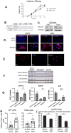
Figure 1
Figure 1
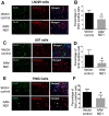
Neuronal transdifferentiation of GBM cells induced by ND1 expression. GBM cells were cultured in a neuron stem cell medium for 10 days before AAV-ND1 transduction. (A) Comparisons of GBM cell infection by the control AAV-mCherry vector and AAV-ND1-mCherry vector. Both vectors showed similar infection rates and efficacy in GBM cultures. (B) Western blotting revealed a high-level expression of ND1 protein in the normal mouse brain; the ND1 level in GBM cell lines U373, LN229, and T98G was noticeably low or barely detectable. A similar expression pattern was seen with β-catenin. (C) AAV-ND1 delivered expression of ND1 in LN229 cells 3 and 14 days after transduction. (D) Immunocytochemical staining 7 days after viral delivery detected the neuronal marker TUJ1 (red) in AAV-ND1 transfected cells, no TUJ1 was seen in vector control cells. (E) The mature neuronal marker NeuN (red) was observed 21 days after ND1 expression. Blue color was the nucleus marker DAPI. (F) Western blot (WB) assays revealed the absence of neuronal marker MAP2, the trophic factor BDNF, and the normal cell marker p53. All these markers significantly increased in ND1-reprogrammed GBM cells. (G) Quantification of the WB test in (F). N = 3, * p < 0.05 vs. control vector’ ** p < 0.01 vs. controls; One-way ANOVA. (H) The TUNEL assay of U87 and T98G cells confirmed the increased DNA damage and apoptotic cell death of ND1-expressing cells. N = 3, *. p < 0.05 vs. control vector, Student unpaired t test. (I) In the MTT assays, AAV-ND1 converted cultures exhibited poorer survival compared to control vector-treated GBM cultures. N = 3, ** p < 0.01 vs. control vector, unpaired t test.
Figure 2
Figure 2
Attenuated proliferation of ND1-reprogrammed GBM cells. Immunocytochemical assays measured the proliferation of GBM cells in different cell lines 3 days after viral transduction. (A,C,E) Representative fluorescent images of three GBM cell lines. Green: BrdU-positive cells; Red: mCherry-labeled transfected cells. (B,D,F) Quantified measurements in the experiments of (A,C,E), respectively. ND1 expression significantly decreased BrdU expression in AAV-ND1 transduced multiple GBM cells. N = 3 cultures, * p < 0.05 vs. control.
Figure 3
Figure 3
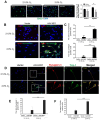
Promoting effects of hypoxia on ND1-induced neuronal conversion of GBM cells. Immunocytochemical assessments 7 days after ND1-transduction of GBM cells performed under 2% and 21% O2, respectively. (A) Fluorescent imaging of BrdU staining and the qualified analysis showed significant reduction of proliferating GBM cells (U373 cell line) reprogrammed after AAV-ND1. * p < 0.05 vs. vector control. (B) Imaging analysis demonstrated the promoting effect of hypoxia on the expression of mature neuronal marker NeuN of LN229 cells. Typical neuronal morphology such as prolonged axonal tracks as seen with these cells. (C) Quantified analysis of the results in (B). ** p < 0.01 vs. vector controls; *** p < 0.005 vs. vector controls; N = 3. (D) The staining of synapsin-1 protein (red) and TUJ-1 (green) in vector control and AAV-ND1 treated U373 cells shown at low and high magnifications. The frame indicates the area of the image with enlarged magnification. (E) Quantified analysis of synapsin-1 expression at low and high O2 levels. ** p < 0.01 vs. vector controls. N = 3. (F) TUNEL staining examined cell death 7 days after virus transduction under 2% O2 and 21% O2 conditions, respectively. The hypoxic condition markedly augmented cell death of ND1-reprogrammed cells. N = 3 assays. *** p < 0.005 vs. controls and reprogrammed cells under 21% O2.
Figure 4
Figure 4
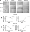
Reduced migrative activity of ND1-converted GBM cells. Wound-healing assay was performed in U87 and T98G cell cultures 3–5 days after ND1-reprogramming. (A) Phase contrast images show wounded areas and different times after the formation of the wounded (empty) area in different groups. (B,C) Time courses of cell migration to cover the empty area and the cell count inside the area at 12, 24, 36, and 48 h after scratch, in U87 cultures (B) and G98G cultures (C). ND1-expressing cells were significantly fewer inside the empty area compared to the number of migrated vector-controlled cells. * p < 0.05 vs. vector controls, N = 3 cultures/group. ANOVA test.
Figure 5
Figure 5
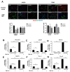
Regulatory roles of the Wnt pathway in ND1-induced GBM reprogramming and cell proliferation. Immunocytochemical staining of ND1 and neuronal markers with the manipulations of the Wnt/β-catenin signaling. (A) Cell proliferation of BrdU staining was assessed 48 h after transduction; GFAP and DAPI were used for cell counting with and without Wnt 3α (100 nM) or XAV939 (20 ng/mL) in vector control and AAV-ND1 cultures. Wnt 3α and XAV939 were co-applied with the virus. Bar graphs underneath are the quantified cell counts of BrdU-positive cells, showing significant inhibitory effects of Wnt 3α and XAV939 in LN229 naïve cultures but not in ND1-expressing cultures. In T98G cells, XAV939 showed inhibitory effects in both vector control and ND1 converted cells, while Wnt 3α increased BrdU-positive cells in ND1 cells. * p < 0.05 and ** p < 0.01 vs. the control vector. (BG) Quantified expressions of reprogramming and neuronal markers 7 days after transduction of LN229 cells, including ND1 (B), GFAP (C), Nestin (D), MAP2 (E), TUJ-1 (F), and Pax 6 (G). Among these markers, Wnt 3α displayed strong inhibitory actions in control GBM cultures and ND1 reprogrammed cells. These data suggest that although the Wnt/β-catenin pathway may have variable regulations on proliferation/cell cycle of GBM cells, the expression/activation of Wnt signaling is required for neuronal transdifferentiation of GBM cells. ** p < 0.01 vs. the control vector.
Figure 6
Figure 6
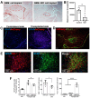
Inhibition of tumor growth by ND1-reprogramming in a GBM mouse model. An orthotopic GBM model was established by implanting U373 cells transduced by control AAV-mCherry or AAV-ND1-mCherry vector into the white matter region of the right hemispheres of adult mice. Tumors were detected as soon as 7–14 days after implantation. (A) In the H&E assay, one or more tumors were identified (dotted line) in and around the injection regions that received control GBM cells, while significantly smaller single tumors or no tumor tissue was visible in mice that received ND1-expressing cells. (B) Significantly decreased tumor size formed by ND1 cells compared to control cells. N = 5/group, * p < 0.05 vs. controls. (C) Immunohistochemical staining of GFAP (blue) and mCherry (red) in the contralateral and transplantation sides of the brain, confirming the seeding of viral transduced cells in the targeted brain region. (D) TUJ-1 staining (green) and mCherry (red) revealed converted neuronal cells in the white matter area. (E) Accumulations of IBA-1positive microglia/microphage (red) in the injection site. In the same region, there were abundant TUJ-1-positive cells (green) indicative of ND1-converted neurons that did not overlay with GFAP (blue) or IBA-1 (red) fluorescence. (F) Quantified analyses of GFAP, IBA-1, and TUJ-1 positive cells. Please note there was basically no TUJ-1 fluorescence in the contralateral side and the control GBM tumor. A drastic increase of TUJ-1 expression was only seen with ND-1 cell transplantation. N = 5–6 mice/group, * p < 0.05 vs. controls, **** p < 0.001 vs. controls.

Similar articles

Cited by

References

    1. Fazeny-Dorner B., Wenzel C., Veitl M., Piribauer M., Rossler K., Dieckmann K., Ungersbock K., Marosi C. Survival and prognostic factors of patients with unresectable glioblastoma multiforme. Anticancer Drugs. 2003;14:305–312. doi: 10.1097/00001813-200304000-00008. - DOI - PubMed
    1. Ostrom Q.T., Price M., Neff C., Cioffi G., Waite K.A., Kruchko C., Barnholtz-Sloan J.S. CBTRUS Statistical Report: Primary Brain and Other Central Nervous System Tumors Diagnosed in the United States in 2016–2020. Neuro Oncol. 2023;25:iv1–iv99. doi: 10.1093/neuonc/noad149. - DOI - PMC - PubMed
    1. Liberski P.P., Kordek R. Ultrastructural pathology of glial brain tumors revisited: A review. Ultrastruct. Pathol. 1997;21:1–31. doi: 10.3109/01913129709023244. - DOI - PubMed
    1. Zheng Y., Carrillo-Perez F., Pizurica M., Heiland D.H., Gevaert O. Spatial cellular architecture predicts prognosis in glioblastoma. Nat. Commun. 2023;14:4122. doi: 10.1038/s41467-023-39933-0. - DOI - PMC - PubMed
    1. Awad A.W., Karsy M., Sanai N., Spetzler R., Zhang Y., Xu Y., Mahan M.A. Impact of removed tumor volume and location on patient outcome in glioblastoma. J. Neuro-Oncol. 2017;135:161–171. doi: 10.1007/s11060-017-2562-1. - DOI - PubMed

Substances