. 2024 May 23;10(11):e31818.
doi: 10.1016/j.heliyon.2024.e31818. eCollection 2024 Jun 15.

IRF8 maintains mononuclear phagocyte and neutrophil function in acute kidney injury

Affiliations

IRF8 maintains mononuclear phagocyte and neutrophil function in acute kidney injury

Na Li et al. Heliyon. .

Abstract

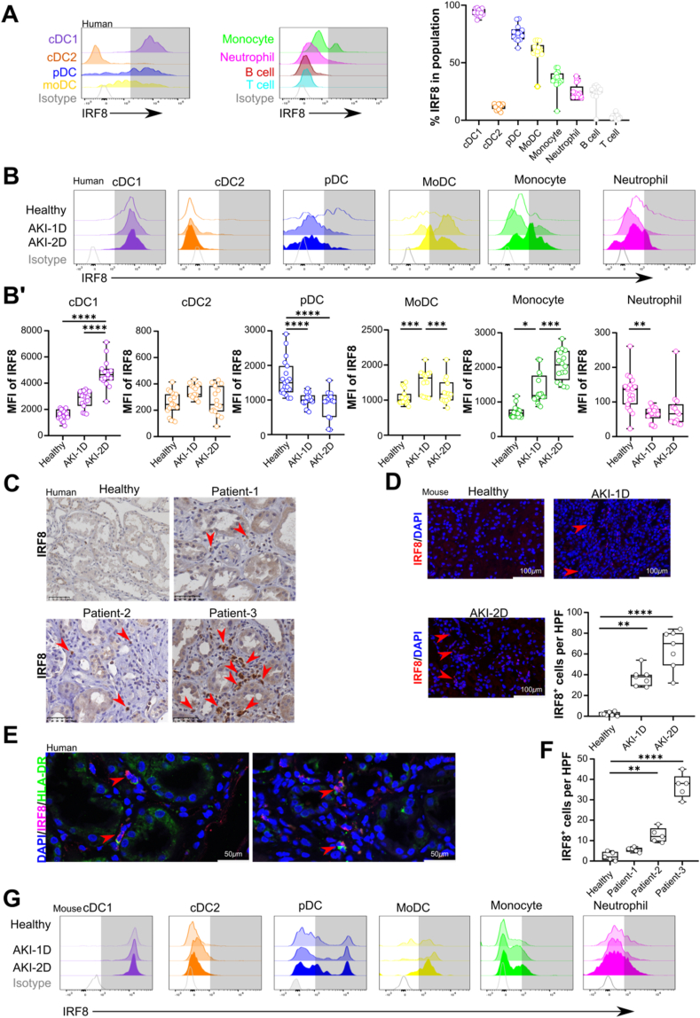
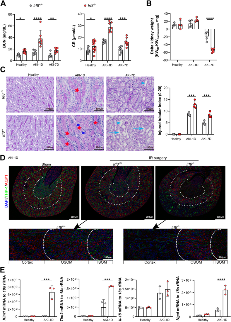
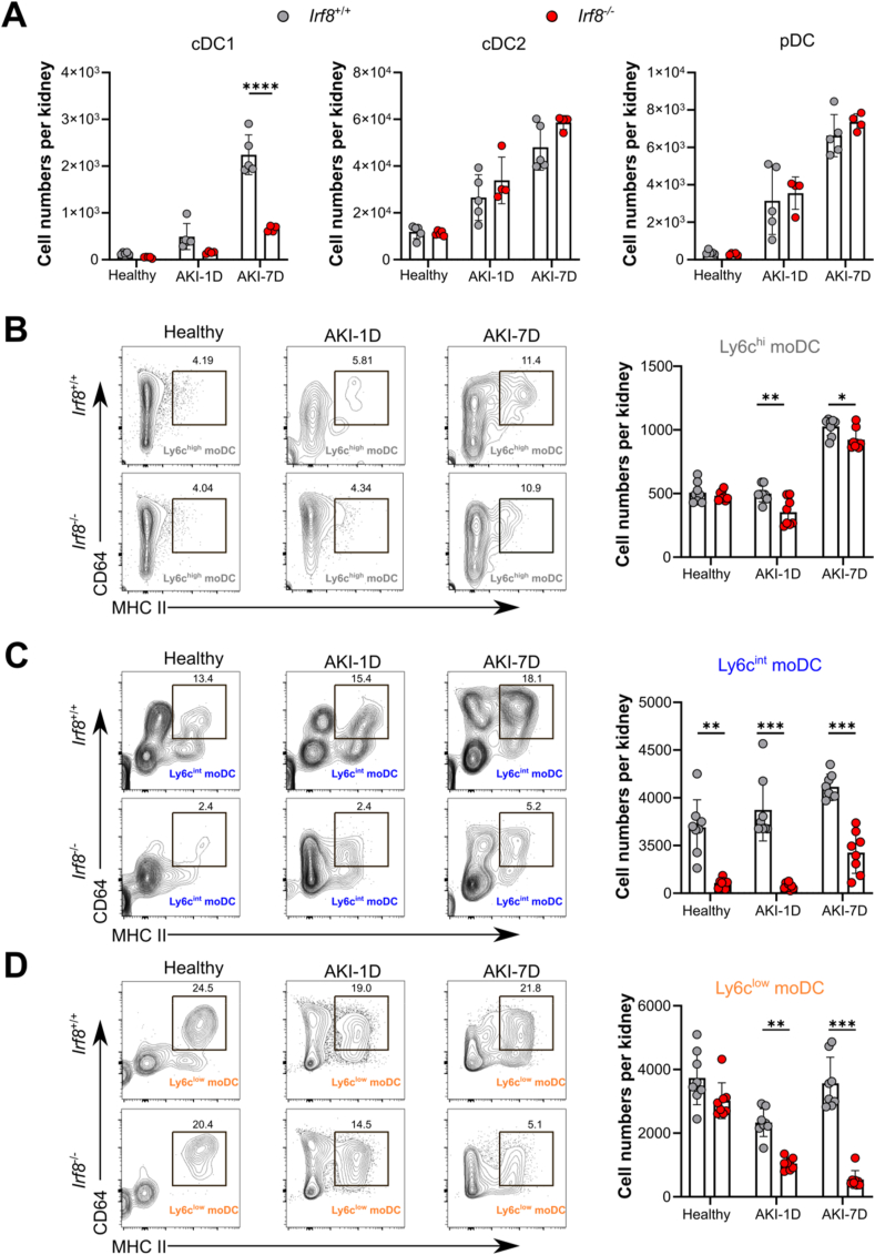
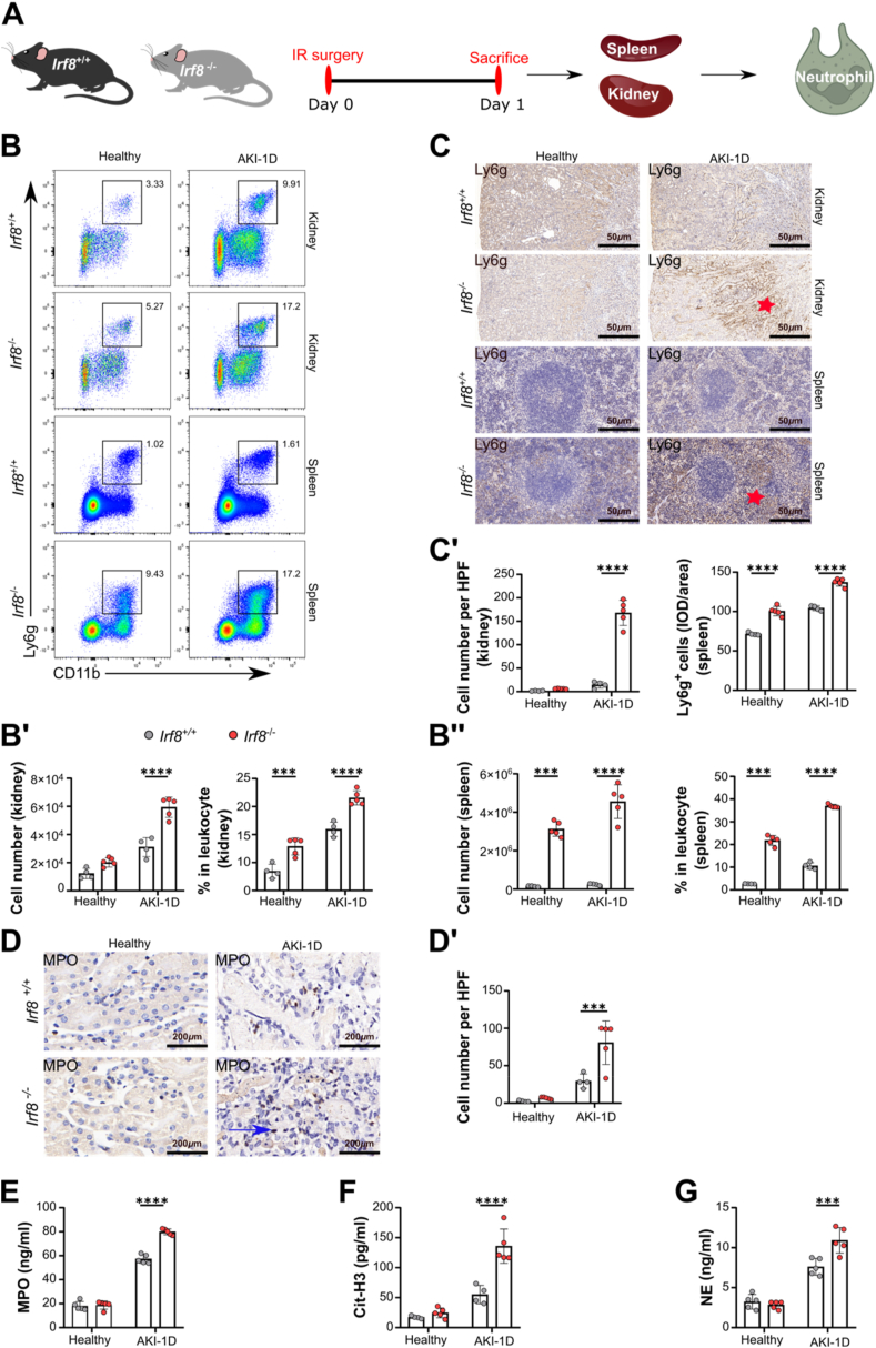
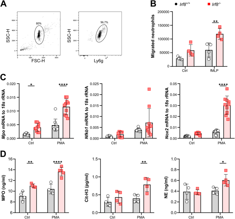
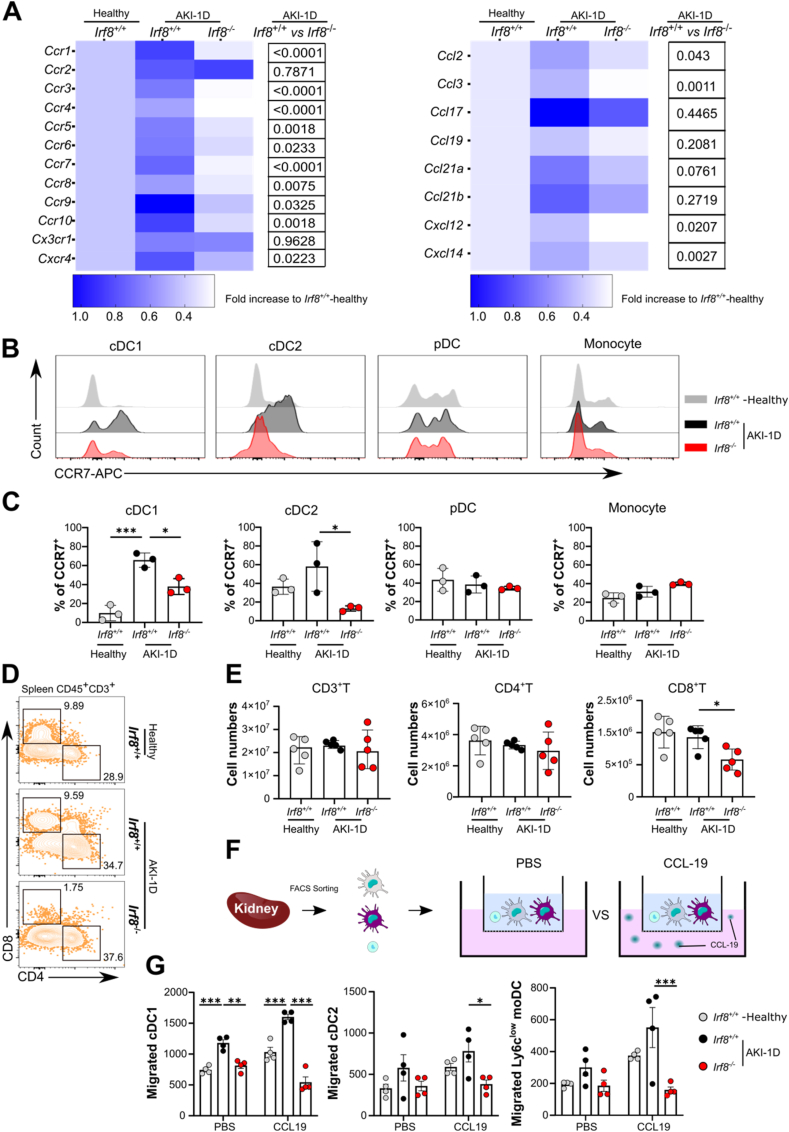
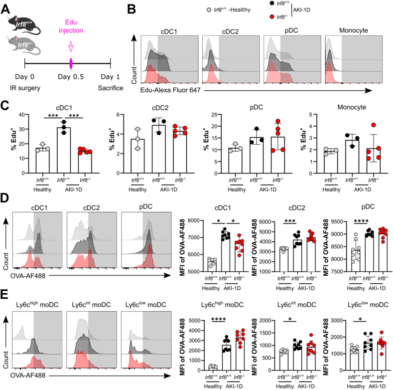
Immune cells are key players in acute tissue injury and inflammation, including acute kidney injury (AKI). Their development, differentiation, activation status, and functions are mediated by a variety of transcription factors, such as interferon regulatory factor 8 (IRF8) and IRF4. We speculated that IRF8 has a pathophysiologic impact on renal immune cells in AKI and found that IRF8 is highly expressed in blood type 1 conventional dendritic cells (cDC1s), monocytes, monocyte-derived dendritic cells (moDCs) and kidney biopsies from patients with AKI. In a mouse model of ischemia‒reperfusion injury (IRI)-induced AKI, Irf8 -/- mice displayed increased tubular cell necrosis and worsened kidney dysfunction associated with the recruitment of a substantial amount of monocytes and neutrophils but defective renal infiltration of cDC1s and moDCs. Mechanistically, global Irf8 deficiency impaired moDC and cDC1 maturation and activation, as well as cDC1 proliferation, antigen uptake, and trafficking to lymphoid organs for T-cell priming in ischemic AKI. Moreover, compared with Irf8 +/+ mice, Irf8 -/- mice exhibited increased neutrophil recruitment and neutrophil extracellular trap (NET) formation following AKI. IRF8 primarily regulates cDC1 and indirectly neutrophil functions, and thereby protects mice from kidney injury and inflammation following IRI. Our results demonstrate that IRF8 plays a predominant immunoregulatory role in cDC1 function and therefore represents a potential therapeutic target in AKI.

Keywords: Acute kidney injury; Dendritic cell; Inflammation; Interferon regulatory factor 8; Monocyte; Neutrophil.


Conflict of interest statement

The authors declare that they have no known competing financial interests or personal relationships that could have appeared to influence the work reported in this paper.

Figures

Fig. 1
Fig. 1
Interferon regulatory factor 8 expression increases in patients and mice with acute kidney injury. (A) Histograms of IRF8 expression in human blood leukocytes from healthy individuals compared with isotype control staining (n = 14 per group). The gating strategy for blood leukocytes is shown in Supplemental Fig. 1. (B) The dynamic expression patterns of IRF8 in blood leukocytes from healthy individuals (n = 14 per group) and patients with AKI (n = 10 per group). (B′) MFI of IRF8 in the blood leukocyte populations in healthy individuals and patients with AKI. (C) Distribution and (F) number of IRF8-positive cells (n = 1 per group) in kidneys from transplantation patients. Red arrows indicate IRF8+ cells. Scale bar, 50 μm. (D) Mouse kidney sections were stained with anti-IRF8 (red) and DAPI (blue), and the numbers of IRF8+ cells are shown (n = 6 per group). Scale bar, 100 μm. Red arrow: IRF8+ cell. (E) Two representative images of co-staining for IRF8 and HLA-DR in kidney specimens from 12 patients pathologically diagnosed with acute tubular injury (n = 12 per group). Red arrow: IRF8+HLA-DR+ cell. Scale bar, 50 μm. (G) Dynamic expression patterns of IRF8 in kidney leukocytes (gated as indicated inSupplemental Fig. 2A) from healthy mice and mice with AKI. The data are shown as the mean ± SD. *p < 0.05, **p < 0.01, ***p < 0.001, and ****p < 0.0001. One-way ANOVA. (For interpretation of the references to color in this figure legend, the reader is referred to the Web version of this article.)
Fig. 2
Fig. 2
Irf8 deletion aggravates renal dysfunction during AKI. Irf8+/+ and Irf8−/− mice were subjected to unilateral ischemic reperfusion (IR) surgery and sacrificed 1 day or 7 days after AKI. The healthy group represents mice that did not undergo IR surgery. (A) Blood urea nitrogen (BUN) levels and serum creatinine (CR) levels (n ≥ 3 per group). (B) Weight loss in the kidneys of the IR group compared to that of the sham group was determined as follows: Delta kidney weight = KWIR – KWcontralateral (n ≥ 3 per group). (C) PAS-stained sections and tubular injury scores were evaluated in high-power fields (n = 4 per group). Scale bar, 100 μm. Red star: cast formation; Blue arrow: cell necrosis. Green arrow: tubular dilation. (D) The upper panel shows images of proximal tubules and distal tubules with immunolabeling for aquaporin-1 (AQP1, red) and Tamm-Horsfall protein-1 (THP-1, green) in the cortex, outer stripe of the outer medulla (OSOM), and inner stripe of the outer medulla (ISOM) (scale bar, 200 μm) from IR kidneys or sham kidneys on day 1 after AKI. Nuclei were counterstained with DAPI (blue). The bottom panel shows magnified IR kidney sections from Irf8+/+ and Irf8−/− mice (scale bar, 100 μm). (E) The mRNA expression of tubular injury markers in the healthy group and AKI-1D group was normalized to 18S rRNA expression (n = 3 per group). Each dot represents one mouse. The data are shown as the mean ± SD. *p < 0.05, **p < 0.01, ***p < 0.001, and ****p < 0.0001. Two-way ANOVA. (For interpretation of the references to color in this figure legend, the reader is referred to the Web version of this article.)
Fig. 3
Fig. 3
Irf8 deletion prevents type I conventional dendritic cell and monocyte-derived dendritic cell infiltration into the kidney during AKI. Irf8+/+ and Irf8−/− mice were subjected to IR surgery. Kidney DCs were collected 1 day or 7 days after AKI and analyzed by flow cytometry according to the strategy illustrated in Supplemental Fig. 2A. (A-D) The absolute numbers of DCs in the kidney after AKI are shown (n > 3 mice per group). Each dot represents one mouse. The data are shown as the mean ± SD. *p < 0.05, **p < 0.01, ***p < 0.001, and ****p < 0.0001. Two-way ANOVA.
Fig. 4
Fig. 4
Irf8 deletion triggers neutrophil recruitment and NET formation during AKI. (A) Study design. Irf8+/+ and Irf8−/− mice were subjected to IR surgery and sacrificed 1 day after AKI. The kidneys and spleens of mice were used for analysis. (B) Flow cytometric analysis of kidney and spleen neutrophils (CD45+CD49CD3eCD19-Ly6g+CD11b+, gated as indicated in Supplemental Fig. 2A). (B′-B″) The percentages and numbers of kidney/spleen neutrophils from healthy controls and mice after AKI (n = 4–5 mice per group). (C–C′) Distribution and cell numbers of Ly6g+ neutrophils in kidneys and spleens (n = 4–5 mice per group) from healthy controls and mice after AKI. (D) Distribution of myeloperoxidase (MPO) in kidneys (n = 4–5 mice per group). Scale bar, 200 μm. (D′) Numbers of MPO+ cells in kidneys. (E-G) Serum was collected from healthy and AKI mice, and the release of MPO, citrullinated histone 3 (Cit-H3), and neutrophil elastase (NE) was determined using ELISA kits (n = 5 mice per group). Each dot represents one mouse. Red star and blue arrow indicate positive cells. The data are shown as the mean ± SD. ***p < 0.001, ****p < 0.0001. Two-way ANOVA. (For interpretation of the references to color in this figure legend, the reader is referred to the Web version of this article.)
Fig. 5
Fig. 5
Irf8 deletion increases neutrophil migration in vitro. (A) Neutrophils were isolated from mouse bone marrow and identified as mature Ly6g+ by flow cytometry with a purity of ∼97 %. (B) The total number of neutrophils isolated from Irf8+/+ and Irf8−/− mice that migrated toward the chemoattractant fMLP was determined by flow cytometry after 3 h (n = 4 mice per group). (C) mRNA expression of Mpo, Nfkb1, and Nox2 in isolated neutrophils subjected to PMA incubation (n ≥ 5 mice per group). (D) The release of Cit-H3, MPO, and NE by PMA-stimulated neutrophils was determined using ELISA kits (n ≥ 4 mice per group). Each dot represents one mouse. The data are shown as the mean ± SD. *p < 0.05, **p < 0.01, and ****p < 0.0001. Two-way ANOVA. Ctrl, control.
Fig. 6
Fig. 6
Irf8 deletion impairs DC trafficking and T-cell priming during AKI. Irf8+/+ and Irf8−/− mice were subjected to IR surgery, and kidney MPCs were analyzed 1 day after AKI. (A) mRNA expression levels of genes encoding CC chemokine receptors (CCRs) and ligands (CCLs) in kidney MPCs were normalized to 18S rRNA expression. The data are illustrated in heatmaps. The color intensity represents the mean expression value of each group. The p values for gene expression differences between the Irf8+/+ and Irf8−/− groups 1 day after AKI were calculated. A p value < 0.05 was considered significant. n = 3–5 mice per group. (B–C) The expression of CCR7 in kidney MPCs was measured by flow cytometry. Kidneys were harvested from Irf8+/+ (black) and Irf8−/− (red) mice 1 day after AKI and from healthy Irf8+/+ mice (gray). Images from three independent experiments and the percentages of CCR7+ cells among each kidney MPC are shown. n = 3 mice per group. (D) Splenic T-cell subsets (gated on CD45+CD19CD3e+ T cells, Supplemental Fig. 2A). (E) Absolute numbers of splenic CD3+ T cells, CD4+ T cells, and CD8+ T cells. n = 5–6 mice per group. (F) Kidney MPCs were isolated 1 day after AKI, and each DC subset was sorted for in vitro Transwell migration assays. (G) The total number of each DC subset that migrated toward the CCL-19 stimulus was quantified. n = 4 mice per group. Each dot represents one mouse. The data are shown as the mean ± SD. *p < 0.05, **p < 0.01, and ***p < 0.001. t-test, one-way ANOVA or two-way ANOVA. (For interpretation of the references to color in this figure legend, the reader is referred to the Web version of this article.)
Fig. 7
Fig. 7
Irf8 deletion impairs the proliferation and antigen uptake of kidney DCs during AKI. (A) Experimental design. Twelve hours after IR surgery, EdU was injected i.p. 12 h before the mice were sacrificed. (B) Histogram plots showing EdU incorporation in kidney MPCs from Irf8+/+ (black) or Irf8−/− (red) mice 1 day after AKI and from healthy Irf8+/+ mice (gray). Representative images of three independent experiments are shown. (C) Percentages of EdU-positive cells in each MPC subset (n = 3–5 mice per group). (D-E) Flow cytometry analysis of antigen uptake by kidney MPCs. Kidneys were harvested from AKI mice and healthy mice. MPCs freshly isolated from kidneys were exposed to ovalbumin-Alexa Fluor 488 (OVA-AF488). Antigen incorporation was visualized by calculating the mean fluorescence intensity (MFI) of OVA-AF488 in each subset (n > 3 mice per group). The data are shown as the mean ± SD. *p < 0.05, ***p < 0.001, and ****p < 0.0001. One-way ANOVA. (For interpretation of the references to color in this figure legend, the reader is referred to the Web version of this article.)

Similar articles

References

    1. Sharfuddin A.A., Molitoris B.A. Pathophysiology of ischemic acute kidney injury. Nature reviews. Nat Rev Nephrol. 2011;7:189–200. - PubMed
    1. Sekine M., Monkawa T., Morizane R., Matsuoka K., Taya C., Akita Y., Joh K., Itoh H., Hayashi M., Kikkawa Y., Kohno K., Suzuki A., Yonekawa H. Selective depletion of mouse kidney proximal straight tubule cells causes acute kidney injury. Transgenic Res. 2012;21:51–62. - PMC - PubMed
    1. Mulay S.R., Linkermann A., Anders H.J. Necroinflammation in kidney disease. JASN (J. Am. Soc. Nephrol.) 2016;27:27–39. - PMC - PubMed
    1. Dong X., Swaminathan S., Bachman L.A., Croatt A.J., Nath K.A., Griffin M.D. Resident dendritic cells are the predominant TNF-secreting cell in early renal ischemia-reperfusion injury. Kidney Int. 2007;71:619–628. - PubMed
    1. Kurts C., Ginhoux F., Panzer U. Kidney dendritic cells: fundamental biology and functional roles in health and disease. Nat. Rev. Nephrol. 2020;16:391–407. - PubMed

LinkOut - more resources