[Preprint]. 2024 May 14:2024.05.14.594147.
doi: 10.1101/2024.05.14.594147.

Stress-induced dysfunction of neurovascular astrocytes contributes to sex-specific behavioral deficits

Affiliations

Stress-induced dysfunction of neurovascular astrocytes contributes to sex-specific behavioral deficits

Justin L Bollinger et al. bioRxiv. .

Abstract

Astrocytes form an integral component of the neurovascular unit, ensheathing brain blood vessels with projections high in aquaporin-4 (AQP4) expression. These AQP4-rich projections facilitate interaction between the vascular endothelium, astrocytes, and neurons, and help stabilize vascular morphology. Studies using preclinical models of psychological stress and post-mortem tissue from patients with major depressive disorder (MDD) have reported reductions in AQP4, loss of astrocytic structures, and vascular impairment in the prefrontal cortex (PFC). Though compelling, the role of AQP4 in mediating stress-induced alterations in blood vessel function and behavior remains unclear. Here, we address this, alongside potential sex differences in chronic unpredictable stress (CUS) effects on astrocyte phenotype, blood-brain barrier integrity, and behavior. CUS led to pronounced shifts in stress-coping behavior and working memory deficits in male -but not female- mice. Following behavioral testing, astrocytes from the frontal cortex were isolated for gene expression analyses. We found that CUS increased various transcripts associated with blood vessel maintenance in astrocytes from males, but either had no effect on- or decreased- these genes in females. Furthermore, CUS caused a reduction in vascular-localized AQP4 and elevated extravasation of a small molecule fluorescent reporter (Dextran) in the PFC in males but not females. Studies showed that knockdown of AQP4 in the PFC in males is sufficient to disrupt astrocyte phenotype and increase behavioral susceptibility to a sub-chronic stressor. Collectively, these findings provide initial evidence that sex-specific alterations in astrocyte phenotype and neurovascular integrity in the PFC contribute to behavioral and cognitive consequences following chronic stress.


Conflict of interest statement

Conflict of Interest The authors declare no conflict of interest.

Figures

Figure 1.
Figure 1.. Chronic unpredictable stress (14-days) induces sex-specific patterns of cognitive and behavioral dysfunction.
A) Adult male and female mice were exposed to 14-days of CUS or were left unstressed. All animals were subjected to behavioral testing (n = 15–20/group). In one cohort, mice received intravenous injections of Alexa Fluor 488-conjugated dextran and tomato lectin, after which brains were processed for confocal microscopy (n = 7–8/group). In a separate cohort, astrocytes were isolated via FACS and astrocyte gene expression was analyzed (n = 8–12/group). B) Average time spent immobile in the forced swim test (FST) prior to- and on CUS day 11. C) Total amount of distance traveled in the open field test (OFT). D) Time spent in the center during the OFT. E) Total amount of time spent exploring objects in the test phase of the temporal object recognition task (TOR). F) Discrimination index in the TOR. G) Schematic of the cognitive T-maze task. H) Time spent exploring both the novel and familiar arm in the T-maze task. I) Total number of alternations in the T-maze task. J) Proportion of spontaneous alternations in the T-maze task. Bars represent mean ± S.E.M. * p<0.05 planned comparison indicated (Sidak’s test).
Figure 2.
Figure 2.. Chronic unpredictable stress (14-days) differentially alters the molecular phenotype of astrocytes in the prefrontal cortex in males and females.
A) A prefrontal cortex-enriched portion of the brain was dissected out and homogenized. Cells were then stained for CD11b and ACSA2 using fluorescently conjugated antibodies and characterized using flow cytometry. CD11b-/ACSA2+ astrocytes were collected using FACS (proportion of gated events relative to the total number of events in each plot is indicated). Representative gating strategy is shown. B) Normalized astrocyte expression of marker genes (Gfap, Vim, S100b), synaptic interaction factors (Lamp1, Mertk, Megf10), and vascular interaction factors (Vegf, Bfgf, Aqp4, Angpt1, Agt) in the frontal cortex. Expression of Gfap, Vim, S100b, and Lamp1 is reflected on the left y-axis, all other genes are reflected on the right y-axis. Bars represent mean ± S.E.M. # p<0.05 main effect (no specific contrasts detected). * p<0.05 planned comparison indicated (Sidak’s test).
Figure 3.
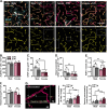
Figure 3.. Chronic stress (14-days) leads to astrocyte atrophy and vascular-associated aquaporin-4 loss in the prefrontal cortex in male, but not female, mice.
A) Representative images of vascular tomato lectin (TL) staining (magenta) and immunohistology for astrocyte GFAP (cyan) and AQP4 (yellow) in the PFC (20× + zoom, scale bar = 100 μm). Dashed quadrilaterals indicate TL+ vessel segments with little- to no AQP4 coverage. B) Number of ALDH1L1+ astrocytes in the PFC per mm2. C) Area of GFAP+ material in the PFC. D) Area of GFAP+ material associated with blood vessels in the PFC. E) Area of astrocyte AQP4+ material relative to vessel area. F) Area of tomato lectin+ vessels in the PFC. G) Left: Representative image of Alexa Fluor 488-conjugated dextran (10,000 MW, pale yellow) and vascular tomato lectin (magenta) in the PFC. Image features three extravasation sites, with one site highlighted in XZ-YZ dimensions. Middle: Number of dextran extravasation sites in the PFC per image. Right: Mean fluorescence intensity of dextran extravasation sites in the PFC (normalized to control males). Bars represent mean ± S.E.M. # p<0.05 main effect (no specific contrasts detected). * p<0.05 planned comparison indicated (Sidak’s test).
Figure 4.
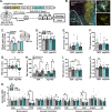
Figure 4.. Knockdown of aquaporin-4 in the prefrontal cortex shifts astrocyte phenotype and induces behavioral susceptibility to a sub-chronic stressor (7-days).
A) Adult male mice received infusions of either AAV5-scrambled-shRNA or AAV5-Aqp4-shRNA into the PFC. Following three weeks of recovery, animals were exposed to 7-days of sub-CUS or were left unstressed. All animals were subjected to behavioral testing (n = 11–16/group). In one cohort, mice received intravenous injections of tomato lectin, after which brains were processed for confocal microscopy (n = 6–8/group). In a separate cohort, astrocytes were isolated via FACS and astrocyte gene expression was analyzed (n = 7–8/group). B) Representative images of AAV5-Aqp4-ShRNA reporter (BFP, yellow) in the PFC. Astrocyte GFAP is shown in cyan, tomato lectin+ vessels are shown in magenta. Dotted line delineates barrier between the forceps minor and PFC. Left image: 10×, scale bar = 200 μm. Right image panel: 60×, scale bar = 20 μm. C) Normalized expression of Aqp4 in dissected PFC or SSCTX (S1J subregion). D) Total amount of distance traveled in the open field test (OFT). E) Time spent in the center during the OFT. F) Total amount of time spent exploring objects in the test phase of the temporal object recognition task (TOR). G) Discrimination index in the TOR. H) Time spent exploring both the novel and familiar arm in the T-maze task. I) Total number of alternations in the T-maze task. J) Proportion of spontaneous alternations in the T-maze task. K) Normalized astrocyte expression of marker genes (Gfap, Vim, S100b), synaptic interaction factors (Lamp1, Mertk, Megf10), and vascular interaction factors (Vegf, Bfgf, Aqp4, Angpt1, Agt) in the frontal cortex. Bars represent mean ± S.E.M. # p<0.05 main effect (no specific contrasts detected). * p<0.05 planned comparison indicated (Sidak’s test).
Figure 5.
Figure 5.. Astrocytes dynamically respond to sub-chronic stress, knockdown of aquaporin-4 leads to astrocyte dystrophy in the prefrontal cortex.
A) Representative images of vascular tomato lectin (magenta) and immunohistology for astrocyte GFAP (cyan) and AQP4 (yellow) in the PFC (20×, scale bar = 100 μm). B) Left: Representative images of immunohistology for ALDH1L1 in the PFC (20×, scale bar = 100 μm). Right: Number of ALDH1L1+ astrocytes in the PFC per mm2. C) Area of GFAP+ material in the PFC. D) Area of GFAP+ material associated with blood vessels in the PFC. E) Total area of AQP4+ material in the PFC. F) Area of astrocyte AQP4+ material relative to vessel area. G) Area of tomato lectin+ vessels in the PFC. # p<0.05 main effect (no specific contrasts detected). * p<0.05 planned comparison indicated (Sidak’s test).

Similar articles

References

    1. Jung JS, Bhat RV, Preston GM, Guggino WB, Baraban JM, Agre P. Molecular characterization of an aquaporin cDNA from brain: candidate osmoreceptor and regulator of water balance. Proc Natl Acad Sci U S A. 1994;91:13052–13056. - PMC - PubMed
    1. Kobayashi H, Minami S, Itoh S, Shiraishi S, Yokoo H, Yanagita T, et al. Aquaporin subtypes in rat cerebral microvessels. Neuroscience Letters. 2001;297:163–166. - PubMed
    1. Badaut J, Lasbennes F, Magistretti PJ, Regli L. Aquaporins in Brain: Distribution, Physiology, and Pathophysiology. J Cereb Blood Flow Metab. 2002;22:367–378. - PubMed
    1. Verkman AS, Binder DK, Bloch O, Auguste K, Papadopoulos MC. Three distinct roles of aquaporin-4 in brain function revealed by knockout mice. Biochimica et Biophysica Acta (BBA) - Biomembranes. 2006;1758:1085–1093. - PubMed
    1. Mathiisen TM, Lehre KP, Danbolt NC, Ottersen OP. The perivascular astroglial sheath provides a complete covering of the brain microvessels: An electron microscopic 3D reconstruction. Glia. 2010;58:1094–1103. - PubMed

Publication types