. 2024 May 7;25(10):5058.
doi: 10.3390/ijms25105058.

The Role of microRNA in the Regulation of Cortisol Metabolism in the Adipose Tissue in the Course of Obesity

Affiliations

The Role of microRNA in the Regulation of Cortisol Metabolism in the Adipose Tissue in the Course of Obesity

Jakub Podraza et al. Int J Mol Sci. .

Abstract

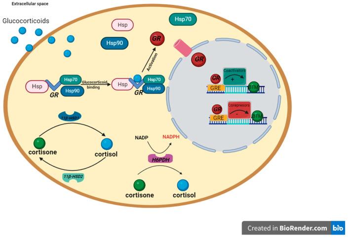
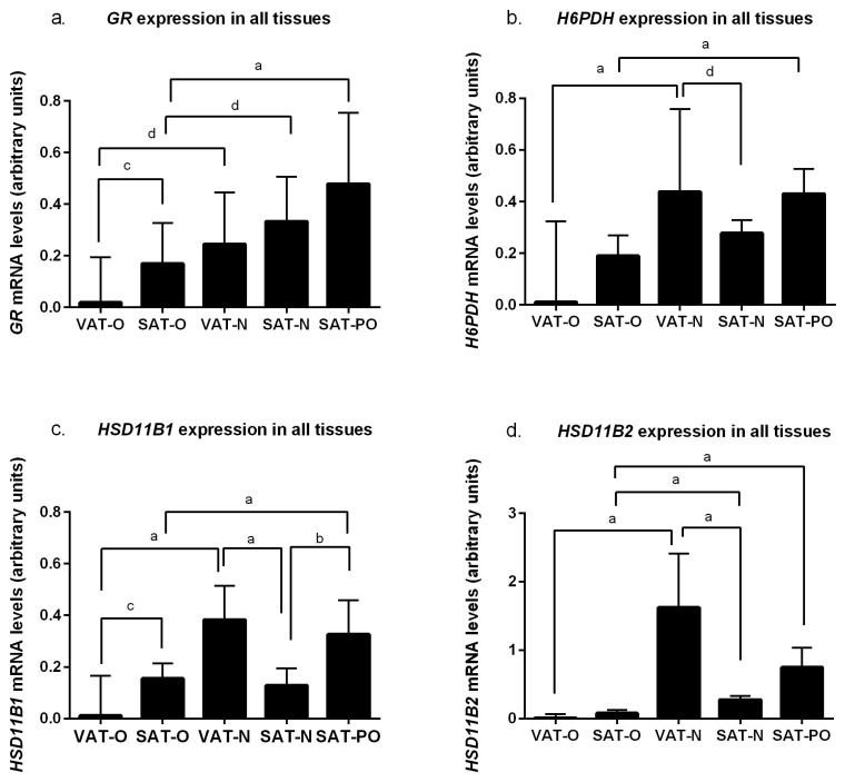
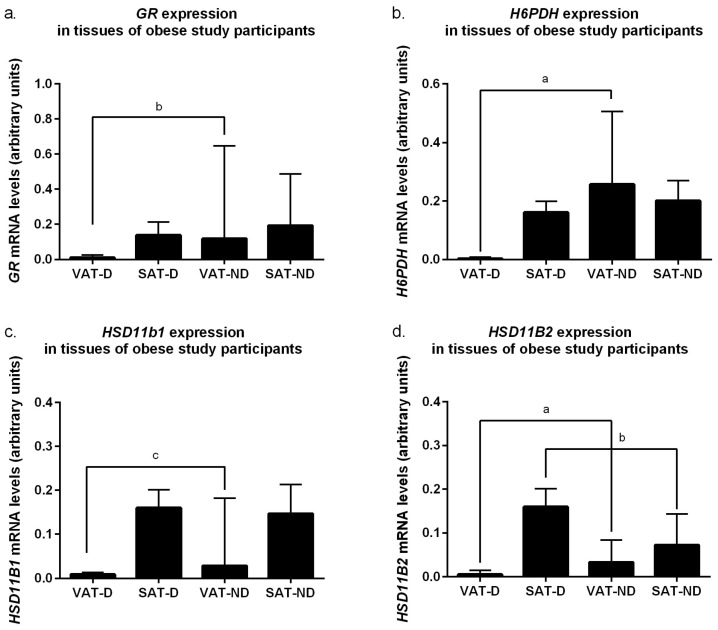
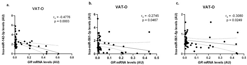
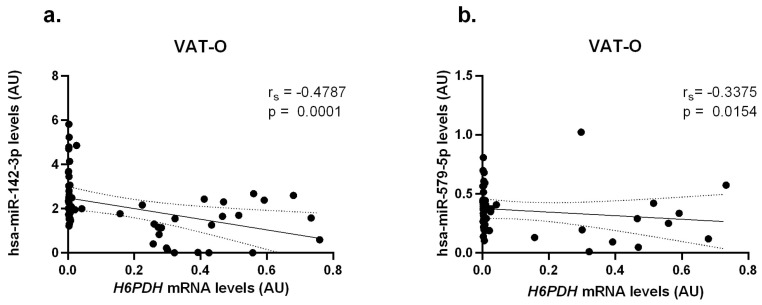
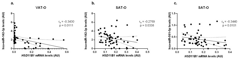
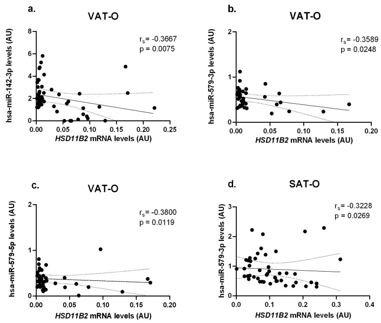
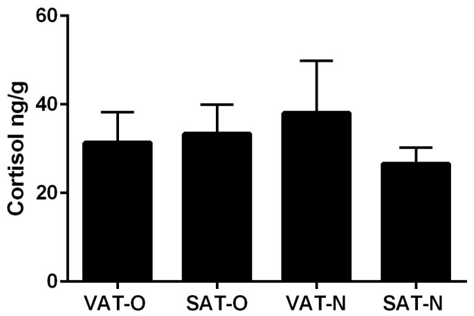
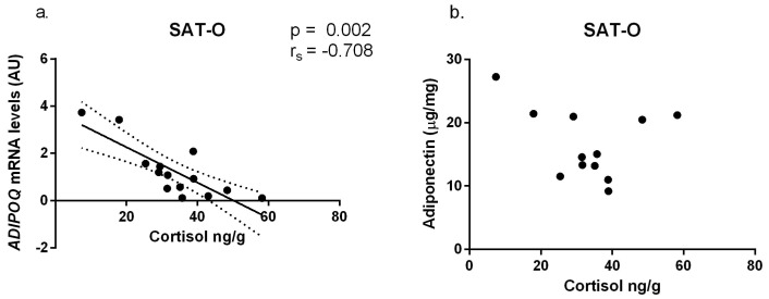
The similarity of the clinical picture of metabolic syndrome and hypercortisolemia supports the hypothesis that obesity may be associated with impaired expression of genes related to cortisol action and metabolism in adipose tissue. The expression of genes encoding the glucocorticoid receptor alpha (GR), cortisol metabolizing enzymes (HSD11B1, HSD11B2, H6PDH), and adipokines, as well as selected microRNAs, was measured by real-time PCR in adipose tissue from 75 patients with obesity, 19 patients following metabolic surgery, and 25 normal-weight subjects. Cortisol levels were analyzed by LC-MS/MS in 30 pairs of tissues. The mRNA levels of all genes studied were significantly (p < 0.05) decreased in the visceral adipose tissue (VAT) of patients with obesity and normalized by weight loss. In the subcutaneous adipose tissue (SAT), GR and HSD11B2 were affected by this phenomenon. Negative correlations were observed between the mRNA levels of the investigated genes and selected miRNAs (hsa-miR-142-3p, hsa-miR-561, and hsa-miR-579). However, the observed changes did not translate into differences in tissue cortisol concentrations, although levels of this hormone in the SAT of patients with obesity correlated negatively with mRNA levels for adiponectin. In conclusion, although the expression of genes related to cortisol action and metabolism in adipose tissue is altered in obesity and miRNAs may be involved in this process, these changes do not affect tissue cortisol concentrations.

Keywords: adipose tissue; cortisol metabolism; glucocorticoid receptor alpha; metabolic inflammation; microRNA; obesity.


Conflict of interest statement

The authors declare no conflicts of interest. The funders had no role in the design of the study; in the collection, analyses, or interpretation of data; in the writing of the manuscript; or in the decision to publish the results.

Figures

Figure 1
Figure 1
The simplified pathway of glucocorticoid receptor signaling and the action of enzymes involved in glucocorticoid metabolism in adipocytes, based on [9,10,11]. In the absence of glucocorticoid, the glucocorticoid receptor (GR) is located in the cytoplasm and forms a complex with chaperones (hsp90, p23, hsp70) as well as immunophillins (FKBP51, FKBP52). Thanks to the actions of these proteins, the GR is maintained in a transcriptionally inactive conformation, which in turn allows the binding of a ligand with high affinity to this receptor. The glucocorticoid receptor alpha (GRα) is the only active receptor for glucocorticoids in human adipose tissue [12]. Physiologically, cortisol is the most common glucocorticoid in humans. In its free, non-globulin-bound form, cortisol crosses the plasma membrane. The availability of this form of cortisol in the cell is controlled by enzymes that are antagonists to each other. 11β-hydroxysteroid dehydrogenase type 1 (11β-HSD1) is responsible for conversion of inactive cortisone to an active cortisol. In contrast, 11β-hydroxysteroid dehydrogenase type 2 (11β-HSD2) catalyzes the opposite reaction, i.e., oxidizes active cortisol into its inactive form [9]. If hexose-6-phosphate dehydrogenase (H6PDH) is present, the equilibrium can favor the activity of 11β-HSD1. H6PDH regenerates NADPH, which increases the activity of 11β-HSD1, and decreases the activity of 11β-HSD2. Once in the cell nucleus, GR binds to its responsive elements (GRE) and thus regulates the expression of target genes.
Figure 2
Figure 2
mRNA levels of genes encoding glucocorticoid receptor alpha (GR), (a); hexose-6-phosphate dehydrogenase (H6PDH), (b); 11β-hydroxysteroid dehydrogenase type 1 (HSD11B1), (c); and 11β-hydroxysteroid dehydrogenase type 2 (HSD11B2), (d) in the visceral (VAT) and subcutaneous (SAT) adipose tissues of the obese individuals, before (O) and after surgically induced weight loss (PO), and in the normal-weight subjects (N). Results normalized to β-actin (ACTB) mRNA concentration are presented as the median with the interquartile range. “a” p < 0.0001; “b” p < 0.001; “c” p < 0.01; “d” p < 0.05.
Figure 3
Figure 3
mRNA levels of genes encoding glucocorticoid receptor alpha (GR), (a); hexose-6-phosphate dehydrogenase (H6PDH), (b); 11β-hydroxysteroid dehydrogenase type 1 (HSD11B1), (c); and 11β-hydroxysteroid dehydrogenase type 2 (HSD11B2), (d) in the visceral (VAT) and subcutaneous (SAT) adipose tissues of obese individuals (O) diagnosed with type 2 diabetes (D) and without diabetes (ND). Results normalized to β-actin (ACTB) mRNA concentration are presented as the median with the interquartile range. “a” p < 0.0001; “b” p < 0.001; “c” p < 0.01.
Figure 4
Figure 4
Correlations between GR mRNA concentrations and hsa-miR-142-3p (a), hsa-miR-561-3p (b), and hsa-miR-561-5p (c) in the visceral adipose tissue of obese study participants (VAT-O). Black dots represent particular participants. Lines represent linear regression analysis with 95% CI interval.
Figure 5
Figure 5
Correlations between H6PDH mRNA concentrations and hsa-miR-142-3p (a) and hsa-miR-579-5p (b) in the visceral adipose tissue of obese study participants (VAT-O). Black dots represent particular participants. Lines represent linear regression analysis with 95% CI interval.
Figure 6
Figure 6
Correlations between HSD11B1 mRNA concentrations and hsa-miR-142-3p (a,b) and hsa-miR-561-5p (c) levels in the visceral (VAT) and subcutaneous (SAT) adipose tissues of obese study participants (O). Black dots represent particular participants. Lines represent linear regression analysis with 95% CI interval.
Figure 7
Figure 7
Correlations of HSD11B2 mRNA concentrations with hsa-miR-142-3p (a), hsa-miR-579-3p (b), and hsa-miR-579-5p (c) levels in the visceral (VAT) and with hsa-miR-579-3p (d) in the subcutaneous (SAT) adipose tissues of obese study participants (O). Black dots represent particular participants. Lines represent linear regression analysis with 95% CI interval.
Figure 8
Figure 8
Cortisol levels in the visceral (VAT) and subcutaneous (SAT) adipose tissues of obese individuals (O) and in normal-weight subjects (N). Results are presented as the median with the interquartile range.
Figure 9
Figure 9
Correlations of ADIPOQ mRNA (a) and adiponectin (b) concentrations with cortisol levels in subcutaneous adipose tissues of obese study participants (SAT-O). Black dots represent particular participants. Lines represent linear regression analysis with 95% CI interval. Adiponectin protein concentrations (µg) are normalized to the tissue protein content (mg).

Similar articles

References

    1. Li J., Papadopoulos V., Vihma V. Steroid biosynthesis in adipose tissue. Steroids. 2015;10:89–104. doi: 10.1016/j.steroids.2015.03.016. - DOI - PubMed
    1. Tchernof A., Mansour M.F., Pelletier M., Boulet M.M., Nadeau M., Luu-The V. Updated survey of the steroid-converting enzymes in human adipose tissues. J. Steroid Biochem. Mol. Biol. 2015;147:56–69. doi: 10.1016/j.jsbmb.2014.11.011. - DOI - PubMed
    1. Haines M.S. Endocrine complications of anorexia nervosa. J. Eat. Disord. 2023;11:24. doi: 10.1186/s40337-023-00744-9. - DOI - PMC - PubMed
    1. Pasquali R., Casanueva F., Haluzik M., van Hulsteijn L., Ledoux S., Monteiro M.P., Salvador J., Santini F., Toplak H., Dekkers O.M. European Society of Endocrinology Clinical Practice Guideline: Endocrine work-up in obesity. Eur. J. Endocrinol. 2020;182:G1–G32. doi: 10.1530/EJE-19-0893. - DOI - PubMed
    1. Goldsammler M., Merhi Z., Buyuk E. Role of hormonal and inflammatory alterations in obesity-related reproductive dysfunction at the level of the hypothalamic-pituitary-ovarian axis. Reprod. Biol. Endocrinol. 2018;16:45. doi: 10.1186/s12958-018-0366-6. - DOI - PMC - PubMed

MeSH terms

Substances