. 2024 Apr 26;13(5):527.
doi: 10.3390/antiox13050527.

Unravelling the Role of Candida albicans Prn1 in the Oxidative Stress Response through a Proteomics Approach

Affiliations

Unravelling the Role of Candida albicans Prn1 in the Oxidative Stress Response through a Proteomics Approach

Victor Arribas et al. Antioxidants (Basel). .

Abstract

Candida albicans Prn1 is a protein with an unknown function similar to mammalian Pirin. It also has orthologues in other pathogenic fungi, but not in Saccharomyces cerevisiae. Prn1 highly increases its abundance in response to H2O2 treatment; thus, to study its involvement in the oxidative stress response, a C. albicans prn1∆ mutant and the corresponding wild-type strain SN250 have been studied. Under H2O2 treatment, Prn1 absence led to a higher level of reactive oxygen species (ROS) and a lower survival rate, with a higher percentage of death by apoptosis, confirming its relevant role in oxidative detoxication. The quantitative differential proteomics studies of both strains in the presence and absence of H2O2 indicated a lower increase in proteins with oxidoreductase activity after the treatment in the prn1∆ strain, as well as an increase in proteasome-activating proteins, corroborated by in vivo measurements of proteasome activity, with respect to the wild type. In addition, remarkable differences in the abundance of some transcription factors were observed between mutant and wild-type strains, e.g., Mnl1 or Nrg1, an Mnl1 antagonist. orf19.4850, a protein orthologue to S. cerevisiae Cub1, has shown its involvement in the response to H2O2 and in proteasome function when Prn1 is highly expressed in the wild type.

Keywords: C. albicans; Cub1; Mnl1; Nrg1; Pirin; Prn1; apoptosis; mitochondria; oxidative stress response; proteasome; proteomics.


Conflict of interest statement

The authors declare no conflicts of interest.

Figures

Figure 1
Figure 1
C. albicans SN250 and prn1∆ cell death and growth in the presence of H2O2. (A) Percentage of propidium iodide (PI)-positive dead cells measured by flow cytometry after 200 min in the presence of 10 mM of H2O2. Results represent the average of three biological replicates. Error bars indicate standard deviation. * p < 0.05, unpaired t-test. (B) Growth curves of both strains in the presence of 6 mM and 7 mM of H2O2. The graph presents the most representative curve of three biological replicates.
Figure 2
Figure 2
Quantitative proteomics assay (DDA-MS) of SN250 and prn1∆ strains in response to 200 min of 10 mM H2O2 treatment. (A) Workflow of quantitative proteomics assay (DDA-MS) to compare SN250 and prn1∆ strains after 200 min 10 mM H2O2 treatment. After cell disruption, protein cell extracts of each strain are digested and quantified; later, peptide samples are analyzed in the mass spectrometer. Images were created with BioRender. (B) Number of quantified proteins and proteins showing significant differences in abundance between the treated and non-treated (control) conditions for each strain. (C) Volcano plots representing proteins with significant changes in abundance. Significant changes in the protein abundance (−log10 q-value > 1.3) after treatment are presented in red for increase or green for decrease. (D) Venn diagram showing common and non-common proteins with significant changes in abundance between strains in response to the treatment.
Figure 3
Figure 3
Heat maps of proteins that changed in abundance after 200 min of 10 mM H2O2 treatment in each strain with respect to the control condition grouped by GO term. (A) Oxidoreductase, (B) protein catabolic process, and (C) translation. Gray gaps indicate proteins not detected in that strain.
Figure 4
Figure 4
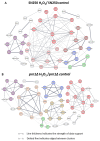
Predicted networks of proteins with increased abundance after H2O2 treatment in the SN250 and prn1∆ strains using STRING software. Line thickness indicates the strength of supporting data. Dotted lines indicate edges between clusters. (A) Clusters: oxidoreductase function (red, dark red, and light red), pre-ribosome and ribosome biogenesis (purple), dehydrogenase complex of the respirasome (blue), and amino acid biosynthesis (green). (B) Clusters: oxidative stress response (red), proteasome regulatory subcomplex (orange), heat shock response (gold), ribosome biosynthesis (purple), mitochondrial oxidoreductase complex (blue), and Rho GTPase regulation (green).
Figure 5
Figure 5
Comparative proteomics analysis between the SN250 and prn1∆ strains in the control condition and after 200 min of 10 mM H2O2 treatment. (A) Number of quantified proteins and those with differences in abundance for each strain. (B) Volcano plots representing proteins with a significantly different abundance between the two strains in each condition. Significant changes in the protein abundance (−log10 q-value > 1.3) are presented in red for prn1Δ strain or green for wild-type SN250 strain.
Figure 6
Figure 6
Effect of Prn1 deletion on cell redox homeostasis, apoptosis, and proteasome activity. (A) Intracellular ROS levels in the SN250 and prn1∆ strains after 200 min of 10 mM H2O2 treatment using the dihydrorhodamine 123 (DHR123) probe. (B) Phosphatidylserine (PS) externalization levels after 50 min of 10 mM H2O2 treatment using Annexin V staining. (C) Chymotrypsin-like proteasome activity after 200 min of 10 mM H2O2 treatment. Results represent the averages of three biological replicates. Error bars indicate standard deviation. * p < 0.05, ** p < 0.01, and *** p < 0.001, unpaired t-test.
Figure 7
Figure 7
Role of the C. albicans protein orthologue to S. cerevisiae Cub1 in the oxidative stress response after H2O2 exposure. (A) Drop growth assay of C. albicans SC5314, prn1∆/PRN1, and cub1∆/CUB1 strains treated with 80 mM of H2O2 for different time intervals. (B) Percentage of PI-positive death cells measured by flow cytometry after 200 min in the presence of 10 mM of H2O2. (C) Chymotrypsin-like proteasome activity after 200 min 10 mM H2O2 treatment. Results represent the averages of three biological replicates. Error bars indicate standard deviation * p < 0.05 and ** p < 0.01, unpaired t-test.

Similar articles

References

    1. Tsui C., Kong E.F., Jabra-Rizk M.A. Pathogenesis of Candida albicans Biofilm. Pathog. Dis. 2016;74:ftw018. doi: 10.1093/FEMSPD/FTW018. - DOI - PMC - PubMed
    1. Talapko J., Juzbašić M., Matijević T., Pustijanac E., Bekić S., Kotris I., Škrlec I. Candida albicans—The Virulence Factors and Clinical Manifestations of Infection. J. Fungi. 2021;7:79. doi: 10.3390/JOF7020079. - DOI - PMC - PubMed
    1. Denning D.W. Global Incidence and Mortality of Severe Fungal Disease. Lancet Infect. Dis. 2024;ahead of print doi: 10.1016/S1473-3099(23)00692-8. - DOI - PubMed
    1. Parums D.V. Editorial: The World Health Organization (WHO) Fungal Priority Pathogens List in Response to Emerging Fungal Pathogens during the COVID-19 Pandemic. Med. Sci. Monit. 2022;28:e939088-1–e939088-3. doi: 10.12659/MSM.939088. - DOI - PMC - PubMed
    1. Yang Z.H., Song Y.G., Li R.Y. A Ten-Year Retrospective Study of Invasive Candidiasis in a Tertiary Hospital in Beijing. Biomed. Environ. Sci. 2021;34:773–788. doi: 10.3967/BES2021.107. - DOI - PubMed

LinkOut - more resources