Review
. 2024 May 7;9(5):278.
doi: 10.3390/biomimetics9050278.

Biomimetic Materials for Skin Tissue Regeneration and Electronic Skin

Affiliations
Review

Biomimetic Materials for Skin Tissue Regeneration and Electronic Skin

Sol Youn et al. Biomimetics (Basel). .

Abstract

Biomimetic materials have become a promising alternative in the field of tissue engineering and regenerative medicine to address critical challenges in wound healing and skin regeneration. Skin-mimetic materials have enormous potential to improve wound healing outcomes and enable innovative diagnostic and sensor applications. Human skin, with its complex structure and diverse functions, serves as an excellent model for designing biomaterials. Creating effective wound coverings requires mimicking the unique extracellular matrix composition, mechanical properties, and biochemical cues. Additionally, integrating electronic functionality into these materials presents exciting possibilities for real-time monitoring, diagnostics, and personalized healthcare. This review examines biomimetic skin materials and their role in regenerative wound healing, as well as their integration with electronic skin technologies. It discusses recent advances, challenges, and future directions in this rapidly evolving field.

Keywords: E-skin; biomimetic; diatom; nanostructure; nature inspired; real-time monitoring; smart bandage; wound dressing; wound healing.


Conflict of interest statement

The authors declare no conflicts of interest.

Figures

Figure 1
Figure 1
Schematic of the stages of wound healing. It goes through four phases: hemostasis, inflammation, tissue regrowth, and remodeling. The processes that occur at each phase are distinct. Reproduced with permission from [3], Copyright 2023 Frontiers (CC BY 4.0).
Figure 2
Figure 2
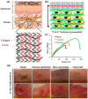
Wound healing mimicking skin. (a) Tissue morphology mimicking skin. (b) Layer-by-layer (L-b-L) cell assembly structure of nanofiber-based skin substitute. (c) Representative stress staining curves of cultured skin substitutes. (d) Full-thickness wound healed with graft over 21 days. (a): Reproduced with permission from [85], Copyright 2020 Elsevier Ltd. (bd): Reproduced with permission from [86], Copyright 2015 Elsevier Ltd.
Figure 3
Figure 3
(a) Schematic of 3D composite scaffold mimicking skin structure. (b) Gap closure in monolayer cells of control and composite scaffolds (PS3) in culture Petri dishes. (a,b): Reproduced with permission from [88], Copyright 2024 Elsevier B.V.
Figure 4
Figure 4
Image of antibacterial mimicking skin. (a) Structure of a double-layer antibacterial scaffold mimicking the epidermis and dermis of the skin. (b) Viability of S. aureus on scaffolds. (c) Morphological changes of S. aureus, E. coli, and MRSA in nanofiber matrix cultures (red arrows indicate morphological changes in the bacterial cell membrane). (d) S. aureus, E. coli, and MRSA colony matrix treated with nanofibers. * p < 0.05 and ** p < 0.01. (a,b): Reproduced with permission from [90], Copyright 2023 American Chemical Society (CC BY 4.0). (c,d): Reproduced with permission from [91], Copyright 2018 American Chemical Society.
Figure 5
Figure 5
(a) Schematic of a nanostructured scaffold with biomimetic and antibacterial properties that mimics ECM. (b) Degree of bacterial inhibition by the nanostructure support. (a,b): (A) drug-free GL, (B) GL/gentamycin(GS)2.5, (C) GL/GS5, (D) GL/GS7.5, (E) GL/GS10, (F) drug-free GL, (G) GL/AgNPs2.5 and (H) GL/AgNPs5. Reproduced with permission from [93], Copyright 2018 Elsevier B.V.
Figure 6
Figure 6
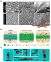
Wound care techniques mimicking animals. (a) SEM images of a lizard’s skin surface, spines, and overall view of the skin surface (top) and a replica lizard skin with a surface made of a bilayer of chitosan and alginate, spines, and ruptured bacterial debris (bottom). (b) Tissue adhesion mechanism of CoSt hydrogel and (c) shape of the hydrogel when bent or twisted. (a): Reproduced with permission from [102], Copyright 2017 Springer Nature. (b,c): Reproduced with permission from [103], Copyright 2022 Wiley-VCH GmbH.
Figure 7
Figure 7
Schematic diagram of inspired mussel and bee hair crevice patches. Reproduced with permission from [109], Copyright 2023 Wiley-VCH GmbH.
Figure 8
Figure 8
Wound management technology mimicking plants. (a) Lotus leaf on hydrophobic surface. (b) Schematic diagram of the asymmetric composite dressing and (c) contact angle image of the dressing layer (right) (a): Reproduced with permission from [112] Copyright 2011 Beilstein-Institut (CC BY 4.0). (b,c): Reproduced with permission from [113], Copyright 2022 American Chemical Society.
Figure 9
Figure 9
Wound management technology mimicking nanostructures. (a) Schematic diagram of biomimetic nanovesicles. (b) Bacterial biofilm and anti-inflammatory therapy of biomimetic nanozyme and IL-4 release mechanism. (c) Schematic diagram of the biomimetic dual nanozyme and (d) regeneration in diabetic infected wounds. (a): Reproduced with permission from [122], Copyright 2023 Elsevier B.V. (b): Reproduced with permission from [123], Copyright 2023 Wiley-VCH GmbH. (c,d): Reproduced with permission from [124], Copyright 2023 Elsevier Inc.
Figure 10
Figure 10
Diatom-mimicking wound care technology. (a) Schematic of a diatom-mimicking polysaccharide adhesive and its adhesive properties (b) in air and (c) in water with biological tissues. (ac): Reproduced with permission from [133], Copyright 2023 American Chemical Society.
Figure 11
Figure 11
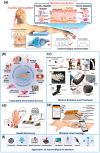
(a) Human tactile perception system. (bf) Various applications of E-skin. (b) Overview of an implantable biomedical device based on Triboelectric Nanogenerators (TENGs). (c) Applications of flexible strain sensors in medical robotics and prosthetics. (d) Real-time bio- and health signal monitoring. (e) Overall design of the wireless smart bandage for chronic wound management. (f) Application of the human–machine interface (a): Reproduced with permission from [159], Copyright 2019 Wiley-VCH Verlag GmbH & Co. KGaA, Weinheim. (b): Reproduced with permission from [160] (CC BY 4.0). (c): Reproduced with permission from [161], Copyright 2022 John Wiley & Sons. (d): Reproduced with permission from [153], (CC BY 4.0). (e): Reproduced with permission from [162], (CC BY 4.0). (f): Reproduced with permission from [163], Copyright 2022 OAE Publishing Inc. (OAE) (CC BY 4.0).
Figure 12
Figure 12
(a) Schematic of the materials and structures of conducting materials. (b) SEM photos of MXenes: accordion-like unexfoliated MXene, MXene nanosheet, and intercalated MXene nanocomposite film and optical photo of the flexible intercalated MXene nanocomposite film. (c) Schematic representation of a piezoelectric and triboelectric nanogenerator on operating on injured skin. (a): Reproduced with permission from [175], Copyright 2019 Wiley-VCH Verlag GmbH & Co. KGaA, Weinheim. (b): Reprinted with permission from [177], Copyright 2022 Elsevier Inc. (c): Reproduced with permission from [79] (CC BY 4.0).

Similar articles

Cited by

References

    1. Dhivya S., Padma V.V., Santhini E. Wound dressings—A review. BioMedicine. 2015;5:22. doi: 10.7603/s40681-015-0022-9. - DOI - PMC - PubMed
    1. Singh S., Young A., McNaught C.-E. The physiology of wound healing. Surgery. 2017;35:473–477. doi: 10.1016/j.mpsur.2017.06.004. - DOI
    1. Hunt M., Torres M., Bachar-Wikstrom E., Wikstrom J.D. Multifaceted roles of mitochondria in wound healing and chronic wound pathogenesis. Front. Cell Dev. Biol. 2023;11:1252318. doi: 10.3389/fcell.2023.1252318. - DOI - PMC - PubMed
    1. Ren H., Zhao F., Zhang Q., Huang X., Wang Z. Autophagy and skin wound healing. Burn. Trauma. 2022;10:tkac003. doi: 10.1093/burnst/tkac003. - DOI - PMC - PubMed
    1. Dorgalaleh A., Daneshi M., Rashidpanah J., Roshani Yasaghi E. An Overview of Hemostasis. In: Dorgalaleh A., editor. Congenital Bleeding Disorders. Springer; Cham, Switzerland: 2018. pp. 3–26.

LinkOut - more resources