Review
. 2024 Apr 26;13(5):298.
doi: 10.3390/biology13050298.

Connexin Gap Junction Channels and Hemichannels: Insights from High-Resolution Structures

Affiliations
Review

Connexin Gap Junction Channels and Hemichannels: Insights from High-Resolution Structures

Maciej Jagielnicki et al. Biology (Basel). .

Abstract

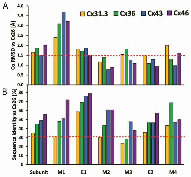
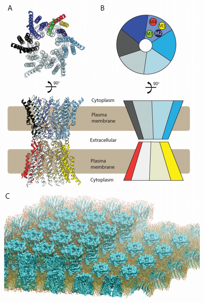
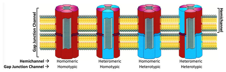
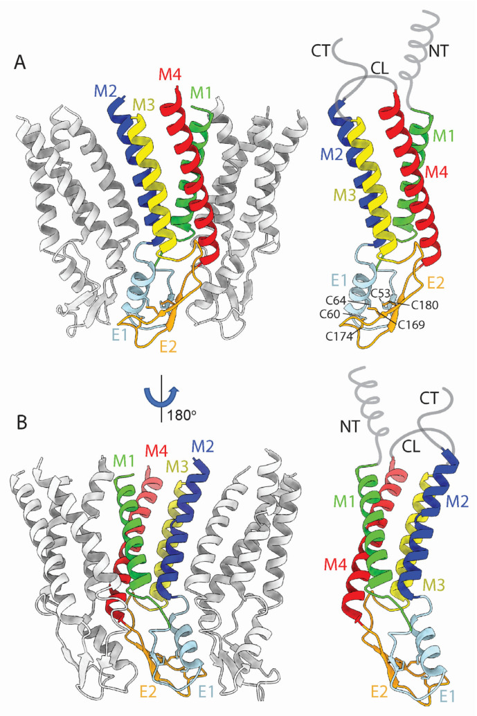
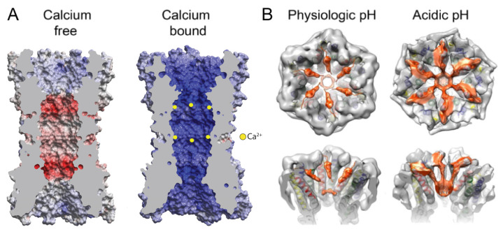
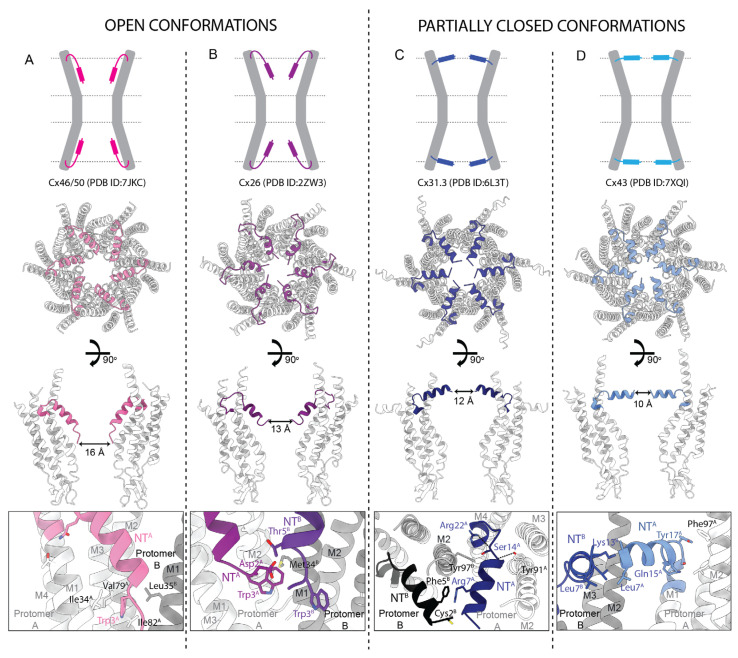
Connexins (Cxs) are a family of integral membrane proteins, which function as both hexameric hemichannels (HCs) and dodecameric gap junction channels (GJCs), behaving as conduits for the electrical and molecular communication between cells and between cells and the extracellular environment, respectively. Their proper functioning is crucial for many processes, including development, physiology, and response to disease and trauma. Abnormal GJC and HC communication can lead to numerous pathological states including inflammation, skin diseases, deafness, nervous system disorders, and cardiac arrhythmias. Over the last 15 years, high-resolution X-ray and electron cryomicroscopy (cryoEM) structures for seven Cx isoforms have revealed conservation in the four-helix transmembrane (TM) bundle of each subunit; an αβ fold in the disulfide-bonded extracellular loops and inter-subunit hydrogen bonding across the extracellular gap that mediates end-to-end docking to form a tight seal between hexamers in the GJC. Tissue injury is associated with cellular Ca2+ overload. Surprisingly, the binding of 12 Ca2+ ions in the Cx26 GJC results in a novel electrostatic gating mechanism that blocks cation permeation. In contrast, acidic pH during tissue injury elicits association of the N-terminal (NT) domains that sterically blocks the pore in a "ball-and-chain" fashion. The NT domains under physiologic conditions display multiple conformational states, stabilized by protein-protein and protein-lipid interactions, which may relate to gating mechanisms. The cryoEM maps also revealed putative lipid densities within the pore, intercalated among transmembrane α-helices and between protomers, the functions of which are unknown. For the future, time-resolved cryoEM of isolated Cx channels as well as cryotomography of GJCs and HCs in cells and tissues will yield a deeper insight into the mechanisms for channel regulation. The cytoplasmic loop (CL) and C-terminal (CT) domains are divergent in sequence and length, are likely involved in channel regulation, but are not visualized in the high-resolution X-ray and cryoEM maps presumably due to conformational flexibility. We expect that the integrated use of synergistic physicochemical, spectroscopic, biophysical, and computational methods will reveal conformational dynamics relevant to functional states. We anticipate that such a wealth of results under different pathologic conditions will accelerate drug discovery related to Cx channel modulation.

Keywords: X-ray crystallography; calcium regulation; channel gating; connexin; electron cryomicroscopy; gap junction channel; gap junction hemichannel; lipid binding; pH regulation.


Conflict of interest statement

The authors declare no conflicts of interest.

Figures

Figure 5
Figure 5
Comparison of structural and sequence homology of Cx isoforms. (A) Cα RMSD-based Cx subunit comparison, with Cx26 [13] (PDB: 5ER7) as a reference structure. The analyses include Cx31.3 [36] (PDB: 6L3T), Cx36 [38] (PDB: 7XNH), Cx43 [39] (PDB: 7F92), and Cx46 [41] (PDB: 7JKC). Dashed orange line denotes Cα RMSD value of 1.5 Å, below which structures are considered identical. Protein alignment performed on Cα atoms in PyMOL [46] using command “super” with parameter “cycles = 0”; RMSD calculation performed on Cα atom pairs in UCSF Chimera [47] using “rmsd” command. (B) Comparison of Cx subunits based on sequence identity, with Cx26 [13] (PDB: 5ER7) as a reference structure. Dashed orange line corresponds to sequence identity of 30%, below which subunits are considered significantly different in amino acid sequence. Analysis was performed with Clustal Omega [48] and includes Cxs sequences listed in (B).
Figure 8
Figure 8
Interactions between Cx channels and bound lipids as revealed by cryoEM. Central panels depict a pair of top and side cutaway views for four representative Cx structures ((A) Cx46/50 (PDB ID:7JKC), (B) Cx43 (PDB ID:7XQF), (C) Cx36 (PDB ID:7XKT), and (D) Cx36 (PDB ID:7XNV)) with interacting lipids depicted in yellow (acyl chains), orange (CHS), purple (POPE), and navy (PG). Red circles denote details of interactions between lipids and Cx protomers; dark grey indicates lipid density; white and light blue two adjacent Cx protomers.
Figure 1
Figure 1
Gap junction structure, nomenclature, and plaque assembly in the membrane. (A) Top and side view of the Cx26 gap junction channel (PDB ID:5ER7). The hemichannel is comprised of 6 protomers, and hemichannels dock end-to-end between apposed cells to form the intercellular wide-pore channel. (B) Schematic representation of a gap junction channel. M1–M4 α-helices are colored green (M1), navy (M2), yellow (M3), and red (M4) in the upper panels of (A,B). Individual protomers are colored in different shades of blue for one hexamer, while the protomers of the hexamer from the adjacent cell are colored from yellow to red. (C) Representation of a gap junction plaque comprising gap junction channels that pack with quasi-hexagonal symmetry.The Cx gene family is diverse, with 21 identified members in the sequenced human genome, and 20 in the mouse (19 of which are orthologs with human Cx). Connexins are commonly named according to their molecular weights (e.g., Cx26 for the 26 kDa isoform), and their molecular masses range between 25 and 60 kDa. Alternatively, Cxs are also classified into five families based on their sequence homology—α, β, γ, δ, and ε, followed by an identifying number (e.g., GJA1 refers to Cx43) [5].
Figure 2
Figure 2
Oligomeric states of gap junction channels and hemichannels assembled from two isoforms (red and blue). HCs can exist as homomeric or heteromeric hexamers, whereas GJCs can exist as homotypic or heterotypic dodecamers. (Adapted from [9]).
Figure 3
Figure 3
Cx protomer structure in the context of the Cx26 hemichannel (PDB ID: 5ER7 [13]). (A) depicts a view within the lipid bilayer showing the perimeter M3 and M4 α-helices of the protomer. (B) depicts a view from the center of the hemichannel showing the pore-lining M1 and M2 α-helices of the protomer. All connexins are predicted to have a topological structure similar to that of Cx26, with four transmembrane domains (M1–M4), two extracellular loops (E1 and E2), a cytoplasmic loop (CL), an amino terminal (NT), and a carboxyl terminal (CT) domain. The positions of disulfide bonds between E1 and E2 are indicated in the right panel of (A). Grey lines represent structurally unresolved CL and CT domains. A region of NT may fold as an α-helix that can have multiple positions.
Figure 4
Figure 4
Docking between hemichannels is the raison d’être of gap junction channels. The subunits in the side view are labelled A to F and A′ to F′, each in the same color. The enlarged views show the hydrogen bonding interactions in E1 (top) and E2 (bottom) that stabilize the dodecameric gap junction channel (Adapted from [30]).
Figure 6
Figure 6
Gating mechanisms of gap junction channels during tissue injury, accompanied by Ca2+ overload and acidic pH. (A) Binding of 12 Ca2+ ions results in a channel pore that is highly electropositive, resulting in the electrostatic block of cation permeation such as K+ [13]. Electrostatic potential surfaces with positive and negative electrostatic potentials are shown in blue and red, respectively (color scale is −15 to +15 kTe−1). The protein interior is grey. Yellow spheres indicate that Ca2+ ions bind between adjacent subunits in each hemichannel. Hemispheric binding at the TMD/ECD interface directly exposes the Ca2+ ions to the aqueous pore, thereby maximizing their electrostatic positivity. (B) Similarly, acidic pH results in steric block of the channel pore by a “ball-and-chain” mechanism, in which the ball is composed of the NT domains. (Adapted from [13,14]).
Figure 7
Figure 7
Distinct conformations of the NT domains resolved in the cryoEM structures of Cx channels. Upper panels—schematic representation of open (A,B) and partially closed (C,D) NT conformations. Central panels—a pair of top and side cutaway views for four corresponding Cx structures with differing NT conformations ((A) Cx46/50 (PDB ID:7JKC), (B) Cx26 (PDB ID:2ZW3), (C) Cx31.3 (PDB ID:6L3T), (D) Cx43 (PDB ID:7XQI)); NTs shown in color, M1–M4 and E1–E2 shown in grey; narrowest pore diameter indicated. Bottom panels—selected details of NT interactions. References: Cx26 [30], Cx31.3 [36], Cx36 [38], Cx43 [39] and Cx46 [41].

Similar articles

Cited by

References

    1. Lucaciu S.A., Leighton S.E., Hauser A., Yee R., Laird D.W. Diversity in connexin biology. J. Biol. Chem. 2023;299:105263. doi: 10.1016/j.jbc.2023.105263. - DOI - PMC - PubMed
    1. Revel J.P., Karnovsky M.J. Hexagonal array of subunits in intercellular junctions of the mouse heart and liver. J. Cell Biol. 1967;33:C7–C12. doi: 10.1083/jcb.33.3.c7. - DOI - PMC - PubMed
    1. Lampe P.D., Laird D.W. Recent advances in connexin gap junction biology. Fac. Rev. 2022;11:14. doi: 10.12703/r/11-14. - DOI - PMC - PubMed
    1. Harris A.L. Connexin channel permeability to cytoplasmic molecules. Prog. Biophys. Mol. Biol. 2007;94:120–143. doi: 10.1016/j.pbiomolbio.2007.03.011. - DOI - PMC - PubMed
    1. Söhl G., Willecke K. An update on connexin genes and their nomenclature in mouse and man. Cell Commun. Adhes. 2003;10:173–180. doi: 10.1080/cac.10.4-6.173.180. - DOI - PubMed