. 2024 May;105(5):001994.
doi: 10.1099/jgv.0.001994.

Role of N-linked glycosylation in porcine reproductive and respiratory syndrome virus (PRRSV) infection

Affiliations

Role of N-linked glycosylation in porcine reproductive and respiratory syndrome virus (PRRSV) infection

Raymond R R Rowland et al. J Gen Virol. 2024 May.

Abstract

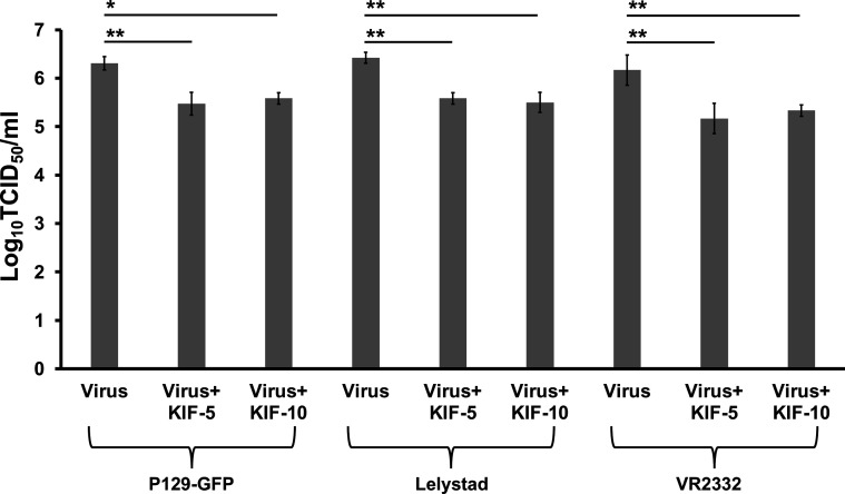
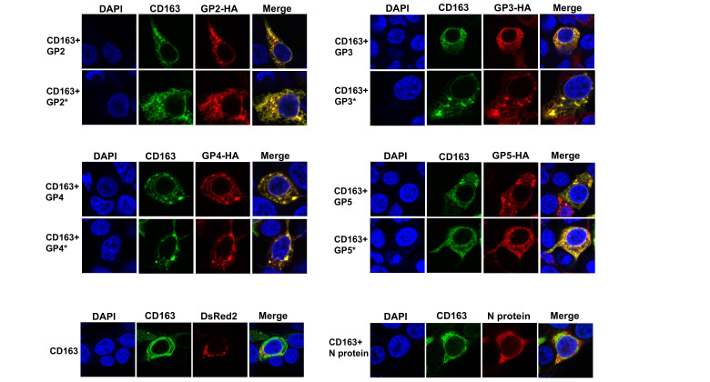
Porcine reproductive and respiratory syndrome (PRRSV) is an enveloped single-stranded positive-sense RNA virus and one of the main pathogens that causes the most significant economical losses in the swine-producing countries. PRRSV is currently divided into two distinct species, PRRSV-1 and PRRSV-2. The PRRSV virion envelope is composed of four glycosylated membrane proteins and three non-glycosylated envelope proteins. Previous work has suggested that PRRSV-linked glycans are critical structural components for virus assembly. In addition, it has been proposed that PRRSV glycans are implicated in the interaction with host cells and critical for virus infection. In contrast, recent findings showed that removal of N-glycans from PRRSV does not influence virus infection of permissive cells. Thus, there are not sufficient evidences to indicate compellingly that N-glycans present in the PRRSV envelope play a direct function in viral infection. To gain insights into the role of N-glycosylation in PRRSV infection, we analysed the specific contribution of the envelope protein-linked N-glycans to infection of permissive cells. For this purpose, we used a novel strategy to modify envelope protein-linked N-glycans that consists of production of monoglycosylated PRRSV and viral glycoproteins with different glycan states. Our results showed that removal or alteration of N-glycans from PRRSV affected virus infection. Specifically, we found that complex N-glycans are required for an efficient infection in cell cultures. Furthermore, we found that presence of high mannose type glycans on PRRSV surface is the minimal requirement for a productive viral infection. Our findings also show that PRRSV-1 and PRRSV-2 have different requirements of N-glycan structure for an optimal infection. In addition, we demonstrated that removal of N-glycans from PRRSV does not affect viral attachment, suggesting that these carbohydrates played a major role in regulating viral entry. In agreement with these findings, by performing immunoprecipitation assays and colocalization experiments, we found that N-glycans present in the viral envelope glycoproteins are not required to bind to the essential viral receptor CD163. Finally, we found that the presence of N-glycans in CD163 is not required for PRRSV infection.

Keywords: N-glycosylation; PRRSV; viral envelope glycoproteins; viral infectivity.


Conflict of interest statement

The authors declare that there are no conflicts of interest.

Figures

Fig. 1.
Fig. 1.. Preparation of monoglycosylated virus. (a) Schematic overview of monoglycosylated virus production and viral glycoproteins with different glycan states. Wild-type (WT) virus with the typical complex and hybrid type N-glycans, high mannose virus containing high mannose type N-glycans, and monoglycosylated virus containing N-acetyl glucosamine (GlcNAc) are shown. (b) Effect of N-glycosylation inhibition on expression and electrophoretic mobility of viral envelope GP5. Western blot of GP5 expression levels of PRRSV-2 (VR-3223 isolate) propagated in MARC-145 cells with KIF at 5–10 µg ml−1 and digested with Endo H or PNGase F. Proteins bands were detected with an anti-GP5 antibody. Densitometry of GP5 bands is shown below the immunoblot. Fold changes are shown relative to lane 1 (virus) and data are mean values± standard deviation (SD) of three independent experiments (NS, not significant). (c) MARC-145 cells were infected with different dilutions of P129-GFP virus (starting MOI=1) in absence or presence of KIF at 5–10 µg ml−1. At 72 h post-infection (hpi), the cells were fixed and visualized under a fluorescence microscope. Similar results were obtained in three independent experiments and representative data is shown. (d) P129-GFP virus (MOI=0.1) was propagated in MARC-145 cells in absence or presence of KIF at 5–10 µg ml−1 for 72 h. Cell supernatants were collected at indicated time points to measure the released viral particles by TCID50 analysis. The TCID50 was calculated by titration on MARC-145 cells. Results are means±standard deviation (SD) values from three independent experiments (*, P<0.05; **, P<0.005; NS, not significant). (e) Effect of Endo H digestion on PRRSV-2 infection. P129-GFP propagated in absence or presence of KIF at 5–10 µg ml−1 for 3 days was treated with Endo H at 37 °C for 1 h. Different dilutions of virus–enzyme mixtures (starting MOI=1) were inoculated onto MARC-145 cells at 37 °C for 1 h. After infection, cells were washed to remove enzyme and unbound virus, and further incubated for 72 h. Virus without enzyme treatment was incubated at 37 °C for 1 h to serve as a temperature stability control. To control for an enzyme effect on cells, Endo H incubated alone at 37 °C for 1 h was mixed with the temperature-stability control virus and the mixture was added to the cells. Cells were then fixed and visualized under a fluorescence microscope. (f) Effect of KIF on PRRSV-1 infection. MARC-145 cells were infected with different dilutions of Lelystad virus (starting MOI=1) in absence or presence of KIF at 5–10 µg ml−1. At 72 hpi, the cells were fixed and stained with PRRSV N protein antibody, followed by Alexa 488-goat anti-mouse IgG (green). Similar results were obtained in three separate experiments and representative data is shown. (g) Lelystad virus (MOI=0.1) was propagated in MARC-145 cells in absence or presence of KIF at 5–10 µg ml−1 for 3 days. Cell supernatants were collected at indicated time points to measure the released viral particles by TCID50 analysis. The TCID50 was calculated as indicated in Fig. 1d. Results are means±standard deviation (SD) values from three independent experiments (*, P<0.05; NS, not significant). (h) Effect of Endo H digestion on PRRSV-1 infection. Lelystad virus propagated in absence or presence of KIF at 5–10 µg ml−1 for 3 days was treated with Endo H at 37 °C for 1 h. Different dilutions of virus–enzyme mixtures (starting MOI=1) were inoculated onto MARC-145 cells at 37 °C for 1 h. After infection, cells were washed to remove enzyme and unbound virus, and further incubated for 72 h. Virus without enzyme treatment was incubated at 37 °C for 1 h to serve as a temperature stability control. To control for an enzyme effect on cells, Endo H incubated alone at 37 °C for 1 h was mixed with the temperature-stability control virus and the mixture was added to the cells. Cells were fixed and stained as indicated above. Similar results were obtained in three separate experiments and representative data is shown. (i) Effect of Endo H digestion on VR2332 infection. The same as (e) but using a VR2332 isolate. Cells were fixed and stained as indicated above. Similar results were obtained in three separate experiments and representative data is shown.
Fig. 2.
Fig. 2.. Effect of KIF on PRRSV infection of macrophages. (a) PAMs were infected with different dilutions of P129-GFP virus, Lelystad virus or VR2332 virus (starting MOI=1) propagated with KIF at 5–10 µg ml−1. At 24 hpi, the cells were fixed and stained as indicated in Fig. 1. P129-GFP infection was visualized directly without antibodies. Virus litres were calculated by titration assay. Results are means±standard deviation (SD) values from three independent experiments (*, P<0.05; **, P<0.005; NS, not significant).
Fig. 3.
Fig. 3.. Effect of N-glycosylation inhibition of CD163 on PRRSV infection. (a) Schematic representation of the N-glycosylation sites of porcine CD163. Ovals and squares identify the SRCR and PST domains, respectively. The cylinder is the transmembrane domain. Seven potential N-glycosylation sites in CD163 are indicated. The N-glycosylation sites were predicted with the NetNGlyc-1.0 server [94]. (b) Left panels. HEK293T cells were transfected with plasmids expressing either CD163-FLAG or a N-glycosylation-deficient CD163 (CD163-FLAG*) and digested with PNGase F. CD163 was detected with anti-FLAG antibody. Middle panels. HEK293T cells were transfected with a plasmid expressing CD163 and treated with tunicamycin (TM) for 16 h before harvesting or digested with PNGase F. CD163 was detected with anti-FLAG antibody. Right panels. Western blots of CD163 expression levels after kifunensine (KIF) treatment for 16 h. Cell lysates were digested with Endo H and immunoblotted with anti-FLAG antibody. In all experiments, GAPDH was used as a loading control. Densitometry of CD163 bands is normalized to the loading control GAPDH. Fold changes are shown relative to empty vector (pCDNA3.1), and results are means±standard deviation (SD) values from three independent experiments (*, P<0.05; **, NS, not significant). CD163-FLAG* refers to N-glycosylation sites removed by replacing Asn with Gln. (c) Western blotting of CD163 expression levels in PAMs cells following N-glycosidases treatment. Cell lysates were digested with digested with Endo H or PNGase F and immunoblotted with anti-CD163 antibody. Bottom panel shows actin loading control. (d) Representative fluorescence images of PRRSV infectivity in HEK293T cells transiently expressing wild-type (WT) or the N-glycosylation-deficient CD163. HEK293T cells were transfected with plasmids expressing mutant or WT CD163 proteins. At 24 h post-transfection, the cells were infected with PRRSV-1 (Lelystad) or PRRSV-2 (VR2332) at MOI of 5 (high MOI) or 0.1 (low MOI). To visualize PRRSV infection, the infected cells were fixed and stained at 72 hpi as in Fig. S5. WT and mutant CD163 proteins were visualized by staining cells with anti-FLAG antibody and then with Alexa 488-goat anti-rabbit IgG (green). Percentages of infected cells for each CD163 variant are shown on the right. Results are means±standard deviation (SD) values from three independent experiments (NS, not significant). (e) Expression of the CD163 N-glycosylation mutant was analysed by Western blotting using anti-FLAG antibody and GAPDH as a loading control. Similar results were obtained in three independent experiments and representative data is shown.
Fig. 4.
Fig. 4.. Effect of Endo H digestion on viral infection of PAMs. P129-GFP (a), Lelystad (b) and VR2332 (c) isolates propagated in absence or presence of KIF at 5–10 µg ml−1 in MARC-145 cells for 3 days were digested with Endo H at 37 °C for 1 h. Different dilutions of virus–enzyme mixtures (starting MOI=1) were inoculated onto PAMs at 37 °C for 1 h. After infection, cells were washed to remove enzyme and unbound virus, and further incubated for 24 h. Virus without enzyme treatment was incubated at 37 °C for 1 h to serve as a temperature stability control. To control for an enzyme effect on cells, Endo H incubated alone at 37 °C for 1 h was mixed with the temperature-stability control virus and the mixture was added to the cells. Cells were then fixed and stained as indicated Fig. 1. P129-GFP infection was visualized directly without antibodies. Similar results were obtained in three separate experiments and representative data is shown. The representative percentages of infected cells for each column are shown on the right. A positive result for infection was recorded as a cell expressing green fluorescence. Five to six fields were randomly selected in each sample to analyse 500–1000 individual cells. Results are means±standard deviation (SD) values from three independent experiments.
Fig. 5.
Fig. 5.. Effect of removal of N-glycans from PRRSV on viral entry and binding to the cell surface. (a) Effect of Endo H digestion on viral entry. PRRSV-2 (P129 strain) propagated in absence or presence of KIF at 5–10 µg ml−1 for 3 days was treated with Endo H at 37 °C for 1 h. Different virus–enzyme mixtures (MOI=1) were inoculated onto MARC-145 cells at 37 °C for 1 h. Cells were then washed to remove enzyme and unbound virus, and further incubated for 9 h. Virus without enzyme treatment was incubated at 37 °C for 1 h to serve as a temperature stability control. To control for an enzyme effect on cells, Endo H incubated alone at 37 °C for 1 h was mixed with the temperature-stability control virus and the mixture was added to the cells. Cells were then fixed and stained with PRRSV N protein antibody, followed by Alexa 594-goat anti-mouse IgG (red). Nuclei were counterstained with DAPI (blue). Similar results were obtained in three separate experiments and representative data is shown. (b) Western blot of GP5 expression levels of P129-GFP propagated in MARC-145 cells with KIF at 5–10 µg ml−1 and digested with Endo H. Proteins bands were detected with an anti-GP5 antibody. Densitometry of GP5 bands is shown below the immunoblot. Fold changes are shown relative to lane 1 (virus) and data are mean values± standard deviation (SD) of three independent experiments (NS, not significant). (c) P129-GFP was propagated in absence or presence of KIF and treated with Endo H as indicated above. Virus–enzyme mixtures (MOI=1) were inoculated onto MARC-145 cells at 4 °C for 1 h and subsequently washed to remove unbound virus. Virus without enzyme treatment was incubated at 37 °C for 1 h to serve as a temperature stability control. To control for an enzyme effect on cells, Endo H incubated alone at 37 °C for 1 h was mixed with the temperature-stability control virus and the mixture was added to the cells. Cells were then fixed, and stained with PRRSV N protein antibody, followed by Alexa 488-goat anti-mouse IgG (green). Nuclei were counterstained with DAPI (blue). Representative zoom images are shown on the bottom. Similar results were obtained in three separate experiments and representative data are shown. (d) The same as C, but after incubation of the virus–enzyme mixtures onto MARC-145 cells and subsequently washing to remove unbound virus, cells were lysed. Cell lysates were analysed with Western blotting and probed with antibodies directed against either the M protein or actin. Densitometry of M protein bands shown below the immunoblots are normalized to the loading control actin. Fold changes are shown relative to Lane one and data are mean values of three independent experiments (NS, not significant). Lane 1, Virus; Lane 2, Virus +Endo H; Lane 3, Virus +KIF-5+Endo H ; Lane 4, Virus +KIF-10+Endo H; Lane 5, Mock.
Fig. 6.
Fig. 6.. Blocking of N-glycosylation of the viral envelope glycoproteins does not disrupt their binding to CD163. (a) Western blotting of N-glycosylation-deficient viral glycoproteins variants expression levels in transfected cells. HEK293T cells were transfected with plasmids expressing GP2, GP3, GP4 and GP5 and treated with TM for 16 h before harvesting or digested with PNGase F or Endo H. Expression profiles of the N-glycosyaltion mutants are also shown. In all experiments, GAPDH was used as a loading control. Densitometry of viral glycoprotein bands are normalized to the loading control GAPDH. Fold changes are shown relative to empty vector (pCDNA3.1), and results are means±standard deviation (SD) values from three independent experiments (*, P<0.05; **, P<0.005; ***, P<0.0005; NS, not significant). (b) Interaction of CD163 with N-glycosylation-deficient PRRSV glycoproteins mutants. HEK293T cells were cotransfected with plasmids encoding HA-tagged individual viral glycoproteins or their N-glycosylation variants, and a plasmid expressing CD163-FLAG protein. Cells were lysed 24 h after transfection and analysed by Western blotting using anti-HA and anti-FLAG antibodies (Input). Subsequently, lysates were immunoprecipitated by using anti-FLAG agarose beads, as described in Methods. To control for background binding of the viral envelope proteins to anti-FLAG beads, we performed similar experiments with HEK293T cells that were cotransfected with plasmids encoding HA-tagged individual viral glycoproteins variants and an empty pCDNA3.1 vector. Anti-FLAG agarose beads were eluted using FLAG peptide, and elutions were analysed by Western blotting using anti-HA and anti-FLAG antibodies (Immunoprecipitation). Similar results were obtained in three independent experiments and representative data is shown. WB, Western blot; IP, Immunoprecipitation.
Fig. 7.
Fig. 7.. Cellular colocalization of CD163 with N-glycosylation-deficient PRRSV glycoproteins. Semiconfluent monolayers of HEK 293 T cells were cotransfected with a plasmid expressing CD163-FLAG and plasmids encoding either wild-type or mutant HA-tagged individual viral glycoproteins. At 24 h post-transfection, cells were fixed and immunostained using anti-FLAG antibody followed by Alexa 488-conjugated goat anti-rabbit IgG (green). The viral glycoproteins were visualized using anti-HA monoclonal antibody, followed by Alexa 594-goat anti-mouse IgG (red). Nuclei were counterstained with DAPI (blue). Representative colocalization (yellow) images are shown. Similar results were obtained in three separate experiments and representative data are shown. As negative controls, cells were cotransfected with either a DsREd-Mito-expressing plasmid or a plasmid expressing the N-protein and a plasmid expressing CD163-FLAG. At 24 h post-transfection, cells were fixed and immunostained as indicated above. Mitochondria were visualized directly without antibodies (red). N protein was immunostained as indicated Fig. 1. Nuclei were counterstained with DAPI (blue). Similar results were obtained in three separate experiments and representative data are shown.

Similar articles

Cited by

References

    1. Snijder EJ, Meulenberg JJ. The molecular biology of arteriviruses. J Gen Virol. 1998;79 (Pt 5):961–979. doi: 10.1099/0022-1317-79-5-961. - DOI - PubMed
    1. Cavanagh D. Nidovirales: a new order comprising Coronaviridae and Arteriviridae. Arch Virol. 1997;142:629–633. - PubMed
    1. Adams MJ, Lefkowitz EJ, King AMQ, Harrach B, Harrison RL, et al. Changes to taxonomy and the International Code of Virus Classification and Nomenclature ratified by the International Committee on Taxonomy of Viruses (2017) Arch Virol. 2017;162:2505–2538. doi: 10.1007/s00705-017-3358-5. - DOI - PubMed
    1. Brinton MA, Gulyaeva AA, Balasuriya UBR, Dunowska M, Faaberg KS, et al. ICTV Virus Taxonomy Profile: Arteriviridae 2021. J Gen Virol. 2021;102 doi: 10.1099/jgv.0.001632. - DOI - PMC - PubMed
    1. Kuhn JH, Lauck M, Bailey AL, Shchetinin AM, Vishnevskaya TV, et al. Reorganization and expansion of the nidoviral family Arteriviridae. Arch Virol. 2016;161:755–768. doi: 10.1007/s00705-015-2672-z. - DOI - PMC - PubMed

Publication types

MeSH terms