Review
. 2024 Jul;11(28):e2401263.
doi: 10.1002/advs.202401263. Epub 2024 May 20.

Microfluidic Biochips for Single-Cell Isolation and Single-Cell Analysis of Multiomics and Exosomes

Affiliations
Review

Microfluidic Biochips for Single-Cell Isolation and Single-Cell Analysis of Multiomics and Exosomes

Chao Wang et al. Adv Sci (Weinh). 2024 Jul.

Abstract

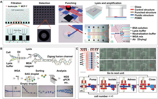
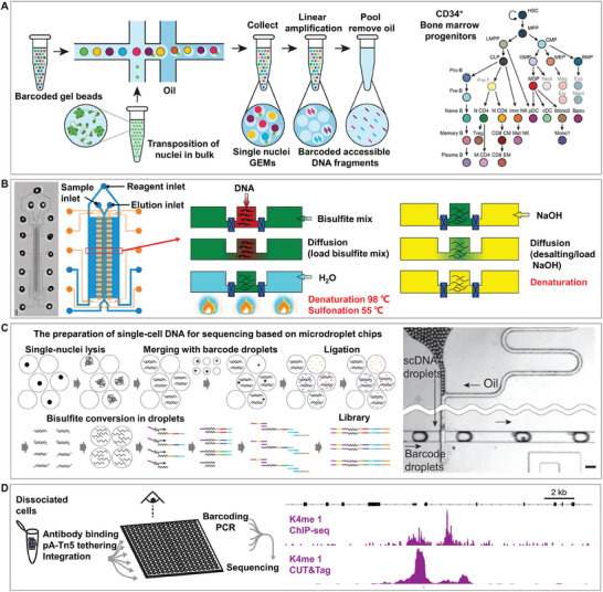
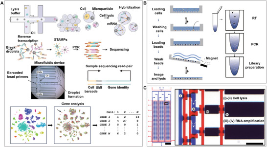
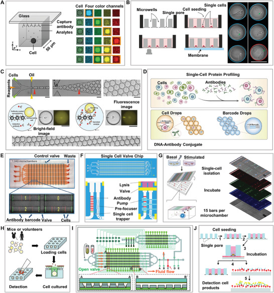
Single-cell multiomic and exosome analyses are potent tools in various fields, such as cancer research, immunology, neuroscience, microbiology, and drug development. They facilitate the in-depth exploration of biological systems, providing insights into disease mechanisms and aiding in treatment. Single-cell isolation, which is crucial for single-cell analysis, ensures reliable cell isolation and quality control for further downstream analyses. Microfluidic chips are small lightweight systems that facilitate efficient and high-throughput single-cell isolation and real-time single-cell analysis on- or off-chip. Therefore, most current single-cell isolation and analysis technologies are based on the single-cell microfluidic technology. This review offers comprehensive guidance to researchers across different fields on the selection of appropriate microfluidic chip technologies for single-cell isolation and analysis. This review describes the design principles, separation mechanisms, chip characteristics, and cellular effects of various microfluidic chips available for single-cell isolation. Moreover, this review highlights the implications of using this technology for subsequent analyses, including single-cell multiomic and exosome analyses. Finally, the current challenges and future prospects of microfluidic chip technology are outlined for multiplex single-cell isolation and multiomic and exosome analyses.

Keywords: isolation quality indicators; microfluidic chips; multiomics and exosome applications; single cells.


Conflict of interest statement

The authors declare no conflict of interest.

Figures

Figure 1
Figure 1
High‐activity micro‐barcode chamber chip. A). Single‐cell printing based on the microchamber chip technology: Gray arrows indicate the direction of single‐cell printing through the microchambers, highlighting the critical stages of single‐cell isolation and gravity deposition. B) High‐throughput microchamber isolation and multiplex protein analysis based on single‐cell printing. Reproduced with permission.[ 32 ] Copyright 2013, American Chemical Society. C) Compared to (B), further increase in throughput is achieved by using shorter microchambers (≈0.48 mm), and enhancement in detection throughput is achieved by combining spatial multiplexing and spectral multiplexing. Adapted with permission.[ 35 ] Copyright 2019, Wiley. D) Assembly of graphene oxide quantum dot (GOQD) biocompatible materials on microchambers to further improve the isolation efficiency and cell activity. Reproduced with permission.[ 37 ] Copyright 2022, Wiley.
Figure 2
Figure 2
High‐efficiency and low‐throughput double‐layer valve chip. A) Methods and principles of single‐cell trapping and releasing based on single‐cell microvalve chips. B) Screening, capture, and release of single cells in different size ranges provide a controllable and precise operation platform for single‐cell research. Reproduced with permission.[ 48 ] Copyright 2021, Elsevier. C) An integrated valve chip for single cell isolation, culture, and analysis to deeply understand the dynamic responses of individual cells to complex immune inputs. Reproduced with permission.[ 49 ] Copyright 2016, Cell Press. D) An integrated valve chip for single‐cell isolation, screening, and recovery supports long‐term monitoring and super‐resolution imaging analysis of single cells. Adapted with permission.[ 50 ] Copyright 2016, Elsevier.
Figure 3
Figure 3
High‐throughput and high‐efficiency microdroplet chip. A) Methods and principles of solid support and support‐free single‐cell separation droplet chips. Single cells exist in individual droplets through an oil‐encapsulating liquid method. B) Large‐scale high‐throughput droplet generation based on microdroplet chips, with droplet size controlled by the flow rates of the dispersed (Qd) and continuous (Qc) phases. This breakthrough overcomes the production throughput limitations of traditional single microfluidic droplet generators, offering new possibilities for high‐throughput droplet production. Adapted with permission.[ 52 ] Copyright 2012, Royal Society of Chemistry. C) Hydrogel droplet support: Encapsulation and culture of bacterial cells in anchored microdroplets highlight the versatility and potential applications of droplet chips for bioanalysis. Adapted with permission.[ 58 ] Copyright 2016, Royal Society of Chemistry. D) Substrate droplet printing: High‐throughput generation of complex arrays of droplets, cells, and microparticles. This technique offers new possibilities and flexibility for the construction of single‐cell and complex assays. Adapted with permission.[ 59 ] Copyright 2017, National Academy of Sciences.
Figure 4
Figure 4
High‐throughput and high‐efficiency microwell chip. A) Methods and principles of the first and second isolation of single cells based on a single‐cell microwell chip. The forces involved in separation include gravity, centrifugal force, hydrodynamic force, and dielectrophoretic force. Microwells used for the second separation are usually larger than those used for the first isolation. B) Separation based on centrifugal force. Adapted with permission.[ 67 ] Copyright 2015, American Chemical Society. C) Separation based on gravity. Adapted with permission.[ 68 ] Copyright 2017, Royal Society of Chemistry. D) Separation based on hydrodynamic force. Reproduced with permission.[ 70 ] Copyright 2013, National Academy of Sciences. E) Separation based on dielectrophoretic force. Adapted with permission.[ 77 ] Copyright 2019, American Chemical Society. F) Gravity‐based secondary separation with gaps. The “gap” refers to the distance between the two chips for secondary separation. Reproduced with permission.[ 79 ] Copyright 2015, Royal Society of Chemistry. G) Gravity‐based secondary separations without gaps. The “gaps” refers to the distance between the two chips for secondary separation. Reproduced with permission.[ 80 ] Copyright 2021, Cold Spring Harbor Laboratory. H) Punch secondary separations based on hydrodynamic force. Reproduced with permission.[ 81 ] Copyright 2015, Royal Society of Chemistry.
Figure 5
Figure 5
Single‐cell isolation chip for genomics. A) Integration of single‐cell isolation, screening, lysis, and DNA amplification detection, using microwell chips offers an effective approach for advanced genetic characterization. Adapted with permission.[ 83 ] Copyright 2015, Royal Society of Chemistry. B) DNA amplification and detection in droplets using passive droplet fusion, leveraging the droplet‐based approach, enables high‐throughput acquisition of contamination‐free and cell‐specific sequence reads. Adapted with permission.[ 86 ] Copyright 2017, Springer Nature. C) The nanoliter volume chamber improves the DNA amplification quality compared to microliter volume utilizing the valve technology and facilitates the efficient analysis of single‐cell genomes. Adapted with permission.[ 91 ] Copyright 2007, Public Library of Science.
Figure 6
Figure 6
Single‐cell isolation chip for epigenomics. A) Single‐cell sequencing assay for transposase‐accessible chromatin (scATAC‐seq) on microdroplet chips facilitates the identification of transcriptional elements and cell differentiation in over 200000 human immune cells within tumor environments. Adapted with permission.[ 101 ] Copyright 2019, Springer Nature. B) Valve microfluidic technology with microfluidic diffusion‐based reduced representation bisulfite sequencing (MID‐RRBS) offers low‐DNA high‐efficiency epigenomic insights. Adapted with permission.[ 97 ] Copyright 2018, Springer Nature. C) Drop‐BS used droplet‐based bisulfite sequencing for scalable single‐cell DNA methylation analysis, processing up to 10000 samples in two days, thereby significantly advancing epigenomics. Adapted with permission.[ 95 ] Copyright 2023, Springer Nature. D) CUT&Tag on microwell chips marks a leap in single‐cell epigenomics by targeting histone modifications for live‐cell sequencing, offering a simpler lower‐reagent workflow than single‐cell chromatin immunoprecipitation sequencing (scChIP‐seq) and boosting single‐cell precision and throughput. Adapted with permission.[ 100 ] Copyright 2019, Springer Nature.
Figure 7
Figure 7
Single‐cell isolation chip for transcriptomics. A) Droplet chip technology for RNA transcription analysis facilitates the high‐throughput examination of gene expression at the single‐cell level, identifying 39 unique cell populations within the mouse retina and establishing an integrated molecular expression map. Reproduced with permission.[ 116 ] Copyright 2015, Cell Press. B) Application of Microwell‐seq technology for the analysis of RNA transcription using a microwell chip, leading to the examination of over 400000 mouse single cells. Reproduced with permission,[ 109 ] Copyright 2018, Cell Press. C) Valve‐based chip for cell lysis and RNA amplification facilitates high‐throughput and accurate reverse transcription‐quantitative polymerase chain reaction (RT‐qPCR) gene expression measurements across hundreds of single cells, significantly lowering the expenses while increasing the sensitivity of detection. Adapted with permission.[ 121 ] Copyright 2011, National Academy of Sciences.
Figure 8
Figure 8
Single‐cell isolation chip for proteomics. A) Multicolor protein detection based on a microwell chip, with detection throughput affected by spectral overlap. Reproduced with permission.[ 126 ] Copyright 2012, National Academy of Sciences. B) Protein dynamic detection based on a microwell chip, where the antibody membrane for protein detection can be replaced. Reproduced with permission.[ 128 ] Copyright 2021, Mdpi. C) Detection of functionalized antibody magnetic beads based on droplet chips. Reproduced with permission.[ 64 ] Copyright 2013, Royal Society of Chemistry. D) Detection of DNA‐antibody magnetic beads based on droplet chips. Reproduced with permission.[ 133 ] Copyright 2017, Springer Nature. E) Secreted protein detection based on the valve chip, marking the first appearance of multiplex barcode chip detection for secreted proteins. Reproduced with permission.[ 39 ] Copyright 2011, Springer Nature. F) Proteome detection based on the valve chip with cleavage units, enabling simultaneous detection of membrane and secreted proteins. Reproduced with permission.[ 136 ] Copyright 2021, Mdpi. G) Single‐cell microchamber chip for the detection of up to 42 secreted proteins using multicolor fluorescence and multiplexing strategies. Reproduced with permission.[ 33 ] Copyright 2015, National Academy of Sciences. H) Secondary isolation based on micromanipulation for monoclonal drug screening. Reproduced with permission.[ 144 ] Copyright 2011, Springer Nature. I) Secondary isolation based on valve structure for monoclonal drug screening. Reproduced with permission.[ 145 ] Copyright 2020, Royal Society of Chemistry. J) Secondary isolation based on film punching for monoclonal drug screening. Reproduced with permission.[ 146 ] Copyright 2019, Royal Society of Chemistry.
Figure 9
Figure 9
Single‐cell isolation chip for exosome analysis. A) Single‐cell exosome analysis based on peelable microwell structures for quantitative monitoring of individual cell behavior. Reproduced with permission.[ 157 ] Copyright 2016, Wiley. B) Single‐cell exosome analysis based on microwell structure with mesh for large‐scale parallel single‐cell transference according to user‐defined criteria. Reproduced with permission.[ 158 ] Copyright 2018, Royal Society of Chemistry. C) Concave microwell structure for the dynamic analysis of single‐cell exosomes, identifying “super secretors” within cell populations. Reproduced with permission.[ 80 ] Copyright 2021, Cold Spring Harbor Laboratory. D) Single‐cell exosome analysis based on the microchamber chip, marking the first multiplex barcode analysis of extracellular vesicle (EV) secretion from single cells, offering new insights on intercellular communication. Reproduced with permission.[ 161 ] Copyright 2020, American Chemical Society.

Similar articles

Cited by

References

    1. Papalexi E., Satija R., Nat. Rev. Immunol. 2018, 18, 35. - PubMed
    1. Mattiazzi Usaj M., Yeung C. H. L., Friesen H., Boone C., Andrews B. J., Cell Syst. 2021, 12, 608. - PMC - PubMed
    1. Zheng H., Pomyen Y., Hernandez M. O., Li C., Livak F., Tang W., Dang H., Greten T. F., Davis J. L., Zhao Y., Mehta M., Levin Y., Shetty J., Tran B., Budhu A., Wang X. W., Hepatology 2018, 68, 127. - PMC - PubMed
    1. Chen X., Litzenburger U. M., Wei Y., Schep A. N., LaGory E. L., Choudhry H., Giaccia A. J., Greenleaf W. J., Chang H. Y., Nat. Commun. 2018, 9, 4590. - PMC - PubMed
    1. Reza K. K., Dey S., Wuethrich A., Jing W., Behren A., Antaw F., Wang Y., Sina A. A. I., Trau M., ACS Nano 2021, 15, 11231. - PubMed

LinkOut - more resources