. 2024 Jul 2;123(13):1763-1780.
doi: 10.1016/j.bpj.2024.05.015. Epub 2024 May 18.

Modeling reveals the strength of weak interactions in stacked-ring assembly

Affiliations

Modeling reveals the strength of weak interactions in stacked-ring assembly

Leonila Lagunes et al. Biophys J. .

Abstract

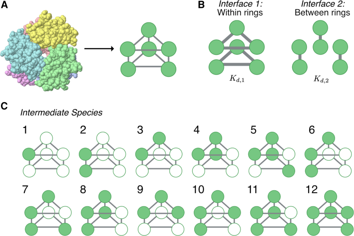
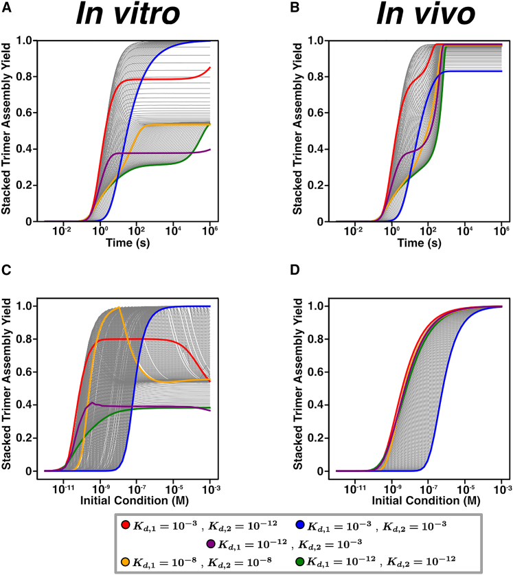
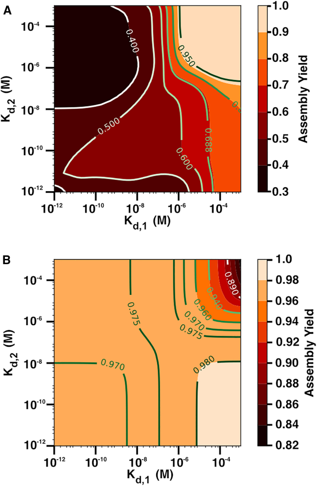
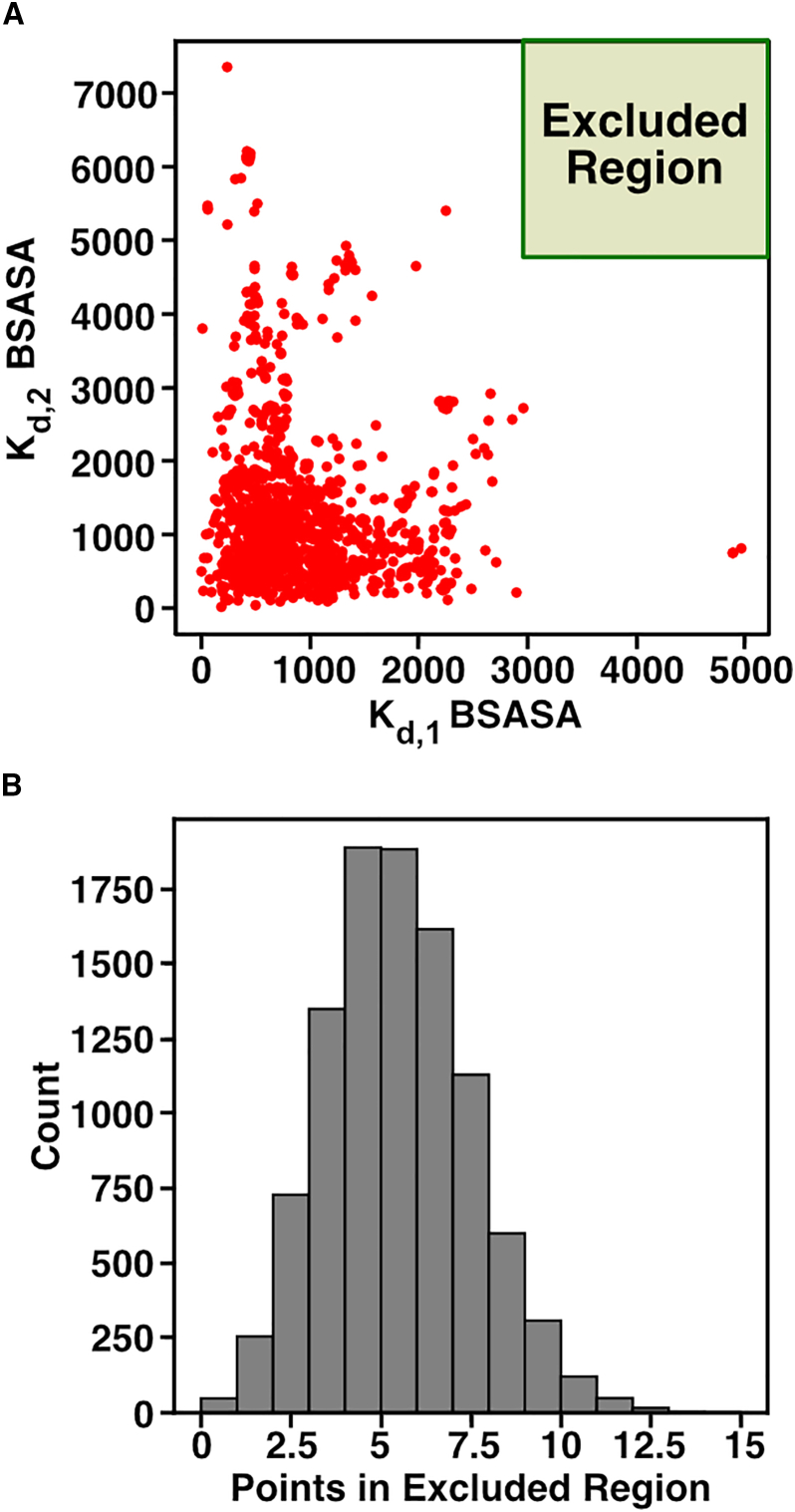
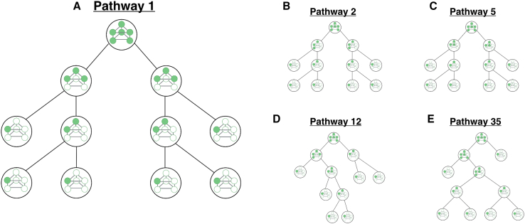
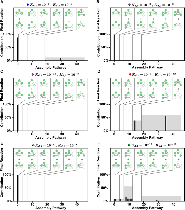
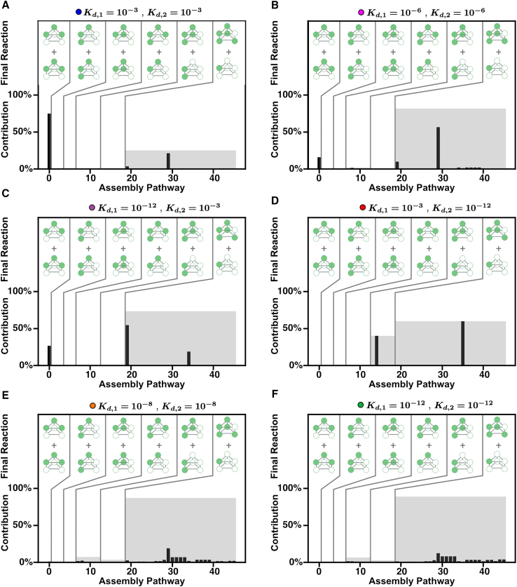
Cells employ many large macromolecular machines for the execution and regulation of processes that are vital for cell and organismal viability. Interestingly, cells cannot synthesize these machines as functioning units. Instead, cells synthesize the molecular parts that must then assemble into the functional complex. Many important machines, including chaperones such as GroEL and proteases such as the proteasome, comprise protein rings that are stacked on top of one another. While there is some experimental data regarding how stacked-ring complexes such as the proteasome self-assemble, a comprehensive understanding of the dynamics of stacked-ring assembly is currently lacking. Here, we developed a mathematical model of stacked-trimer assembly and performed an analysis of the assembly of the stacked homomeric trimer, which is the simplest stacked-ring architecture. We found that stacked rings are particularly susceptible to a form of kinetic trapping that we term "deadlock," in which the system gets stuck in a state where there are many large intermediates that are not the fully assembled structure but that cannot productively react. When interaction affinities are uniformly strong, deadlock severely limits assembly yield. We thus predicted that stacked rings would avoid situations where all interfaces in the structure have high affinity. Analysis of available crystal structures indicated that indeed the majority-if not all-of stacked trimers do not contain uniformly strong interactions. Finally, to better understand the origins of deadlock, we developed a formal pathway analysis and showed that, when all the binding affinities are strong, many of the possible pathways are utilized. In contrast, optimal assembly strategies utilize only a small number of pathways. Our work suggests that deadlock is a critical factor influencing the evolution of macromolecular machines and provides general principles for understanding the self-assembly efficiency of existing machines.


Conflict of interest statement

Declaration of interests The authors declare no competing interests.

Figures

Figure 1
Figure 1
Stacked-trimer model schematic. (A) An example stacked trimer (X-ray structure from PDB: 2FO3) on the left is depicted with each monomer in a unique color. Any such stacked trimer can be represented as a graph with edges representing the noncovalent bonds between the six proteins. Each protein has three binding interfaces (bonds are represented by the solid gray lines). (B) Each protein can form two types of bonds, with proteins on the same ring (interface 1: within) and with proteins from the opposing ring (interface 2: between). Each bond type has associated Kd values, Kd,1 and Kd,2, respectively. (C) A list of all the intermediate species that can be formed during stacked-trimer assembly in our model. To see this figure in color, go online.
Figure 2
Figure 2
Dynamics of stacked-trimer assembly. (A and B) Time-course plots for (A) in vitro and (B) in vivo model with different Kd,1 and Kd,2 parameter values. Initial monomer concentration is 4×10⁶ M, and for the in vivo model the degradation rate δ is set to 2.8×10⁴ s−1. (C and D) Assembly yield curves with respect to increasing initial monomer concentration with different Kd,1 and Kd,2 parameter values. Same color scheme as in (A) and (B) for both in vitro and in vivo models. Gray lines represent cases for which Kd,1 and Kd,2 increase from 103 M to 1012 M. To see this figure in color, go online.
Figure 3
Figure 3
Assembly yield for different values of Kd,1 and Kd,2. Heatmaps showing assembly yield after 24 h for (A) in vitro and (B) in vivo models with fixed initial monomer concentration of 4×10⁶ M and where the binding affinities Kd,1 and Kd,2 increase from 1012 M to 103M. Note that all results for the in vivo case correspond to steady-state yields. Contour lines are shown to visualize assembly yield values. For the in vivo model, δ=2.8×10⁴ s−1. To see this figure in color, go online.
Figure 4
Figure 4
Buried solvent-accessible surface area (BSASA) in PDB stacked-trimer structures. (A) We identified stacked trimers from the PDBePISA database and collected the BSASA for interfaces between and within rings for each. We used the BSASA within and between rings as a proxy for Kd,1 and Kd,2, respectively. (B) From the stacked trimers in (A), we resampled the data using a permutation test and determined how many cases had Kd,1 and Kd,2 BSASA in the excluded region (green rectangle in A). The histogram shows the total cases where Kd,1 and Kd,2 BSASA were in the green rectangle (counts) based on 10,000 trials. Note that out of the 10,000 trials, only 48 had zero cases where Kd,1 and Kd,2 BSASA were in the excluded region (green rectangle). To see this figure in color, go online.
Figure 5
Figure 5
Example assembly pathways for a stacked trimer. Each panel shows an assembly pathway to form a stacked trimer. While we have enumerated all 46 pathways, five examples are shown here. The pathways begin at the bottom of the graphic and end with the stacked trimer at the top of the graphic. (A) Pathway 1 has a final step in which two trimeric rings bind to form a stacked trimer. (B) Pathway 2 is one pathway in which the final step is a reaction between two trimers (which in this case are not rings). (C) Pathway 5 represents an alternative to pathway 2 in (B) with the same final step. (D) Pathway 12 is one pathway for which the final step is the reaction between a tetramer and a dimer to form the stacked trimer. (E) Pathway 35 is one pathway in which the final reaction is to add a single monomer to a pentamer to form the stacked trimer. To see this figure in color, go online.
Figure 6
Figure 6
Assembly pathway contributions for different combinations of Kd,1 and Kd,2 in the in vitro model. (AF) Each panel shows all the assembly pathways, enumerated as in Fig. 4, and their calculated pathway contribution. Pathways are grouped based on the final assembly reaction as indicated by the graphics in the top of each graph labeled as “Final Reaction.” The black bar is the individual pathway contribution, and the gray region represents the sum of the pathway contributions for all the pathways in the group. To see this figure in color, go online.
Figure 7
Figure 7
Assembly pathway contributions for different combinations of Kd,1 and Kd,2 in vivo model. (AF) Each panel shows all the assembly pathways, enumerated as in Fig. 4, and their calculated pathway contribution. Pathways are grouped based on the final assembly reaction as indicated by the graphics in the top of each graph, labeled as “Final Reaction.” The black bar is the individual pathway contribution, and the gray region represents the sum of the pathway contributions for all the pathways in the group. To see this figure in color, go online.

Similar articles

References

    1. Yuan S., Zhou G., Xu G. Translation machinery: the basis of translational control. J. Genet. Genomics. 2024;51:367–378. - PubMed
    1. Budenholzer L., Cheng C.L., Hochstrasser M., et al. Proteasome Structure and Assembly. J. Mol. Biol. 2017;429:3500–3524. - PMC - PubMed
    1. Stan G., Lorimer G.H., Thirumalai D. Friends in need: How chaperonins recognize and remodel proteins that require folding assistance. Front. Mol. Biosci. 2022;9 - PMC - PubMed
    1. Prywes N., Phillips N.R., Savage D.F., et al. Rubisco Function, Evolution, and Engineering. Annu. Rev. Biochem. 2023;92:385–410. - PubMed
    1. Zhou A., Rohou A., Rubinstein J.L., et al. Structure and conformational states of the bovine mitochondrial ATP synthase by cryo-EM. Elife. 2015;4 - PMC - PubMed

Publication types