Review
. 2024 May 15:17:11786469241248287.
doi: 10.1177/11786469241248287. eCollection 2024.

A Review of the Evidence for Tryptophan and the Kynurenine Pathway as a Regulator of Stem Cell Niches in Health and Disease

Affiliations
Review

A Review of the Evidence for Tryptophan and the Kynurenine Pathway as a Regulator of Stem Cell Niches in Health and Disease

Benjamin Sebastian Summers et al. Int J Tryptophan Res. .

Abstract

Stem cells are ubiquitously found in various tissues and organs in the body, and underpin the body's ability to repair itself following injury or disease initiation, though repair can sometimes be compromised. Understanding how stem cells are produced, and functional signaling systems between different niches is critical to understanding the potential use of stem cells in regenerative medicine. In this context, this review considers kynurenine pathway (KP) metabolism in multipotent adult progenitor cells, embryonic, haematopoietic, neural, cancer, cardiac and induced pluripotent stem cells, endothelial progenitor cells, and mesenchymal stromal cells. The KP is the major enzymatic pathway for sequentially catabolising the essential amino acid tryptophan (TRP), resulting in key metabolites including kynurenine, kynurenic acid, and quinolinic acid (QUIN). QUIN metabolism transitions into the adjoining de novo pathway for nicotinamide adenine dinucleotide (NAD) production, a critical cofactor in many fundamental cellular biochemical pathways. How stem cells uptake and utilise TRP varies between different species and stem cell types, because of their expression of transporters and responses to inflammatory cytokines. Several KP metabolites are physiologically active, with either beneficial or detrimental outcomes, and evidence of this is presented relating to several stem cell types, which is important as they may exert a significant impact on surrounding differentiated cells, particularly if they metabolise or secrete metabolites differently. Interferon-gamma (IFN-γ) in mesenchymal stromal cells, for instance, highly upregulates rate-limiting enzyme indoleamine-2,3-dioxygenase (IDO-1), initiating TRP depletion and production of metabolites including kynurenine/kynurenic acid, known agonists of the Aryl hydrocarbon receptor (AhR) transcription factor. AhR transcriptionally regulates an immunosuppressive phenotype, making them attractive for regenerative therapy. We also draw attention to important gaps in knowledge for future studies, which will underpin future application for stem cell-based cellular therapies or optimising drugs which can modulate the KP in innate stem cell populations, for disease treatment.

Keywords: IDO-1; TDO2; Tryptophan metabolism; aryl hydrocarbon receptor; cancer stem cells; hematopoietic stem cells; human diseases; induced pluripotent stem cells; kynurenine pathway; mesenchymal stromal cells; neural stem cells.


Conflict of interest statement

The author(s) declared the following potential conflicts of interest with respect to the research, authorship, and/or publication of this article: Prof. Bruce Brew is a member of the International Journal for Tryptophan Research journal editorial board. The other co-authors declare that they have no conflict of interest.

Figures
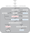
Figure 1.
Figure 1.
Schematic diagram of the Kynurenine pathway. Tryptophan (TRP) is primarily metabolised by the KP (~95%), with only a small amount converted into serotonin and melatonin (~3%), tryptamine or utilised in protein synthesis (~1%). The metabolism of TRP through the KP is initiated by transformation to N’formylkynurenine followed by kynurenine (KYN). This first step is regulated by the enzymes indoleamine-2,3-dioxygenase (IDO-1 and 2) and tryptophan-2,3-dioxygenase (TDO2). N’formylkynurenine is degraded by arylformamidase (AFMID) to the main metabolite KYN. KYN is then converted to either anthranilic acid (AA), kynurenic acid (KYNA) or 3-hydroxykynurenine (3-HK). These conversions are controlled by the kynureninase (KYNU), kynurenine aminotransferases (KATs) and kynurenine 3-monooxygenase (KMO) enzymes, respectively. There is some evidence that KYNA can be further transformed into quinaldic acid, likely via dihydroxylation, although the majority remains unmetabolised., 3-HK is converted to either 3-hydroxyanthranilic acid (3-HAA) by the action of KYNU, or to xanthurenic acid by KAT enzymes. 3-HAA is also formed though hydroxylation of AA. 3-HAA is an antioxidant (i.e. protective) towards certain types of reactive oxygen species (ROS), - but when exposed to intracellular metal ions (e.g. copper II+) it can be a pro-oxidant, forming ROS such as OH. 3-HAA is further metabolised into 2-amino-3-carboxymuconic-semialdehyde by the enzyme 3-hydroxyanthranilate-3,4-dioxygenase (3-HAO), or cinnabarinic acid via autoxidation., 2-amino-3-carboxymuconic-semialdehyde is catabolised down two pathways, being (1) metabolised to 2-aminomuconate-6-semialdehyde by 2-amino-3-carboxymuconate-semialdehyde decarboxylase (ACMSD), and then non-enzymatically to (neuroprotective) picolinic acid (PIC), or (2) non-enzymatically to (neurotoxic) quinolinic acid (QUIN), which is further metabolised toward the essential cofactor nicotinamide adenine dinucleotide (NAD) by quinolinate phosphoribosyltransferase (QPRT; several subsequent reactions are needed to produce NAD). Importantly, the physiological function and effects on cells of all the metabolites in the KP as a result of TRP metabolism are not same. There are both neuroprotective (KYNA, PIC, and cinnabarinic acid) and neurotoxic (3-HK and QUIN) metabolites. In the figure neuroprotective metabolites are marked in blue whereas neurotoxic metabolites are marked in red.
Figure 2.
Figure 2.
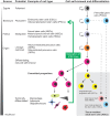
Schematic diagram demonstrating the hierarchy of stem cells occurring in the body during development, and their progressive fate restriction before terminal differentiation into cell types. A zygote is a fertilized egg that has totipotent capability, facilitating differentiation to any cell type and a whole organism. The zygote further develops to form a blastocyst which consists of trophoblast, blastocoel and inner cell mass (ICM). The ICM can give rise to pluripotent embryonic stem cells (ESCs), which after further rounds of symmetric division (indicated by semicircle arrows) can give rise to primordial germ cells (PGCs), which are precursors of gametes and can generate pluripotent Very Small Embryonic-Like Stem Cells (VSELs) which persist into adulthood, express markers of ESCs and are typically quiescent. ESCs are capable of differentiation into any differentiated cell type via progressive fate restriction (asymmetric cell division) to specific, multipotent stem cells, including hematopoietic stem cells (HSCs), mesenchymal stromal cells (MSCs) and neural stem cells (NSCs). These multipotent cells arise during development, can differentiate into limited specialized cells within the specific lineage, and are retained as tissue-resident adult stem cells (ASCs; grey shaded region at the bottom right). ASCs are undifferentiated cells derived from adult tissues or organs, which are characteristically quiescent, reflecting their innate characteristics of genomic stability underlying their long life, typically proliferating or mobilising only when required for replacement of cells lost over the lifetime of the organism or when tissues are injured or diseased. Lineage-restricted precursor cells are committed to differentiating into cell types of a specific lineage, but display reduced multipotency. Examples include neuron and glia-restricted precursor cells, which are derived from NSCs. Committed progenitors tend to differentiate into specific cell types such as macrophages, adipocytes and oligodendrocytes. Examples include oligodendrocyte progenitor cells (OPCs), which are uniformly distributed throughout the parenchyma of the adult CNS and retain the ability to proliferate and terminally differentiate into mature oligodendrocytes, and neuroblasts, which are very primitive, committed neurons. The final cell type considered – induced pluripotent stem cells (iPSC) can be generated from somatic cells (green arrow, intended to demonstrate that this process works against the natural flow of stem cell progressive fate restriction and lineage commitment), via genetic or more recently, non-genetic means. Solid lines with arrows represent progressive fate restriction to defined types of stem cells as development proceeds, whereas dotted lines represent different types of stem cells of varying differentiation capacity. This figure was created using the CorelDraw graphics suite.
Figure 3.
Figure 3.
Schematic diagram demonstrating the hierarchy of bone marrow-derived HSCs occurring in the body during development and throughout adulthood, and their progressive fate restriction before terminal differentiation into myeloid and lymphoid-derived cell types. HSCs are the only cells within the niche that are capable of both multipotent differentiation (into all blood cell types) and self-renewal. Multipotent progenitors can be further subdivided based on their proliferation type. Long term self-renewing HSCs are quiescent cells that self-renew indefinitely to maintain the pool of blood cells needed throughout life due to turnover of differentiated cells. Short term HSCs are progeny generated from long term HSCs, and tend to be highly proliferative and generate multipotent progenitors that have lost their self-renewal capability but ultimately produce the repertoire of differentiated blood cells depicted. Figure adapted and altered from the original source and used under creative commons BY 4.0 license (http://creativecommons.org/licenses/by/4.0).
Figure 4.
Figure 4.
Schematic diagram of the key features of KP metabolism in stem cell types covered in this review. A saggital plane through a pregnant human female is shown in the centre of the schematic, in order to depict the localisation and known functions of the KP in pluripotent, embryonic, stem and precursor cells from the different tissues and organs depicted, which are then enlarged in adjacent expanded boxes either side to illustrate the major known KP phenotype in those cells. In each box, a cartoon representation of the major reactions in the KP is shown in the top left corner of each expanded box, confirmed metabolites produced shown in green text, and therefore enzymatic pathways likely to be active indicated by green arrows. Where studies have not investigated KP expression in that cell type, the pathways are shown in gray. Where studies have shown conflicting evidence for example, between mouse and human or between primary cells and cell lines, this is indicated in orange. TRP is shown in black as the key metabolite from which all downstream metabolites are derived. AhR is shown with a green background (to represent proven expression in that cell type and where relevant, functionality with respect to KP ligands) versus red (uncertain/not investigated). A plus ‘+’ inside of a circle represents pathways or outcomes that are positively enhanced, while a minus ‘−’ sign indicates negative/reduced pathways/outcomes. A ‘T’ symbol is used when referring to the actions of an inhibitor. ‘1’ and ‘2’ in circles represent separate outcomes as indicated and referred to in the heading of each box. Question marks appear where unresolved questions arise or where more evidence would be helpful to substantiate the phenotype. Summary of individual boxes: Human CDSCs inhibit T lymphocyte growth via IDO-1 mediated TRP depletion, however the expression of other KP enzymes and metabolites produced is currently unknown, as is the activation of AhR. iPSCs, can be isolated from somatic cells like skin. Their growth is regulated by extracellular TRP concentration, where high basal IDO-1 expression promotes production of metabolites including n-formylkynurenine which upon secretion is pro-proliferative (and may act in an autocrine or paracrine manner), while AhR is not involved and NAD synthesis is not altered. While many metabolites have been detected, expression levels of their corresponding enzymes has not been investigated. KP expression or AhR function in EPCs is unknown, however in vitro evidence shows that KYN can regulate their proliferation, and circulating EPC numbers reduced when a selective IDO-1 inhibitor was given to mice, suggesting KP metabolites are protective, while AhR function is unknown. HSCs synthesise little KYN basally, however synthesis significantly rises with IFN-γ treatment., KP metabolites KYN, KYNA, and 3-HAA potently inhibit HSC proliferation in vitro. Whether this occurs via AhR (confirmed expressed in HSCs) is unclear. qPCR analysis has proven mouse NSCs express major KP enzymes and respond to IFN-γ however secreted metabolites have not investigated and enzyme expression varies in for example, cell lines. KYNA negatively regulates iPS-derived NSC proliferation and promotes neurogenesis via AhR., CSCs synthesise high NAD levels from the salvage pathway needed for proliferation, while activation of interplay between KP activation in CSCs, secretion of metabolites and AhR activation via secreted ligand KYN in T cells is important for immune evasion. Inhibiting AhR signaling does not always give a beneficial outcome in all types of cancer, as discussed in the text. Placental MSCs are immunosuppressive by limiting T cell proliferation through TRP depletion via IDO-1, though much about their KP enzyme expression beyond that is not known. Bone marrow MSCs express most KP enzymes when examined by PCR. ESCs are regulated by KP activation through IDO-1, and activation of AhR by KP metabolites to maintain self-renewal and pluripotent phenotype, while KAT-2 activity and production of metabolite 2-AAA is a biomarker for ectodermal differentiation. TDO2 is not basally expressed but can be induced by a neurodevelopmental toxin, while KMO is not expressed., Expression/detection of many KP enzymes and metabolites is currently unknown. Source: Figure was created with Biorender.com. Abbreviations: AA, anthranilic acid; 2-AAA, 2-aminoadipic acid; 3-HAA, 3-hydroxyanthranilic acid; AhR, aryl hydrocarbon receptor; CDSC’s, cardiac stem cells; CSCs, cancer stem cells; EPCs, endothelial progenitor cells; ESCs, embryonic stem cells; HSCs, hematopoietic stem cells; IDO-1, indoleamine-2,3-dioxygenase; IFN-γ, interferon-gamma; iPSCs, induced pluripotent stem cells; KAT-2, kynurenine aminotransferase-2; KYN, kynurenine; KYNA, kynurenic acid; MSCs, mesenchymal stromal cells; NAD, nicotinamide adenine dinucleotide; n-FK, n-formylkynurenine; NSC, neural stem cell; PIC, picolinic acid; TRP, tryptophan; QUIN, quinolinic acid.

Similar articles

Cited by

References

    1. Jones SP, Guillemin GJ, Brew BJ. The kynurenine pathway in stem cell biology. Int J Tryptophan Res. 2013;6:57-66. doi:10.4137/IJTR.S12626 - DOI - PMC - PubMed
    1. Croitoru-Lamoury J, Lamoury FM, Caristo M, et al.. Interferon-gamma regulates the proliferation and differentiation of mesenchymal stem cells via activation of indoleamine 2,3 dioxygenase (IDO). PLoS One. 2011;6:e14698. doi:10.1371/journal.pone.0014698 - DOI - PMC - PubMed
    1. Jones SP, Franco NF, Varney B, et al.. Expression of the kynurenine pathway in human peripheral blood mononuclear cells: implications for inflammatory and neurodegenerative disease. PLoS One. 2015;10:e0131389. doi:10.1371/journal.pone.0131389 - DOI - PMC - PubMed
    1. Braidy N, Grant R, Adams S, Brew BJ, Guillemin GJ. Mechanism for quinolinic acid cytotoxicity in human astrocytes and neurons. Neurotox Res. 2009;16:77-86. doi:10.1007/s12640-009-9051-z - DOI - PubMed
    1. Braidy N, Grant R, Brew BJ, Adams S, Jayasena T, Guillemin GJ. Effects of kynurenine pathway metabolites on intracellular NAD synthesis and cell death in human primary astrocytes and neurons. Int J Tryptophan Res. 2009;2:61-69. doi:10.4137/ijtr.s2318 - DOI - PMC - PubMed