. 2024 May 15;14(1):62.
doi: 10.1186/s13578-024-01244-2.

Microbiota-derived 3-phenylpropionic acid promotes myotube hypertrophy by Foxo3/NAD+ signaling pathway

Affiliations

Microbiota-derived 3-phenylpropionic acid promotes myotube hypertrophy by Foxo3/NAD+ signaling pathway

Penglin Li et al. Cell Biosci. .

Abstract

Background: Gut microbiota and their metabolites play a regulatory role in skeletal muscle growth and development, which be known as gut-muscle axis. 3-phenylpropionic acid (3-PPA), a metabolite produced by colonic microorganisms from phenylalanine in the gut, presents in large quantities in the blood circulation. But few study revealed its function in skeletal muscle development.

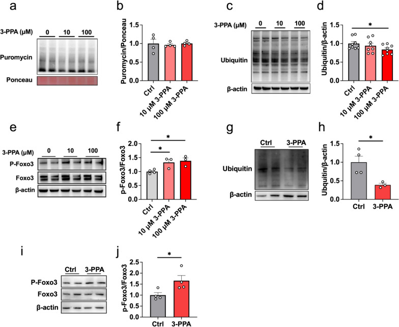
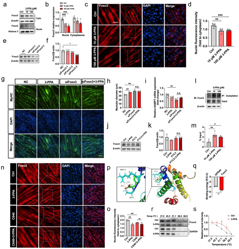
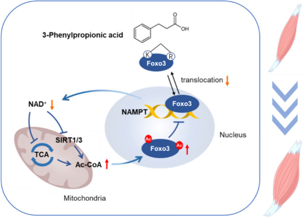
Results: Here, we demonstrated the beneficial effects of 3-PPA on muscle mass increase and myotubes hypertrophy both in vivo and vitro. Further, we discovered the 3-PPA effectively inhibited protein degradation and promoted protein acetylation in C2C12 and chick embryo primary skeletal muscle myotubes. Mechanistically, we supported that 3-PPA reduced NAD+ synthesis and subsequently suppressed tricarboxylic acid cycle and the mRNA expression of SIRT1/3, thus promoting the acetylation of total protein and Foxo3. Moreover, 3-PPA may inhibit Foxo3 activity by directly binding.

Conclusions: This study firstly revealed the effect of 3-PPA on skeletal muscle growth and development, and newly discovered the interaction between 3-PPA and Foxo3/NAD+ which mechanically promote myotubes hypertrophy. These results expand new understanding for the regulation of gut microbiota metabolites on skeletal muscle growth and development.

Keywords: 3-Phenylpropionic acid; Acetylation; Gut microbiota metabolites; Muscle hypertrophy; NAD+.


Conflict of interest statement

The authors declare that they have no competing interests.

Figures

Fig. 1
Fig. 1
Effect of 3-PPA on muscle mass and C2C12 myotubes hypertrophy. a Body weight gain in 6 week-old male mice with7 weeks’ treatment of 0.5%3-PPA (n = 10). b Lean mass and fat mass of mice after 6 weeks’ treatment of 3-PPA by QMR (n = 10). c Gastrocnemius weight of mice after 7  weeks’ treatment (n = 10). d–e The laminin immunofluorescent staining (d) and frequency histogram of gastrocnemius muscle fiber cross-sectional area statistical analysis (e) in C57BL/6 male mice after 3 weeks 3-PPA treatment (n = 3). f Schematic representation of C2C12 treated with 3-PPA for 2 days. g–h Immunofluorescence images (g) and statistics of myotube diameter (h) of C2C12 myotubes in 3-PPA treatment for 2 days (n = 4). i–j Immunofluorescence images (i) and statistics of myotube diameter (j) of chick embryo primary skeletal muscle cells in 3-PPA treatment for 2 days (n = 3)
Fig. 2.
Fig. 2.
3-PPA attenuated protein degradation instead of protein synthesis in C2C12 myotubes. a–b Immunoblots (a) and quantification (b) of the puromycin-labeled protein expression of C2C12 myotubes with 3-PPA treatment for 2 days (n = 8). c–d Immunoblots (c) and quantification (d) of total ubiquitination protein expression of C2C12 myotubes with 3-PPA treatment for 2 days (n = 4). e–f Immunoblots (e) and quantification (f) of P-Foxo3 and Foxo3 protein expression of C2C12 myotubes with 3-PPA treatment (n = 3). g–h Immunoblots (g) and quantification (h) of the total ubiquitination protein expression of chick embryo primary skeletal muscle myotubes with 3-PPA treatment for 2 days (n = 3-4). (i–j) Immunoblots (i) and quantification (j) of P-Foxo3 and Foxo3 protein expression in chick embryo primary skeletal muscle myotubes (n = 4)
Fig. 3
Fig. 3
Protein acetylation modification mediates the effect of 3-PPA in myotube hypertrophy. a–d The glucose content (a), LD content (b), ATP content (c), ROS content (d) in C2C12 myotubes with 3-PPA treatment for 2 days (n = 3 to 4). (e) Mitochondria biogenesis and function related gene mRNA expression in C2C12 myotubes with 3-PPA treatment for 2 days (n = 4 to 8). f Mitochondria autophagy related gene mRNA expression in C2C12 myotubes with 3-PPA treatment for 2 days (n = 4). g Tricarboxylic acid cycle related gene mRNA expression in C2C12 myotubes with 3-PPA treatment for 2 days (n = 6). h Relative Acetyl-CoA content in C2C12 myotubes with 3-PPA treatment for 2 days (n = 6). ij Immunoblots (i) and quantification (j) of Pan-acetylation protein expression of C2C12 myotubes with 3-PPA treatment for 2 days (n = 3). k–l Immunofluorescence images (k) and statistics of myotube diameter (l) of C2C12 myotubes in C646 and 3-PPA co-treatment (n = 3). m The ratio of NAD+/NADH in in C2C12 myotubes with 3-PPA treatment for 2 days (n = 3). n mRNA expression of NANAT2 and NAMPT in C2C12 myotubes with 3-PPA treatment for 2 days (n = 6). o mRNA expression of SIRT1and SIRT3 in C2C12 myotubes with 3-PPA treatment for 2 days (n = 6)
Fig. 4
Fig. 4
Foxo3/NAD+ pathway is required for 3-PPA to increase protein acetylation modification. a–b Immunoblots (a) and quantification (b) of Foxo3 protein expression in nuclei and cytoplasm of C2C12 myotubes with 3-PPA treatment for 2 days (n = 4). c–d Immunofluorescence images (c) and statistics (d) of relative nuclei Foxo3 protein in C2C12 myotubes in 3-PPA treatment for 2 days (n = 3). e–f Immunoblots (e) and quantification (f) of Foxo3 protein expression in C2C12 myotubes with different siRNA sequence treatment (n = 3 to 4). g–h Immunofluorescence images (g) and statistics (h) of myotube diameter in C2C12 myotubes with siFoxo3 and 3-PPA co-treatment (n = 3). i mRNA expression of NAMPT in C2C12 myotubes with si Foxo3 and 3-PPA co-treatment (n = 4). j–k Immunoblots (j) and quantification (k) of Foxo3 protein expression in C2C12 myotubes with P7C3 and 3-PPA co-treatment (n = 3). (l-m) Immunoprecipitation (l) and statistics (m) of Ace- Foxo3 in C2C12 myotubes with 3-PPA treatment for 2 days (n = 4). (n–o) Immunofluorescence images (n) and statistics (o) of Foxo3 of C2C12 myotubes in C646 and 3-PPA co-treatment (n = 3). (p) Binding region of 3-PPA and Foxo3 protein. (q) Calculated binding energy of Foxo3 and GPR40. r–s Immunoblots (r) and quantification (s) of relative Foxo3 protein expression in CETSA assay by 3-PPA treatment (n = 3)
Fig. 5
Fig. 5
Schematic diagram of 3-PPA’s function on myotube hypertrophy

Similar articles

References

    1. Baskin KK, Winders BR, Olson EN. Muscle as a “mediator” of systemic metabolism. Cell Metab. 2015;21(2):237–248. doi: 10.1016/j.cmet.2014.12.021. - DOI - PMC - PubMed
    1. Li P, Feng J, Jiang H, Feng X, Yang J, Yuan Y, et al. Microbiota derived d-malate inhibits skeletal muscle growth and angiogenesis during aging via acetylation of cyclin a. EMBO Rep. 2024;25(2):524–543. doi: 10.1038/s44319-023-00028-y. - DOI - PMC - PubMed
    1. Kang SH, Lee HA, Kim M, Lee E, Sohn UD, Kim I. Forkhead box O3 plays a role in skeletal muscle atrophy through expression of E3 ubiquitin ligases MuRF-1 and atrogin-1 in cushing’s syndrome. Am J Physiol Endocrinol Metab. 2017;312(6):E495–e507. doi: 10.1152/ajpendo.00389.2016. - DOI - PubMed
    1. Gellhaus B, Böker KO, Gsaenger M, Rodenwaldt E, Hüser MA, Schilling AF, et al. Foxo3 knockdown mediates decline of myod1 and myog reducing myoblast conversion to myotubes. Cells. 2023 doi: 10.3390/cells12172167. - DOI - PMC - PubMed
    1. Soulez M, Tanguay PL, Dô F, Dort J, Crist C, Kotlyarov A, et al. ERK3-MK5 signaling regulates myogenic differentiation and muscle regeneration by promoting FoxO3 degradation. J Cell Physiol. 2022;237(4):2271–2287. doi: 10.1002/jcp.30695. - DOI - PubMed