[Preprint]. 2024 May 5:2024.05.03.592485.
doi: 10.1101/2024.05.03.592485.

Cysteine Rich Intestinal Protein 2 is a copper-responsive regulator of skeletal muscle differentiation

Affiliations

Cysteine Rich Intestinal Protein 2 is a copper-responsive regulator of skeletal muscle differentiation

Odette Verdejo-Torres et al. bioRxiv. .

Abstract

Copper (Cu) is an essential trace element required for respiration, neurotransmitter synthesis, oxidative stress response, and transcriptional regulation. Imbalance in Cu homeostasis can lead to several pathological conditions, affecting neuronal, cognitive, and muscular development. Mechanistically, Cu and Cu-binding proteins (Cu-BPs) have an important but underappreciated role in transcription regulation in mammalian cells. In this context, our lab investigates the contributions of novel Cu-BPs in skeletal muscle differentiation using murine primary myoblasts. Through an unbiased synchrotron X-ray fluorescence-mass spectrometry (XRF/MS) metalloproteomic approach, we identified the murine cysteine rich intestinal protein 2 (mCrip2) in a sample that showed enriched Cu signal, which was isolated from differentiating primary myoblasts derived from mouse satellite cells. Immunolocalization analyses showed that mCrip2 is abundant in both nuclear and cytosolic fractions. Thus, we hypothesized that mCrip2 might have differential roles depending on its cellular localization in the skeletal muscle lineage. mCrip2 is a LIM-family protein with 4 conserved Zn2+-binding sites. Homology and phylogenetic analyses showed that mammalian Crip2 possesses histidine residues near two of the Zn2+-binding sites (CX2C-HX2C) which are potentially implicated in Cu+-binding and competition with Zn2+. Biochemical characterization of recombinant human hsCRIP2 revealed a high Cu+-binding affinity for two and four Cu+ ions and limited redox potential. Functional characterization using CRISPR/Cas9-mediated deletion of mCrip2 in primary myoblasts did not impact proliferation, but impaired myogenesis by decreasing the expression of differentiation markers, possibly attributed to Cu accumulation. Transcriptome analyses of proliferating and differentiating mCrip2 KO myoblasts showed alterations in mRNA processing, protein translation, ribosome synthesis, and chromatin organization. CUT&RUN analyses showed that mCrip2 associates with a select set of gene promoters, including MyoD1 and metallothioneins, acting as a novel Cu-responsive or Cu-regulating protein. Our work demonstrates novel regulatory functions of mCrip2 that mediate skeletal muscle differentiation, presenting new features of the Cu-network in myoblasts.

Keywords: Crip2; copper; myogenesis; transcriptional regulation.


Figures
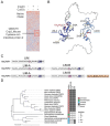
Figure 1.
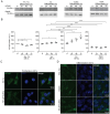
Figure 1.. Phylogenetic analyses of the CRIP family members and appearance of the Cu+-binding sites.
A. Synchrotron based X-Ray fluorescence analyses coupled to protein sequencing by mass spectrometry of native PAGE gels from whole cell extracts of differentiating (24 h) primary myoblasts showed the presence of mCrip2 in a section that showed enriched signal of copper (indicated by red box). B. AlphaFold-predicted structure of mCrip2. The N-terminal LIM domain is colored dark blue and the C-terminal LIM domain is colored light blue. The conserved Zn-binding residues are shown as balls and sticks, while the conserved histidine residues located near the Zn2 sites are colored red. C. Metal-binding motifs found in human CRIPs. Individual LIM domains are shown as boxes and labeled. Individual Zn-finger motifs are shaded with grey and labeled. The CRIP-specific HxxX motif is highlighted in yellow. In addition to having duplicated LIM domains, CRIP2 and CRIP3 proteins contain conserved histidine residue adjacent to Cys53 and Cys172 (purple shading). Human CRIP3 contains a histidine residue next to Cys35 in the Zn2 sites that are not found in CRIP1 or CRIP2 (pink shading). D. Maximum likelihood tree of CRIPs with 2 two LIM domains. “A-H” refers to conservation of human CRIP2 His36 and “B-H” refers to conservation of human CRIP2 His173.
Figure 2.
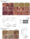
Figure 2.. Biochemical characterization of hsCRIP2 as a Cu+-binding protein.
A. Recombinant human hsCRIP2 protein purification; representative Coomassie Brilliant Blue (CBB) gel and western blots of hsCRIP2 protein detected using an anti-Crip2 and anti-Strep-tag antibodies. Determination of the Cu+ dissociation constant (KD) of hsCRIP2 using the BCS competition assay; spectrophotometric titration of 25 μM BCS and 10 μM Cu+ with increasing concentrations of hsCRIP2. The arrow indicates the decrease in absorbance at 483 nm upon each protein addition. hsCRIP2-Cu+ dissociation constants were calculated based on the stoichiometry determinations, assuming two Cu+ sites (B) or four Cu+ sites (C) per hsCRIP2 molecule. D. Cyclic Voltammograms obtained for a) Au/CRIP2, b) Au/CRIP2-Cu and c) Au-Cys/CRIP2-Cu, measured in a potential window of −500 to 500 mV using a scan rate of 100 mV/s with 0.1 M phosphate buffer pH 7.2 as supporting electrolyte.
Figure 3.
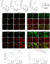
Figure 3.. Effect of Cu supplementation in mCrip2 expression and localization in proliferating and differentiating primary myoblasts.
A. B. Representative western blots of mCrip2 protein levels in proliferating (48 h) and differentiating (24, 48, 72 h) myoblasts in the presence or absence of insulin, CuSO4, and the Cu-chelator TEPA. Anti-Gapdh antibody was used as a loading control. C. Densitometric quantification of mCrip2 bands in proliferating and differentiating primary myoblasts shown in A. N=5, *P < 0.05; **P < 0.01. Representative confocal microscopy images showing the cytosolic and nuclear localization of mCrip2 (GREEN) in proliferating (D) and differentiating (E) primary myoblasts. Nuclei were counterstained with DAPI (blue). N=6.
Figure 4.
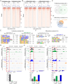
Figure 4.. CRISPR/Cas9-mediated knock-out of mCrip2 in primary myoblasts.
(A) Location of the three sgRNA designed to delete mCrip2 by CRISPR/Cas9 system in primary myoblasts. The three constructs were targeted to intron/exon boundaries. (B) Representative western blot (top) and (C) densitometric quantification (bottom) of mCrip2 protein levels in proliferating (48 h) and differentiating (24 h) primary myoblasts. An anti-vinculin antibody was used as a loading control. N=6; ****P < 0.0001. (D) CRISPR/Cas9-mediated deletion of mCrip2 does not impact proliferation of primary myoblasts. Proliferation curves comparing wild type, empty vector (EV) control, and mCrip2 depleted (sgRNA1, sgRNA2 and sgRNA3) primary myoblasts. No significant differences in growth were found between the five strains. The Cu-dependent increase in proliferation rate previously reported (Vest et al., 2018) is maintained in mCrip2 KO myoblasts. Data represent the mean of 3 biologically independent experiments +/− SE. Statistical analyses compared each strain grown in the presence (100 μM) vs. absence of CuSO4 in the culture media. *P < 0.05; **P < 0.01; ***P < 0.001; ****P < 0.0001. (E) Representative light micrographies of proliferating myoblasts immunostained for Pax7 at 48 h cultured with or without 100 μM CuSO4; N=3.
Figure 5.
Figure 5.. CRISPR/Cas9-mediated KO of mCrip2 impairs differentiation of cultured primary myoblasts.
(A) Representative light micrographs of differentiating myoblasts immunostained for myogenin after 48 h of inducing differentiation in the presence or absence of insulin and Cu. (B) Calculated fusion index for controls and three different clones of myoblasts transduced with mCrip2 sgRNAs. Data represent three independent biological experiments +/− SE. N=3; **P < 0.01; ***P < 0.001; ****P < 0.0001. (C) The differentiation defect of mCrip2 KO myoblasts is rescued by reintroducing hsCRIP2 gene. Representative western blot of primary myoblasts transduced with either empty vector (EV), the mCrip2 sgRNA3 and the KO mCrip2 cells transduced with hsCRIP2 construct. Samples were obtained for 48 h after inducing differentiation. A specific anti-Crip2 antibody depicts the levels of protein and vinculin was used as loading control. (D) Representative light micrographs of differentiating myoblasts immunostained for myogenin (E) Calculated fusion index for EV control, KO cells and recovered cells with wild type hsCRIP2. Data represents 3 independent biological experiments +/− SE. ****P < 0.0001.
Figure 6.
Figure 6.. Changes in the cellular distribution of Cu in myoblasts depleted of mCrip2.
Nuclear and cytosolic Cu content of proliferating (A) differentiating (B) primary myoblasts determined by AAS. Data represent the distribution of the data obtained from three independent biological experiments; *P < 0.05; **P < 0.01; ***P < 0.001. (C) Representative western blot showing the purity of the subcellular fractions. Lamin A/C, and α-Tubulin were used as controls to show the separation of nuclear and cytoplasmic fractions. Myoblasts depleted of mCrip2 present elevated levels of labile Cu+ and Cu2+ pools. Live-cell confocal microscopy images of wild type primary myoblasts and KO for mCrip2 supplemented or not with CuSO4. Cu+ imaging was performed by incubating the cells with 5 μM CS1 (Cu+, green) and CD649.2 (Cu2+, red) for 10 min at 25°C. Wild type proliferating (D) and differentiating myoblasts (E). Primary myoblasts with CRISPR/Cas9-mediated deletion of mCrip2 under proliferation (F) and differentiating conditions (G). Quantification of the fluorescence of live-cell imaging for Cu+ with the CS1 probe (H) and Cu2+ with the CD649.2 probe (I) in proliferating myoblasts and differentiating myoblasts. N=3, *P < 0.05; ***P < 0.001; ****P < 0.0001.
Figure 7.
Figure 7.. mCrip2 binding to chromatin in proliferating and differentiating myoblasts.
Representative heat maps from peak calling by Sparse Enrichment Analysis (SEACR) for CUT&RUN. IgG controls and mCrip2 binding are depicted. (A) Proliferating WT primary myoblasts were supplemented or not with 100 μM CuSO4. (B) Differentiating (24 h) WT primary myoblasts were supplemented or not with insulin of 30 μM CuSO4. Overlap of CUT&RUN peaks of mCrip2 across the genome observed in proliferating (C) and differentiating (D) cells in the presence or absence of Cu and insulin. See complete set of genes in Supp. Table 4. Main changes of mCrip2 motif-binding dependent on Cu supplementation in proliferating and differentiating primary myoblasts. Novel consensus DNA-binding motifs identified from peak calling by Sparse Enrichment Analysis (SEACR) for CUT&RUN within mCrip2 peaks in proliferating cells (E) and differentiating myoblasts (F) supplemented or not with Cu. The top five most significant motifs enriched, including the DNA logo, its corresponding TF, and its P value are shown. See Supp. table 4 for complete list of peaks. Representative genome browser tracks of CUT&RUN experiments examining mCrip2 binding to the MyoD1 (G), Mt1 (H) and Mt2 (I) promoters in proliferating and differentiating myoblasts (upper panels). MyoD1, M1 and Mt2 promoters were selected as a representative locus for validation by ChIP-qPCR (lower panels). Plots represent data obtained from 5 independent biological experiments.
Figure 8.
Figure 8.. Changes in gene expression dependent on mCrip2 knockout.
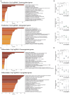
GO term analysis of differentially expressed, down- and up-regulated genes induced by the KO of mCrip2 (sgRNA3) and steady state mRNA expression of selected genes validated by qPCR in proliferating (A,B) and differentiating (C,D) myoblasts. Cut-off was set at 2.0 of the −log(adjusted P value). See Supp. Table 5 for the complete list of genes. N = 3 All values are reported as means ± SEM. Significance was determined by two-way ANOVA; *p < 0.05, **p < 0.01, ***p < 0.001 and ****p <0.0001 compared to control cells.
Figure 9.
Figure 9.. Depletion of mCrip2 prevents the Cu-dependent induction of metallothioneins.
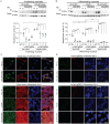
Representative western blot (upper panel) and quantification (bottom panel) of Metallothionein 1 (Mt) expression in control and mCrip2 KO proliferating (48 h; A) and differentiating (24 h; B) primary myoblasts. Gapdh was used as loading control. Representative confocal microscopy of Metallothionein 1 (Mt1, green) and mCrip2 (red) expression of empty vector (C) and the mCrip2 sgRNA3 myoblast clone (D) cultured under the same conditions as in A. Nuclei were stained with DAPI (blue). The images depicted are representative of 3 independent biological experiments. Size bar = 10 μm.

Similar articles

References

    1. Festa R. A., and Thiele D. J. (2011) Copper: an essential metal in biology. Current biology : CB 21, R877–883 - PMC - PubMed
    1. Fraústo da Silva J. J. R., and Williams R. J. P. (2001) The biological chemistry of the elements : the inorganic chemistry of life, Oxford University Press, Oxford
    1. Csiszar K. (2001) Lysyl oxidases: a novel multifunctional amine oxidase family. Progress in nucleic acid research and molecular biology 70, 1–32 - PubMed
    1. Moriya M., Ho Y. H., Grana A., Nguyen L., Alvarez A., Jamil R., Ackland M. L., Michalczyk A., Hamer P., Ramos D., Kim S., Mercer J. F., and Linder M. C. (2008) Copper is taken up efficiently from albumin and alpha2-macroglobulin by cultured human cells by more than one mechanism. American journal of physiology. Cell physiology 295, C708–721 - PMC - PubMed
    1. Chen L., Li N., Zhang M., Sun M., Bian J., Yang B., Li Z., Wang J., Li F., Shi X., Wang Y., Yuan F., Zou P., Shan C., and Wang J. (2021) APEX2-based Proximity Labeling of Atox1 Identifies CRIP2 as a Nuclear Copper-binding Protein that Regulates Autophagy Activation. Angew Chem Int Ed Engl 60, 25346–25355 - PubMed

Publication types