Review
. 2024 Apr 30;24(9):2865.
doi: 10.3390/s24092865.

Biosensor-Enhanced Organ-on-a-Chip Models for Investigating Glioblastoma Tumor Microenvironment Dynamics

Affiliations
Review

Biosensor-Enhanced Organ-on-a-Chip Models for Investigating Glioblastoma Tumor Microenvironment Dynamics

Gayathree Thenuwara et al. Sensors (Basel). .

Abstract

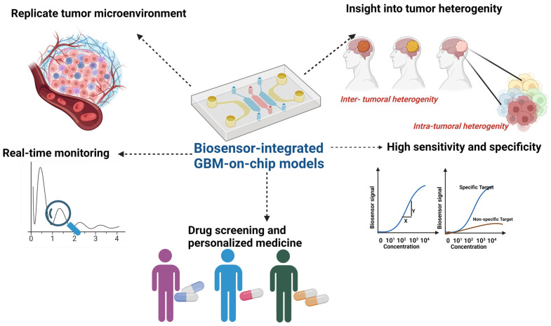
Glioblastoma, an aggressive primary brain tumor, poses a significant challenge owing to its dynamic and intricate tumor microenvironment. This review investigates the innovative integration of biosensor-enhanced organ-on-a-chip (OOC) models as a novel strategy for an in-depth exploration of glioblastoma tumor microenvironment dynamics. In recent years, the transformative approach of incorporating biosensors into OOC platforms has enabled real-time monitoring and analysis of cellular behaviors within a controlled microenvironment. Conventional in vitro and in vivo models exhibit inherent limitations in accurately replicating the complex nature of glioblastoma progression. This review addresses the existing research gap by pioneering the integration of biosensor-enhanced OOC models, providing a comprehensive platform for investigating glioblastoma tumor microenvironment dynamics. The applications of this combined approach in studying glioblastoma dynamics are critically scrutinized, emphasizing its potential to bridge the gap between simplistic models and the intricate in vivo conditions. Furthermore, the article discusses the implications of biosensor-enhanced OOC models in elucidating the dynamic features of the tumor microenvironment, encompassing cell migration, proliferation, and interactions. By furnishing real-time insights, these models significantly contribute to unraveling the complex biology of glioblastoma, thereby influencing the development of more accurate diagnostic and therapeutic strategies.

Keywords: biosensor; cellular dynamics; diagnostic strategies; glioblastoma; microenvironment; organ-on-a-chip; precision medicine; real-time monitoring; therapeutic approaches.


Conflict of interest statement

The authors declare no conflicts of interest.

Figures

Figure 1
Figure 1
Glioblastoma tumor microenvironment.
Figure 2
Figure 2
Dynamic interplay of immune cells in glioblastoma: facilitating tumor progression and evading immune surveillance (created with Biorender).
Figure 3
Figure 3
Crosstalk between astrocytes and glioblastoma cells in the tumor microenvironment (created with Biorender).
Figure 4
Figure 4
Extrinsic and intrinsic targets within glioblastoma (GBM) biology are vital for tumor-on-chip models. Extrinsic targets involve elements of the tumor microenvironment such as extracellular matrix (ECM) components, immune cells (e.g., macrophages and T cells), stromal cells (e.g., fibroblasts and endothelial cells), cytokines, chemokines, angiogenic factors, hypoxic conditions, acidic pH, extracellular vesicles, growth factors (e.g., VEGF and EGF), and matrix metalloproteinases (MMPs). Intrinsic targets include intra- and intercellular interactions/functions like oncogenic signaling pathways (e.g., EGFR, PI3K/AKT, and MAPK), tumor suppressor genes (e.g., p53 and PTEN), DNA repair mechanisms, epigenetic modifications (e.g., DNA methylation, histone modifications), cell cycle regulators (e.g., cyclins and CDKs), apoptotic pathways, cellular metabolism pathways, cancer stem cells (CSCs), drug efflux pumps (e.g., ABC transporters), and immune checkpoint molecules (e.g., PD-1 and CTLA-4). Incorporating these targets into GBM-on-chip models facilitates comprehensive studies elucidating their roles in tumor progression, drug response, and therapeutic resistance, contributing significantly to our understanding and treatment of GBM (created with Biorender).
Figure 5
Figure 5
Workflow for creating a glioblastoma tumor microenvironment in microfluidic chips. This figure illustrates the step-by-step process for preparing a glioblastoma tumor microenvironment using traditional microfluidic chips. Starting from chip design and fabrication, the workflow progresses through cell culture, seeding, preparation of extracellular matrix (ECM), fluidic control, introduction of chemical stimuli, imaging, analysis, and long-term culture and monitoring. Each step is crucial for accurately mimicking the complex tumor microenvironment and enables researchers to study tumor biology, drug responses, and therapeutic interventions in a controlled laboratory setting (created with Biorender).
Figure 6
Figure 6
Schematic representation of the 3D bioprinting process for creating the tumor microenvironment of glioblastoma. This illustration depicts the step-by-step procedure involved in 3D bioprinting to simulate the tumor microenvironment typical of glioblastoma. The process encompasses cell selection and culture, biomaterial preparation, bioprinting setup, and post-printing processes. Initially, relevant cell types, including glioblastoma cells, stromal cells, and endothelial cells, are cultured separately to achieve optimal confluence and viability. Subsequently, a suitable bioink, capable of supporting cell viability and mimicking the extracellular matrix composition, is chosen and combined with the cultured cells. The bioprinter is then set up, with the cell-laden bioink loaded into cartridges or syringes. Guided by computer-aided design models, the bioprinter deposits layers of bioink onto a substrate or scaffold in a controlled, layer-by-layer fashion. Printing parameters, such as nozzle size, speed, and pressure, are adjusted to ensure the desired resolution and cell viability. Post-printing processing involves allowing the construct to undergo crosslinking or gelation for stabilization, followed by culture under controlled conditions to promote tissue maturation. The resulting bioprinted model reflects the intricate architecture and cellular composition of the glioblastoma microenvironment, facilitating characterization and analysis for research and therapeutic applications (created with Biorender).
Figure 7
Figure 7
Biosensor detection of GBM biomarkers: sequential process. The process by which biosensors detect biomarkers associated with GBM begins with the recognition of specific biomarkers present within the GBM microenvironment. These biomarkers include proteins, nucleic acids, metabolites, extracellular vesicles, cell surface markers, angiogenic factors, and immune checkpoint molecules. Following biorecognition, a signal is either generated or modulated, which subsequently undergoes transmission and detection by the biosensor system. This systematic sequence of events, involving biorecognition, signal transmission, and detection, facilitates the identification and quantification of GBM biomarkers. Ultimately, this process yields valuable insights into GBM tumor biology, aiding in the assessment of diagnosis, prognosis, and treatment response (created with Biorender).
Figure 8
Figure 8
Integrated biosensors in GBM-on-chip models revolutionize glioblastoma research by enabling real-time monitoring of tumor dynamics and treatment responses. This schematic illustrates the multifaceted benefits of biosensor-integrated GBM-on-chip models, including the replication of tumor microenvironmental cues, real-time monitoring of biomarker expression and cellular responses, high sensitivity and specificity in biomarker detection, facilitation of drug screening and personalized medicine approaches, insights into tumor heterogeneity, and reduced reliance on animal models for preclinical studies. These advancements pave the way for improved understanding of GBM biology and the development of more effective therapeutic strategies (created with Biorender).

Similar articles

References

    1. Lin S., Xu H., Zhang A., Ni Y., Xu Y., Meng T., Wang M., Lou M. Prognosis Analysis and Validation of m6A Signature and Tumor Immune Microenvironment in Glioma. Front. Oncol. 2020;10:541401. doi: 10.3389/fonc.2020.541401. - DOI - PMC - PubMed
    1. Thenuwara G., Curtin J., Tian F. Advances in diagnostic tools and therapeutic approaches for gliomas: A comprehensive review. Sensors. 2023;23:9842. doi: 10.3390/s23249842. - DOI - PMC - PubMed
    1. Wen P.Y., Packer R.J. The 2021 WHO Classification of Tumors of the Central Nervous System: Clinical Implications. Neuro-Oncology. 2021;23:1215–1217. doi: 10.1093/neuonc/noab120. - DOI - PMC - PubMed
    1. Fisher J.P., Adamson D.C. Current FDA-Approved Therapies for High-Grade Malignant Gliomas. Biomedicines. 2021;9:324. doi: 10.3390/biomedicines9030324. - DOI - PMC - PubMed
    1. Lapointe S., Perry A., Butowski N.A. Primary brain tumours in adults. Lancet. 2018;392:432–446. doi: 10.1016/s0140-6736(18)30990-5. - DOI - PubMed

Grants and funding

This research received no external funding.

LinkOut - more resources