. 2024 Apr 23;25(9):4608.
doi: 10.3390/ijms25094608.

Herpes Simplex Virus ICP27 Protein Inhibits AIM 2-Dependent Inflammasome Influencing Pro-Inflammatory Cytokines Release in Human Pigment Epithelial Cells (hTert-RPE 1)

Affiliations

Herpes Simplex Virus ICP27 Protein Inhibits AIM 2-Dependent Inflammasome Influencing Pro-Inflammatory Cytokines Release in Human Pigment Epithelial Cells (hTert-RPE 1)

Anna Caproni et al. Int J Mol Sci. .

Abstract

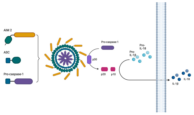
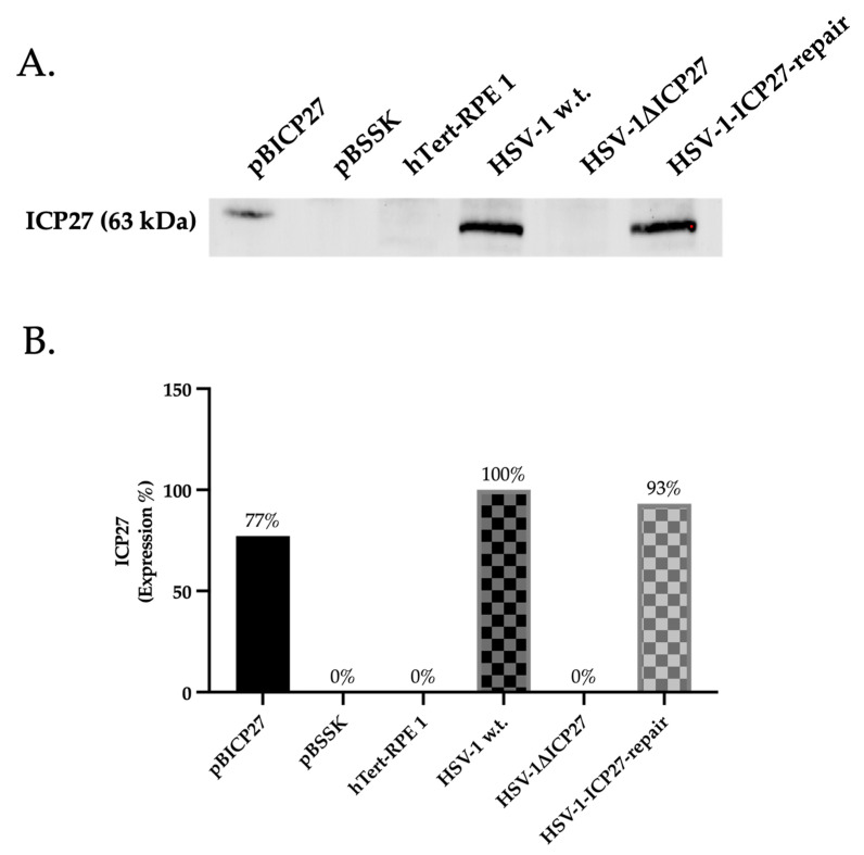
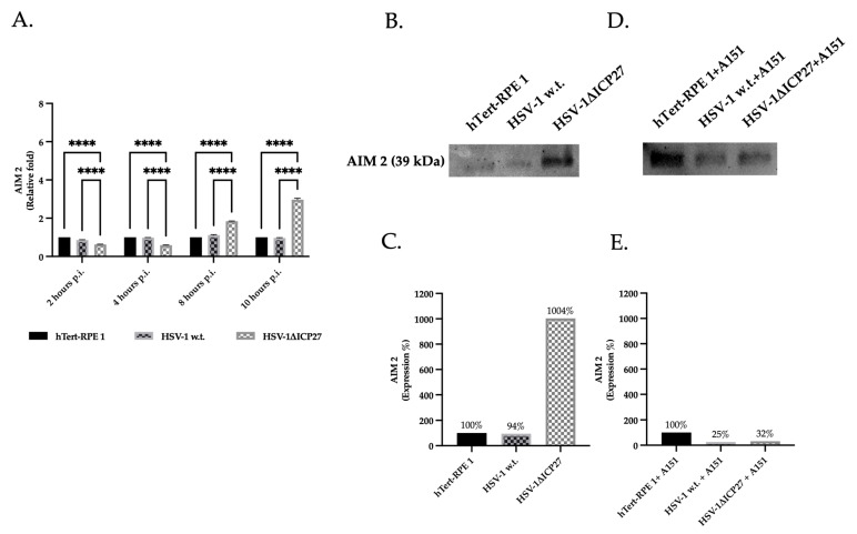
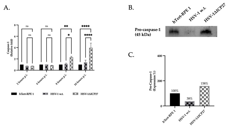
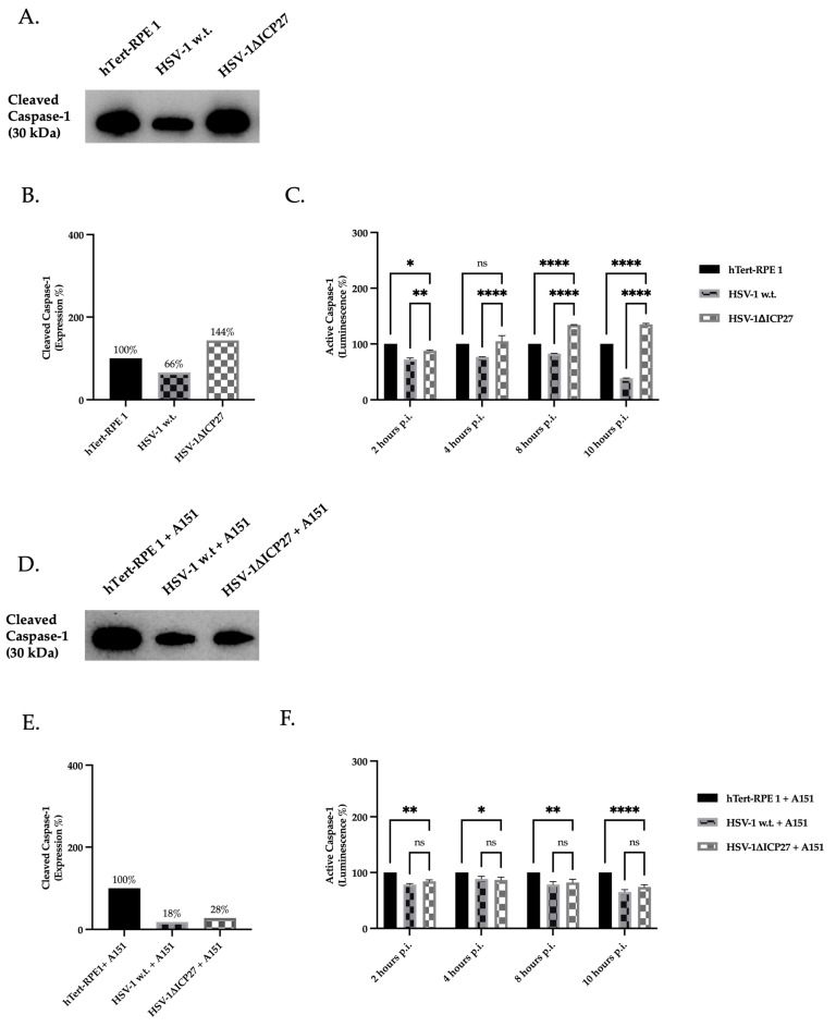
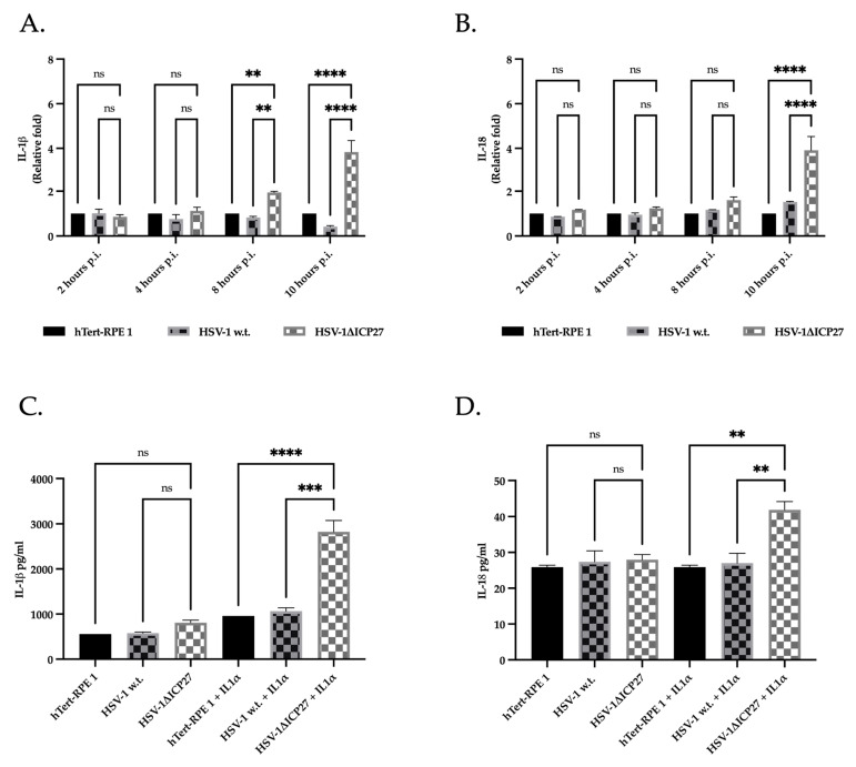
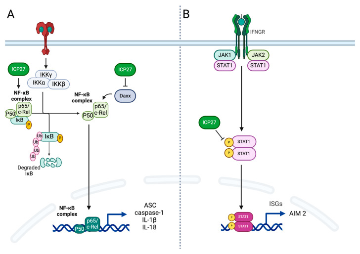
Although Herpes simplex virus type 1 (HSV-1) has been deeply studied, significant gaps remain in the fundamental understanding of HSV-host interactions: our work focused on studying the Infected Cell Protein 27 (ICP27) as an inhibitor of the Absent-in-melanoma-2 (AIM 2) inflammasome pathway, leading to reduced pro-inflammatory cytokines that influence the activation of a protective innate immune response to infection. To assess the inhibition of the inflammasome by the ICP27, hTert-immortalized Retinal Pigment Epithelial cells (hTert-RPE 1) infected with HSV-1 wild type were compared to HSV-1 lacking functional ICP27 (HSV-1∆ICP27) infected cells. The activation of the inflammasome by HSV-1∆ICP27 was demonstrated by quantifying the gene and protein expression of the inflammasome constituents using real-time PCR and Western blot. The detection of the cleavage of the pro-caspase-1 into the active form was performed by using a bioluminescent assay, while the quantification of interleukins 1β (IL-1β) and 18 (IL-18)released in the supernatant was quantified using an ELISA assay. The data showed that the presence of the ICP27 expressed by HSV-1 induces, in contrast to HSV-1∆ICP27 vector, a significant downregulation of AIM 2 inflammasome constituent proteins and, consequently, the release of pro-inflammatory interleukins into the extracellular environment reducing an effective response in counteracting infection.

Keywords: host–virus interaction; inflammasome; innate immunity; viruses.


Conflict of interest statement

The authors declare no conflicts of interest.

Figures

Figure 1
Figure 1
The oligomerization of AIM 2, ASC, and pro-caspase-1 proteins results in the formation of the heptosome after the AIM 2 sensor recognizes viral DNA that has been released into the cytoplasm. Proteins within the inflammasome aggregate, causing pro-caspase-1 to be processed into its temporary form, p30. The CARD domain (p10) is removed during self-cleavage of p30, releasing the active caspase p20. Interleukins 1β and 18 are converted into their active form by active caspase-1 and released into the extracellular milieu. The figure was generated using Biorender.com.
Figure 2
Figure 2
Evaluation of presence/absence of ICP27 protein. (A) Western blot assay for ICP27 protein in cells transfected with pBICP27 and pBSSK and in cells infected with HSV-1 w.t., HSV-1ΔICP27, and HSV-1-ICP27-repair at 24 h post-transfection or infection. (B) Densitometric quantification of ICP27 levels was performed using stain-free blotting, and the results were reported as a percentage calculated from the ratio of normalized protein levels to total protein (Supplementary Materials—Figures S1 and S2). Relative percentages correspond to the proportions of HSV-1 w.t. (100%) infected cells. Detection of total proteins and band was performed using the ChemidocTMMP Imaging System (Biorad, Segrate (MI), Italy) and normalization was performed using Image Lab Software (6.0.0, Biorad, Segrate (MI), Italy).
Figure 3
Figure 3
Gene and protein expression of AIM 2 sensor protein. (A) Real-time PCR of untreated hTert-RPE 1, HSV-1 w.t., and HSV-1ΔICP27-infected cells (M.O.I. of 3) at 2, 4, 8, and 10 h.p.i. Relative fold changes are related to the levels in untreated cells (hTert-RPE 1). Values are averages of SEM ± from three biological replicates. Two-way ANOVA was performed. ****, p < 0.0001. (B,D) Western blot assay for AIM 2 in both untreated (B) and pre-treated with AIM 2 inhibitor A151 (D) hTert-RPE 1, HSV-1 w.t., and HSV-1ΔICP27-infected cells (M.O.I. of 3) at 10 h.p.i. (C,E) Densitometric quantification of AIM 2 levels was performed using stain-free blotting, and the results were reported as a percentage calculated from the ratio of normalized protein levels to total protein (Supplementary Materials: Figures S3–S6). Relative percentages correspond to the proportions of untreated (100%) cells. Detection of both total protein (stain-free blot) and detected bands was performed using the ChemidocTMMP Imaging System (Biorad) and normalization was performed using Image Lab Software (Biorad).
Figure 4
Figure 4
Gene and protein expression of ASC adaptor protein. (A) Real-time PCR of untreated hTert-RPE 1, HSV-1 w.t., and HSV-1-ΔICP27-infected cells (M.O.I. of 3) at 2, 4, 8, and 10 h.p.i. Relative fold changes are related to the levels in untreated cells (hTert-RPE 1). Values are averages SEM ± from three biological replicates. Two-way ANOVA was performed. ns p ≥ 0.1, ***, p < 0.001. (B) Western blot assay for ASC (22 kDa) in both untreated hTert-RPE 1, HSV-1 w.t., and HSV-1ΔICP27-infected cells (M.O.I. of 3) at 10 h.p.i. (C) Densitometric quantification of ASC levels was performed using stain-free blotting, and the results were reported as a percentage calculated from the ratio of normalized protein levels to total protein (Supplementary Materials—Figures S7 and S8). Relative percentages correspond to the proportions of untreated (100%) cells. Detection of both total protein (stain-free blot) and detected bands was performed using the ChemidocTMMP Imaging System (Biorad) and normalization was performed using Image Lab Software (Biorad).
Figure 5
Figure 5
Gene and protein expression of pro-caspase-1. (A) Real-time PCR of untreated hTert-RPE 1, HSV-1 w.t., and HSV-1ΔICP27-infected cells (M.O.I. of 3) at 2, 4, 8, and 10 h.p.i. Relative fold changes are related to the levels in untreated cells (hTert-RPE 1). Values are averages SEM ± from three biological replicates. Two-way ANOVA was performed. ns p ≥ 0.1, * p < 0.1, ** p < 0.01, ****, p < 0.0001. (B) Western blot assay for pro-caspase-1 (45 kDa) in untreated hTert-RPE 1, HSV-1 w.t., and HSV-1ΔICP27-infected cells (M.O.I. of 3) at 10 h.p.i. (C) Densitometric quantification of pro-caspase-1 levels was performed using stain-free blotting, and the results were reported as a percentage calculated from the ratio of normalized protein levels to total protein (Supplementary Materials—Figures S9 and S10). Relative percentages correspond to the proportions of untreated (100%) cells. Detection of both total protein (stain-free blot) and detected bands was performed using the ChemidocTMMP Imaging System (Biorad), and normalization was performed using Image Lab Software (Biorad).
Figure 6
Figure 6
Protein expression and enzymatic activity of caspase-1. (A,D) Western blot assay for transient caspase-1 (30 kDa) in both untreated (A) and pre-treated with AIM 2 inhibitor A151 (D) hTert-RPE 1, HSV-1 w.t., and HSV-1ΔICP27-infected cells (M.O.I. of 3) at 10 h.p.i. (B,E) Densitometric quantification of caspase-1 levels was performed using stain-free blotting, and the results were reported as a percentage calculated from the ratio of normalized protein levels to total protein (Supplementary Materials–Figures S11–S14). Relative percentages correspond to the proportions of untreated (100%) cells. Detection of both total protein (stain-free blot) and detected bands was performed using the ChemidocTMMP Imaging System (Biorad) and normalization was performed using Image Lab Software (Biorad). (C,F) Bioluminescent assay for active caspase-1 in untreated (C) and pre-treated with AIM 2 inhibitor A151 (F) hTert-RPE 1, HSV-1 w.t., and HSV-1ΔICP27-infected cells (M.O.I. of 3) at 2, 4, 8, and 10 h.p.i. Values are average SEM ± from three biological replicates. Two-way ANOVA was performed. ns p ≥ 0.1, *, p < 0.1, **, p < 0.01, ****, p < 0.0001.
Figure 7
Figure 7
Gene expression and quantification of released IL-1β and IL-18. (A,B) Real-time PCR of untreated hTert-RPE 1, HSV-1 w.t., and HSV-1ΔICP27-infected cells (M.O.I. of 3) at 2, 4, 8, and 10 h.p.i for IL-1β (A) and IL-18 (B). Relative fold changes are related to the levels in untreated cells (hTert-RPE 1). Values are averages SEM ± from three biological replicates. Two-way ANOVA was performed. ns p ≥ 0.1, **, p < 0.01, ****, p < 0.0001. (C) ELISA assay for IL-1β in both untreated or stimulated with IL-1α hTert-RPE 1, HSV-1 w.t., and HSV-1ΔICP27-infected cells (M.O.I. of 3) at 10 h.p.i. Values are average SEM ± from three biological replicates. Two-way ANOVA was performed. ns p ≥ 0.1, ***, p < 0.001. ****, p < 0.0001. (D) ELISA assay for IL-18 in both untreated or stimulated with IL-1α hTert-RPE 1, HSV-1 w.t., and HSV-1ΔICP27-infected cells (M.O.I. of 3) at 10 h.p.i. Values are average SEM ± from three biological replicates. Two-way ANOVA was performed. ns p ≥ 0.1, **, p < 0.01.
Figure 8
Figure 8
Role of ICP27 protein in immune evasion: (A) ICP27 is required for the inhibition of NF-kB by stabilizing the NF-kB inhibitor IkB and by repressing the sumoylation of Daxx factor that consists of the inhibition of the transcriptional activity of NfkB essential for transcription of ASC, caspase-1, IL-1β, and Il-18 genes. (B) ICP27 downregulates STAT1 phosphorylation and prevents the accumulation of STAT1 in the nucleus, which prevents transcription of the AIM 2 gene.

Similar articles

References

    1. Thellman N.M., Triezenberg S.J. Herpes simplex virus establishment, maintenance, and reactivation: In vitro modeling of latency. Pathogens. 2017;6:28. doi: 10.3390/pathogens6030028. - DOI - PMC - PubMed
    1. Menendez C.M., Carr D.J.J. Defining nervous system susceptibility during acute and latent herpes simplex virus-1 infection. J. Neuroimmunol. 2017;308:43–49. doi: 10.1016/j.jneuroim.2017.02.020. - DOI - PMC - PubMed
    1. Zhu S., Viejo-Borbolla A. Pathogenesis and virulence of herpes simplex virus. Virulence. 2021;12:2670–2702. doi: 10.1080/21505594.2021.1982373. - DOI - PMC - PubMed
    1. Mustafa M., Illzam E.M., Muniandy R.K., Sharifah A.M., Nang M.K., Ramesh B. Herpes simplex virus infections, Pathophysiology and Management. IOSR J. Dent. Med. Sci. 2016;15:85–91. doi: 10.9790/0853-150738591. - DOI
    1. Kumar S.P., Chandy M.L., Shanavas M., Khan S., Suresh K.V. Pathogenesis and life cycle of herpes simplex virus infection-stages of primary, latency and recurrence. J. Oral. Maxillofac. Surg. Med. Pathol. 2016;28:350–353. doi: 10.1016/j.ajoms.2016.01.006. - DOI

MeSH terms

Grants and funding

This research received no external funding.

LinkOut - more resources