. 2024 May 7;55(1):58.
doi: 10.1186/s13567-024-01312-y.

The haemagglutinin-neuraminidase protein of velogenic Newcastle disease virus enhances viral infection through NF-κB-mediated programmed cell death

Affiliations

The haemagglutinin-neuraminidase protein of velogenic Newcastle disease virus enhances viral infection through NF-κB-mediated programmed cell death

Xiaolong Lu et al. Vet Res. .

Abstract

The haemagglutinin-neuraminidase (HN) protein, a vital membrane glycoprotein, plays a pivotal role in the pathogenesis of Newcastle disease virus (NDV). Previously, we demonstrated that a mutation in the HN protein is essential for the enhanced virulence of JS/7/05/Ch, a velogenic variant NDV strain originating from the mesogenic vaccine strain Mukteswar. Here, we explored the effects of the HN protein during viral infection in vitro using three viruses: JS/7/05/Ch, Mukteswar, and an HN-replacement chimeric NDV, JS/MukHN. Through microscopic observation, CCK-8, and LDH release assays, we demonstrated that compared with Mukteswar and JS/MukHN, JS/7/05/Ch intensified the cellular damage and mortality attributed to the mutant HN protein. Furthermore, JS/7/05/Ch induced greater levels of apoptosis, as evidenced by the activation of caspase-3/8/9. Moreover, JS/7/05/Ch promoted autophagy, leading to increased autophagosome formation and autophagic flux. Subsequent pharmacological experiments revealed that inhibition of apoptosis and autophagy significantly impacted virus replication and cell viability in the JS/7/05/Ch-infected group, whereas less significant effects were observed in the other two infected groups. Notably, the mutant HN protein enhanced JS/7/05/Ch-induced apoptosis and autophagy by suppressing NF-κB activation, while it mitigated the effects of NF-κB on NDV infection. Overall, our study offers novel insights into the mechanisms underlying the increased virulence of NDV and serves as a reference for the development of vaccines.

Keywords: NF-κB; Newcastle disease virus; haemagglutinin–neuraminidase; programmed cell death; viral infection.


Conflict of interest statement

The authors declare that they have no competing interests.

Figures

Figure 1
Figure 1
Schematic representation of the NDVs utilized in this study, along with the cytopathic damage induced by these NDVs. A A schematic representation illustrating the process of constructing the NDVs. The JS/MukHN strain is derived from JS/7/05/Ch and incorporates the Mukteswar-type HN gene through an NDV reverse genetics system. The HN proteins of the two parental viruses exhibited variations at six distinct aa sites: Asn 19 Ser, Ala 29 Thr, Met 145 Thr, Val 266 Ile, Ala 494 Asp, and Glu 495 Lys. Additionally, there is an aa mutation (Pro 438 Ser) in the NP protein in one of the parental viruses. The aa mutation in the NP protein of JS/7/05/Ch is indicated by an orange triangle, while its mutant HN protein is highlighted in an orange box. B The automated mode of SWISS-MODEL and PyMol 2.4.0 software were utilized to generate homology models of the HN proteins, employing a matching PDB file (ID: 1e8v.1. A) as previously described [20]. The model NDV strains express diverse HN proteins, and four differential aa sites (Met145Thr, Val266Ile, Ala494Asp, and Glu495Lys) between the prototypic and mutant HN proteins lead to modifications in the three-dimensional protein structure. The spatial locations of the four mutations are depicted in the zoom-in picture, and the distinct aa sites are emphasized using various colours. C The morphology of the infected CEFs was visualized under a microscope (100×). Scale bar: 50 μm. D The viability of infected CEF cells was determined with a CCK-8 detection kit. (E) LDH release levels were quantified in the cell culture supernatants of the NDV-infected cells. ***p < 0.001; ****p < 0.0001. The significance of the Mukteswar- and JS/MukHN-infected groups was compared to that of the JS/7/05/Ch-infected group.
Figure 2
Figure 2
Assessment of cell apoptosis triggered by NDVs. A The scatter plots depict the flow cytometric assessment of phosphatidylserine (PS) translocation following Annexin V and PI staining in both mock-infected and NDV-infected CEFs at 24 and 48 hpi. The 4 quadrants represent the number of cells in each quadrant. The Q4 quadrant signifies the early stages of apoptosis, while the Q2 quadrant represents late-stage apoptosis and necrosis. The Q3 quadrant is indicative of viable cells. B The effects of NDV infection on cell apoptosis were analysed by a DNA ladder assay. Agarose gel electrophoretic patterns showing DNA fragmentation in mock-infected and NDV-infected CEF cells at an MOI of 1. DNA ladders of approximately 180–200 bp of fragmented DNA were visualized by staining with ethidium bromide in a 1.0% agarose gel. C The effects of NDV infection on cell apoptosis were analysed by TUNEL assay. After the CEFs were subjected to mock infection or infection with NDV at an MOI of 1 for 12 or 24 h, a quantitative analysis of TUNEL-positive cells was conducted across groups based on a minimum of ten images for each well. The TUNEL-positive cell content was determined by quantifying the number of TUNEL-positive cells relative to the total number of CEFs. Scale bar: 100 μm. D The expression levels of apoptotic proteins were detected by Western blotting. Western blotting analysis was performed to determine the protein levels of pro-caspase-3, -8, and -9 in CEFs after mock infection or infection with NDV at an MOI of 1 for 24 h. The data were analysed using ImageJ and are presented as the ratio of pro-caspase protein to the specific ACTB protein. *p < 0.05; **p < 0.01; **** p < 0.0001; ns indicates no significance.
Figure 3
Figure 3
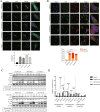
Evaluation of cell autophagy induced by NDVs. A Determination of autophagosome formation mediated by NDVs. CEF cells transfected with a GFP-LC3 plasmid were exposed to NDVs at an MOI of 1 for 24 h. Subsequently, the cells were examined using fluorescent microscopy to detect the GFP-LC3 puncta. A positive control was established through rapamycin treatment, while a negative control involved DMSO treatment. DAPI (blue) was used for nuclear DNA staining. Scale bar: 20 μm. B Determination of autolysosome formation mediated by NDVs. DF-1 cells, genetically modified to express the GFP-RFP-LC3 gene, were subjected to NDV infection at an MOI of 1 for 24 h. Autophagosomes, represented by orange puncta, and autolysosomes, indicated by red puncta, were observed using a confocal microscope. DAPI (blue) was utilized to stain nuclear DNA. Scale bar: 20 μm. The percentage of red puncta was calculated as follows: red puncta (%) = (number of red puncta/(number of red puncta + number of orange puncta)) × 100%. C Quantification of autophagy-related protein expression levels after NDV infection at different doses and for different durations. CEF cells were subjected to mock infection or infection with NDVs at multiple MOIs and time intervals. LC3-II lipidation and p62 degradation in cell lysates were evaluated by Western blotting. The data analysed with ImageJ are presented as the ratio of LC3-II to LC3-I and the ratio of p62 to ACTB. D The mRNA expression levels of autophagy-related genes in NDV-infected cells. CEF cells were infected with NDV at an MOI of 1 or mock infected for 24 h. Total RNA was extracted from the CEF cells, reverse transcribed, and subjected to qRT‒PCR to measure the relative mRNA expression of autophagy-related genes. *p < 0.05; **p < 0.01; ***p < 0.001; ****p < 0.0001.
Figure 4
Figure 4
Impact on cell viability and virus replication after inhibition of apoptosis and autophagy in NDV-infected cells. CEF cells were incubated with either Z-VAD-FMK (20 µM) for A and B or CQ (50 µM) for C and D, while DMSO served as the control. After a 1 h incubation period, the cells were exposed to NDV at an MOI of 1. Subsequently, the cell lysates and supernatants were analysed to assess cell viability (A and C) and viral titres (B and D) at 12, 24, 36, and 48 hpi. *p < 0.05; **p < 0.01; ***p < 0.001; ****p < 0.0001.
Figure 5
Figure 5
Assessment of NF-κB activity triggered by NDVs. A NF-κB activity induced by NDVs was assessed using a dual luciferase reporter assay system. CEF cells were co-transfected with luciferase plasmids in 24-well plates, with the Renilla luciferase plasmid serving as a mock control. Subsequently, the transfected cells were exposed to NDVs at an MOI of 1 for 24 h. The relative NF-κB activity in the mock group was considered to be 1. B Analysis of the nuclear translocation efficiency of the NF-κB p65 protein. CEF cells were infected with NDVs at an MOI of 1 for 24 h in 12-well plates, and the proteins were then extracted from the cytoplasm and nucleus. The extracted proteins were examined for NF-κB p65 protein levels through Western blot analysis. The cytoplasmic and nuclear proteins were quantified by the GAPDH and histone H3 proteins, respectively. The data analysed with ImageJ are presented as the ratio of p65 to GAPDH in the cytoplasm and the ratio of p65 to histone H3 in the nucleus. Muk indicates Mukteswar, JS indicates JS/7/05/Ch, and JM indicates JS/MukHN. C and D Assessment of IκB-α protein expression levels after NDV infection in a time- and dose-dependent manner. CEF cells were exposed to NDVs at multiple time intervals (0, 12, 18, and 24 h) and MOIs (0, 0.01, 0.1, and 1 MOI). Then, the protein expression levels of IκB-α were examined by Western blotting. The data analysed with ImageJ are presented as the ratio of IκB-α to ACTB. ***p < 0.001.
Figure 6
Figure 6
Effects on apoptosis, autophagy, and viral infection after inhibition of NF-κB in NDV-infected cells. The NF-kB inhibitor PDTC was used to inhibit NF-kB activity. After preincubation of CEF cells with 60 μM PDTC for 1 h, the cells were infected with NDVs at an MOI of 1 for the indicated time. DMSO treatment served as the control. A The cell lysate was collected for the determination of p62 degradation, LC3-II/I conversion, and caspase-3 activation at 24 hpi. The data analysed with ImageJ are presented as the ratios of p62 to ACTB, LC3-II to LC3-I, and pro-caspase-3 to ACTB. Muk indicates Mukteswar, JS indicates JS/7/05/Ch, and JM indicates JS/MukHN. B The cell supernatants were analysed to assess viral titres at 12, 24, 36, and 48 hpi through a TCID50 assay. C The cell lysates were analysed to assess cell viability at 12, 24, 36, and 48 hpi through a CCK-8 assay. *p < 0.05; **p < 0.01.
Figure 7
Figure 7
Schematic diagram illustrating how the mutant HN protein modulates cell apoptosis and autophagy through the NF-κB pathway during NDV infection. When NDV harbouring the prototypic HN protein infects cells, it can trigger effective degradation of the IκB-α protein, facilitating p65/RelA nuclear translocation and activating NF-κB. This process attenuates cell apoptosis and autophagy but boosts virus replication and cell survival. Conversely, the mutant HN protein weakens the degradation of IκB-α and the nuclear translocation of p65/RelA, thereby inhibiting NF-κB activation during NDV infection. This decrease in NF-κB activation leads to increased apoptosis and autophagy. The mutant HN protein amplifies the effects of apoptosis and autophagy on virus replication and cell survival while impeding the adverse impacts of NF-κB on virus replication and cell survival.

Similar articles

References

    1. Yan C, Liu H, Jia Y, Prince-Theodore DW, Yang M, Addoma Adam FE, Ren J, Cao X, Wang X, Xiao S, Zhang S, Yang Z. Screening and mechanistic study of key sites of the hemagglutinin-neuraminidase protein related to the virulence of Newcastle disease virus. Poult Sci. 2020;99:3374–3384. doi: 10.1016/j.psj.2020.04.014. - DOI - PMC - PubMed
    1. Amarasinghe GK, Ayllon MA, Bao Y, Basler CF, Bavari S, Blasdell KR, Briese T, Brown PA, Bukreyev A, Balkema-Buschmann A, Buchholz UJ, Chabi-Jesus C, Chandran K, Chiapponi C, Crozier I, de Swart RL, Dietzgen RG, Dolnik O, Drexler JF, Durrwald R, Dundon WG, Duprex WP, Dye JM, Easton AJ, Fooks AR, Formenty PBH, Fouchier RAM, Freitas-Astua J, Griffiths A, Hewson R, et al. Taxonomy of the order Mononegavirales: update 2019. Arch Virol. 2019;164:1967–1980. doi: 10.1007/s00705-019-04247-4. - DOI - PMC - PubMed
    1. Anis Z, Morita T, Azuma K, Ito H, Ito T, Shimada A. Histopathological alterations in immune organs of chickens and ducks after experimental infection with virulent 9a5b newcastle disease virus. J Comp Pathol. 2013;149:82–93. doi: 10.1016/j.jcpa.2012.09.011. - DOI - PubMed
    1. Xu X, Qian J, Qin L, Li J, Xue C, Ding J, Wang W, Ding W, Yin R, Jin N, Ding Z. Chimeric Newcastle disease virus-like particles containing DC-binding peptide-fused haemagglutinin protect chickens from virulent Newcastle disease virus and H9N2 Avian influenza virus challenge. Virol Sin. 2020;35:455–467. doi: 10.1007/s12250-020-00199-1. - DOI - PMC - PubMed
    1. Mirza AM, Deng R, Iorio RM. Site-directed mutagenesis of a conserved hexapeptide in the paramyxovirus hemagglutinin-neuraminidase glycoprotein: effects on antigenic structure and function. J Virol. 1994;68:5093–5099. doi: 10.1128/jvi.68.8.5093-5099.1994. - DOI - PMC - PubMed